Liver Research

Open journal

ISSN 2379-4038

Non-Alcoholic Fatty Liver Disease: The Effect of Bile Acids and Farnesoid X Receptor Agonists on Pathophysiology and Treatment

Quratulain Khalid, Ian Bailey and Vinood B. Patel*

Vinood B. Patel, PhD, PGCertHE, FHEA

Senior Lecturer in Clinical Biochemistry, Department of Biomedical Sciences, Faculty of Science and Technology, University of Westminster
London W1W 6UW, UK; Tel. 0207 911 5000; E-mail: v.b.patel@westminster.ac.uk

INTRODUCTION

Bile acids produced in the liver as an end product of cholesterol catabolism were originally categorised as physiological detergents that facilitated the metabolism of dietary lipids and lipid soluble vitamins (A, D, E and K), and the hepatobiliary secretion of endogenous metabolites and xenobiotics.1-4 However, recent interest in bile acids over the past few years has shed new light on their roles in both the synthetic and regulatory metabolic pathways, pertaining to lipid, carbohydrate and cholesterol regulation acting as indispensable signalling molecules co-ordinating these network of biological processes.5

Whilst bile acids (Chenodeoxycholic acid (CDCA), Deoxycholic acid (DCA), Lithocholic acid (LCA), Cholic Acid (CA)) can negatively feedback their own production,6 they can also act as endogenous ligands for nuclear receptors to facilitate this regulation.3,4,7 The nuclear receptor, Farnesoid X Receptor (FXR; NR1H4) was the first bile acid receptor discovered,8 followed by other nuclear receptors in the NR1I subfamily, namely Constitutive Androstane Receptor (CAR; NR1I3), Pregnane X Receptor (PXR; NR1I2) and Vitamin D Receptor (VDR; NR1I1).4,9

In terms of nuclear receptor activation, PXR and VDR are stimulated by lithocholic acid (EC50 of approximately 100 nM), which is a hydrophobic bile acid derived from the 7-dehydroxylation of CDCA by intestinal bacteria.2,4 FXR though can be stimulated to varying degrees by most bile acids (CDCA>LCA=DCA>CA), but with the highest potency by CDCA, with an EC50 of approximately 10 µM.2 Conversely, constitutive androstane receptor is not triggered directly by bile acids but nonetheless is vital for controlling detoxification and transport of bile acids.10,11 Of all the nuclear receptors, VDR is expressed broadly across different tissue types. However, FXR and PXR are found abundantly, mainly in tissues in direct contact with bile acids for example in the intestine and liver.2

FXR is now considered as a “master regulator of bile acid metabolism” as it is involved in all phases of the biosynthetic pathway,2 affecting gene expression of ileal bile acid binding protein, small heterodimer partner, phospholipid transfer protein, ABC transporters and apolipoprotein C-II.2,12-14 Activation of these nuclear receptors leads to a reduction in bile acid synthesis, promotion of lipid oxidation, drug metabolism and transport, as well as affecting cholesterol metabolism.14 Conversely dysregulation of bile acid metabolism has a significant impact on inflammatory and metabolic disorders, such as Non-alcoholic fatty liver disease (NAFLD), diabetes, and obesity.2,11,15 This review aims to highlight recent advances in bile acid nuclear receptor activation, as well as the therapeutic potential of bile acids and their derivatives for the treatment of NAFLD.

BILE ACID SYNTHETIC PATHWAYS

The metabolism of bile acids is tightly controlled, where approximately 95% of the 3g bile acid body pool that is secreted into the intestine is reabsorbed via the enterohepatic circulation, with a small amount excreted in the faeces (200-600 mg/day). Bile acids that are lost are replaced by denovo hepatic synthesis5 derived from cholesterol catabolism (approximately 500 mg).16

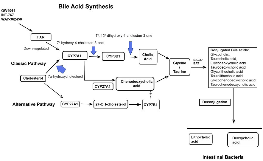
Hepatic bile acid synthesis begins from cholesterol catabolism involving a 17 step enzymatic pathway.2,14,16 The synthesis of bile acids is a complex process catalysed by several cytochrome P450 enzymes2 and involves two major bile acid biosynthetic pathways, the classic and alternative pathway.4 In the classic pathway, cholesterol is converted to 7α-hydroxycholesterol by the microsomal cytochrome P450 enzyme, Cholesterol 7α-hydroxylase (CYP7A1), which is also the rate-limiting step.7 In humans, the immediate by product of these pathways are the primary bile acids, Cholic Acid (CA) and Chenodeoxycholic acid (CDCA).14 The proportion of CA to CDCA synthesised is approximately equal and is regulated by the microsomal enzyme sterol 12-hydroxylase (CYP8B1) (Figure 1). The alternative pathway is carried out by mitochondrial sterol 27-hydroxylase (CYP27A1). This pathway occurs in the liver, macrophages and several tissue types in the body and predominantly synthesise CDCA.17 The import of cholesterol to the inner mitochondrial membrane, via soluble cholesterol binding protein is thought to be the rate-limiting step in the alternative pathway.18

Figure 1: Bile acid synthesis.

LROJ-1-106Fig 1

Catabolism of cholesterol produces the primary bile acids Cholic Acid (CA) and Chenodeoxycholic acid (CDCA) through one of two pathways in the liver. Key regulated enzymes in both pathways are shown. In the classic pathway, Cholesterol-7α-hydroxylase (CYP7A1) catalyses the first rate-limiting step to convert cholesterol to 27-hydroxycholesterol. However, sterol 27α-hydroxylase (CYP27A1) initiates the alternative pathway. Oxysterols, such as sterol 7α-hydroxylase (CYP7B1) produced in peripheral tissues are transported to hepatocytes and converted to CDCA and CA in the alternative pathway. CA synthesis is tightly regulated by 7α-hydroxylase (CYP8B1) in the classic pathway. In the intestine, CA and CDCA are conjugated with Glycine (G) or Taurine (T) by the enzymes Bile Acid Transferase (BAT) and Bile acid coenzyme A synthase (BACS). Some conjugated bile acids such as glycocholic acid, taurocholic acid, glycodeoxycholic acid, taurodeoxycholic acid, glycolithocholic acid, taurolithocholic acid, glycochenodeoxycholic acid and taurochenodeoxycholic acidare de-conjugated and subsequently dehydroxylated at the 7α-position by bacterial enzymes and converted to the secondary bile acids, Deoxycholic acid (DCA) and Lithocholic acid (LCA) respectively. Farnesoid X receptor is activated by agonists such as GW4064, INT-767 and WAY-362450 leading to reduced expression of CYP7A1 and lower levels hepatic bile acids.2,4,5

Subsequently, bile acids are conjugated to amino acids, typically taurine or glycine via the enzymes bile acid transferase and bile acid coenzyme A synthase, with the proportion of glycine to taurine conjugates estimated as 3 to 1.17,19 Conjugation of bile acids is an important process as it prepares the bile acids for effective detoxification, enhances their amphipathicity and solubility properties, which consequently leads to impermeability to cell membranes and reduced bile acid toxicity. However, some conjugated bile acids are deconjugated in the intestine by anaerobic bacteria converting CA and CDCA via 7α-dehydroxylase to the secondary bile acids, Lithocholic acid (LCA) and Deoxycholic acid (DCA), respectively (Table 1). Both of these bile acids are thought to have toxic properties, with DCA causing colon cancer for example.20 Whilst most DCA is reabsorbed in the colon and LCA is excreted in faeces, around 4% of LCA is transported to the liver where it is conjugated by amidation and sulfation and subsequently excreted into bile. Furthermore, sulfation of hydrophobic bile acids by sulfotransferase 2A1 is a key route for bile acid detoxification.17,19

Table 1: Principle Bile Acids.

Primary

Secondary

Conjugated

Cholic acid (CA)

Deoxycholic acid (DCA)

Glycocholic acid

Chenodeoxycholic acid (CDCA)

Lithocholic Acid (LCA) Urosdeoxycholic acid (UDCA)

Taurocholic acid Glycodeoxycholic acid Taurodeoxycholic acid Glycolithocholic acid Taurolithocholic acid

Glycochenodeoxycholic acid

Taurochenodeoxycholic acid

Main bile acids found in the liver and intestine.21,22

BILE ACID TRANSPORT IN THE ENTEROHEPATIC CIRCULATION

Increased bile acid concentration in the hepatocytes is the driving force of phosphatidylcholine, cholesterol and bile acid transport through the basolateral membrane, and removal of bile acids at the canalicular membrane into the biliary system.23 Conjugated bile acids are actively transported by the canalicular bile salt export pump and stored in the gallbladder.24,25 Furthermore, mutations in the bile salt export pump gene leads to progressive familial cholestatic syndrome, with the accumulation of toxic hydrophobic bile acids leading to cirrhosis in some cases.26 The proportion of these molecules, phospholipids, cholesterol and bile acids is tightly controlled by forming mixed micelles in the bile to (i) boost cholesterol solubility, (ii) decrease bile acid toxicity in the bile duct, and (iii) during digestion facilitate the uptake of nutrients into enterocytes. Excess cholesterol and/or hydrophobic bile salts causes saturated bile accumulation which can consequently lead to the formation of cholesterol gallstones in the biliary system and gall bladder.

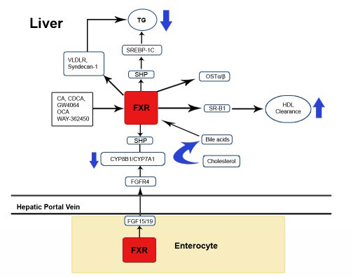
At the brush border membrane of the ileum 95% of bile acids are actively reabsorbed by the sodium dependent transporter. Upon absorption, bile acids bind to the ileal bile acid binding protein and diffuse across the basolateral membrane for release into the blood via the heterodimeric organic solute transporters.27 Completing this cycle, uptake of reabsorbed bile acids to the liver is mediated by Na+ dependent taurocholate cotransport peptide located in the sinusoidal membrane. Sinusoidal membranes function to efflux bile acids into the circulatory system and therefore express bile acid efflux transporters such as multidrug resistance protein.4 During cholestasis it has been recognised that these sinusoidal membranes are triggered and could play a fundamental role in the protection of liver injury when bile acids accumulate excessively in hepatocytes.2 Recent evidence suggests bile acids have the ability to initiate the production of the metabolic hormone fibroblast growth factor 15/19 (FGF-15 in mice, FGF-19 homolog in humans) in the ileocyte through the action of a functional FXR domain. Fibroblast growth factor 15/19 is transported to the liver where it subsequently binds to its cognate tyrosine kinase receptor. This results in the activation of the c-Jun N-terminal kinases 1/2 signalling pathway and down-regulation of CYP7A1 and bile acid synthesis (Figure 2). Interestingly, it is reported that cognate tyrosine kinase receptor β-klotho null mice have raised CYP7A1 mRNA levels and a larger bile acid pool, and that bile acids and cytokines (TNF-α) are also capable of triggering the c-Jun N-terminal kinases 1/2 signalling pathway and as a result down-regulate CYP7A1 Mrna.28

Figure 2: Overview of FXR regulation.

LROJ-1-106Fig2

Bile acids Cholic Acid (CA) and cChenodeoxycholic acid (CDCA) and FXR agonists such as GW4064, OCA and WAY-362450 activate hepatic and intestinal FXR to regulate-genes vital for bile acid metabolism. The activation of hepatic FXR lowers plasma cholesterol and Triglyceride (TG) synthesis via several pathways. For instance Short Heterodimer Protein (SHP)-dependent inhibition of Sterol Regulatory Element Binding Protein-1c (SREBP-1c) leads to the suppression of hepatic TG. Furthermore FXR suppresses bile acid synthesis by reducing CYP7A1 and CYP8B1 expression via SHP. Stimulation of Very Low Density Lipoprotein Receptor (VLDLR) and syndecan-1 also promotes the clearance of TG lipoproteins, and similarly Scavenger receptor class B1 (SR-B1) promotes the clearance of High Density Lipoprotein (HDL) in the liver. In the enterocytes bile acids bind to FXR and as a result elevate the expression of two transporters, organic solute transporter alpha and beta (OSTα, OSTβ) that facilitate bile acid transport into the hepatic portal vein. Induction of intestinal FXR increases the expression of fibroblast growth factor 15/19 (FGF15/19) into the hepatic portal vein. The subsequent binding of FGF15/19 to the hepatic cell-surface receptor fibroblast growth factor receptor 4 (FGFR4) triggers the JNK pathway leading to the suppression of CYP7A1 and CYP8B1 hence a decrease in bile acid synthesis.3,29

THERAPEUTIC APPROACH OF BILE ACIDS FOR NON-ALCOHOLIC FATTY LIVER DISEASE

At present, NAFLD is the most common form of chronic liver disease worldwide and is generally associated with clinical features of the metabolic syndrome.30 The incidence is significantly increased in diabetics (up to 63%) and in the morbidly obese.31 Given the epidemic of obesity attributable to the content of fat in modern diet, it is estimated that 42 million children were obese in 2010 and this figure is expected to rise to almost 60 million by 2020.32 These projections foresee a continued worsening trend in obesity and chronic liver disease, thus alternative therapeutic options are needed.

Generally the spectrum of liver pathology covers steatosis to the more severe conditions Non-alcoholic steatohepatitis (NASH), fibrosis and cirrhosis.33 Although the precise molecular mechanisms underlying the progression of NAFLD remains unclear,34 the accumulation of triglycerides is proposed in the early stages of NAFLD/NASH, whereas insulin resistance, oxidative stress and inflammation are all important contributing factors in disease progression.35,36 Inflammation is characterised by c-Jun N-terminal kinases activation34 and reactive oxygen species production derived from the metabolism of excessive free fatty acids via microsomal cytochrome P450A oxidation, peroxisomal β-oxidation, and hepatic mitochondrial dysfunction.37,38

To date there are two major categories of NAFLD therapies: (i) lifestyle intervention (weight loss, physical exercise) and (ii) pharmaceutical therapies (insulin sensitizers, lipid-lowering agents).33 Considering insulin resistance plays a core role in the pathogenesis of NASH, glitazones and peroxisome proliferator-activated receptor γ agonists used in the treatment of type 2 diabetes have been comprehensively studied.39 In particular, Thiazolidinedione (TZD), an insulin sensitizer has been shown to improve hepatic biochemical and histological parameters in patients.40 Similarly, treatment with the TZD pioglitazone led to a reduction in aspartate aminotransferase (AST) levels by 30- 58%, improved hepatic insulin sensitivity, and reversed steatosis in NASH patients.39 TZDs also reduced the expression of genes linked with inflammation such as interleukin-6 and TNF-α, and possessed regulatory properties through proliferator-activated receptor γ activation of adipokines, the latter possessing crucial roles in the pathogenesis of NAFLD.41 Furthermore, in diabetic patients with NASH, a phase II double blind trial demonstrated that pioglitazone significantly decreased steatosis, inflammation and ballooning necrosis.42

Thiazolidinedione (TZD), an insulin sensitizer has been shown to improve hepatic biochemical and histological parameters in patients.40 Similarly, treatment with the TZD pioglitazone led to a reduction in aspartate aminotransferase (AST) levels by 30- 58%, improved hepatic insulin sensitivity, and reversed steatosis in NASH patients.39 TZDs also reduced the expression of genes linked with inflammation such as interleukin-6 and TNF-α, and possessed regulatory properties through proliferator-activated receptor γ activation of adipokines, the latter possessing crucial roles in the pathogenesis of NAFLD.41 Furthermore, in diabetic patients with NASH, a phase II double blind trial demonstrated that pioglitazone significantly decreased steatosis, inflammation and ballooning necrosis.42

However, there is variable data from TZDs studies regarding regression of fibrosis.39 A meta-analysis showed that pioglitazone, but not rosiglitazone reduced fibrosis.43 However, a large trial comparing ‘Pioglitazone versus Vitamin E for the treatment of non-diabetic patients with NASH’ showed improved histology in NASH patients who received pioglitazone but no significant improvement in fibrosis stage for either treatment.44 Despite the promising results of these pioglitazone studies, the adverse effects of TZD such as weight gain, increased bladder cancer risk and cardiovascular morbidity has limited the use of pioglitazone in patients with NASH.45 Although vitamin E (400 IU/day) demonstrated some benefit to NASH subjects as shown by reduced inflammation,44,46 other reports suggest vitamin E is associated with cardiac morbidity including heart failure and haemorrhagic stroke.47

Thus targeting insulin resistance and oxidative stress is crucial but not adequate for effectively treating NASH, signifying the need for wider hepato-protective agents. Emerging evidence suggests beneficial properties of bile acids and their derivatives in the treatment of NAFLD by regulating lipid and glucose pathways, reducing inflammation and lowering hepatic triglyceride levels.3 Agonists have been developed targeting FXR receptors as a therapeutic approach in NAFLD as discussed below.

ROLE OF FXR IN NAFLD

Evidence of the vital role that FXR plays in NAFLD has been observed in FXR null mice. Here FXR null mice present with hepatic steatosis, inflammation, elevated bile acid levels, hyperlipidaemia and fibrosis.48 Furthermore, in FXR deficient mice fed a 1% cholesterol diet, mice exhibited severe muscle wastage, raised hepatic cholesterol content and a 23-fold greater increase in hepatic triglyceride. Additionally, excessive levels of bile acids correlated to 30% mortality in FXR deficient mice by day 7, which was attributable to liver failure.49 Gut derived lipopolysaccharide also plays a crucial role in the pathogenesis of NASH.50 Increased lipopolysaccharide stimulates nuclear factor kappa β which acts to recruit inflammatory cells, thus promoting inflammation, fibrosis and carcinogenesis in advanced NAFLD.51 In FXR null mice, inflammation was characterised by increased TNF-α and interleukin-1β levels, high circulating and hepatic bile acid levels as well as spontaneous hepatocellular carcinoma at 12 months.52 Although these studies indicate a causal link between elevated bile acids and inflammation, FXR activation has been shown to suppress the nuclear factor kappa β pathway, as well as the inflammatory cytokines and cyclooxygenase-2 in hepatocytes.53

FXR activation also regulates carbohydrate metabolism.54 Administration of cholic acid to C57BL/6J mice led to a decrease in fasting glucose concentration and reduced expression of the phosphoenolpyruvate carboxykinase gene in the liver, and also in HepG2 cells incubated with chenodeoxycholic acid.36,55 Other studies found that administration of the FXR agonist GW4064 to streptozotocin-induced diabetic rat or adenovirusmediated activation of hepatic FXR reduced phosphoenolpyruvate expression and reversed hyperglycaemia and normalised glycogen storage.55,56 Thus, FXR is thought to regulate gluconeogenesis through its key enzyme phosphoenolpyruvate carboxykinase.57 These studies suggest activated FXR ameliorates lipid and glucose metabolism and prevents inflammation, thus such properties of FXR make it a novel therapeutic approach in NAFLD treatment.

FXR AGONISTS IN NAFLD AND NASH

A variety of studies have targeted FXR to modulate the metabolism of lipids, carbohydrates and bile acids in NASH,58 with the synthetic agonist GW4064 widely studied. Here, FXR deficient mice show both systemic and hepatic insulin resistance, which can be normalised by giving the FXR agonist GW4064.59 In a diabetic mouse model lacking leptin receptors, treatment with GW4064 for 5 days led to a substantial decrease in the plasma levels of glucose and triglycerides.58 Whereas administration of GW4064 normalised steatosis and serum triglycerides levels in aged mice possibly due to the reduction in endoplasmic reticulum stress.60 Finally, GW4064 reduced bile acid synthesis, and increased bile acid export in a model of cholestasis.61 In alternate models of NASH, C57BL/6 mice fed a methionine and choline deficient diet and treated with the FXR agonist WAY-362450 for 4 weeks showed a decline in serum AST and ALT levels, improved liver histology and decreased inflammatory cell infiltration and fibrosis.29 These studies indicate that FXR agonists may be useful for the treatment of NASH and related liver disorders by normalising carbohydrate, lipid and bile acid metabolism.

Obeticholic acid (OCA) is a synthetic bile acid analogue, also known as INT-747 and the 6α-ethyl derivative of CDCA, and was originally identified in 2002 for its hepatoprotective and anti-cholestatic properties in a model of cholestasis, protecting hepatocytes against acute necrosis triggered by LCA.62 Interestingly, OCA synthesised through the addition of the ethyl group to CDCA exhibits 100-fold greater agonistic activity than CDCA and this potency has been confirmed by the analysis of the co-crystal structure of the FXR ligand binding domain.63

OCA has been studied in a rabbit model of the metabolic syndrome and in the Zucker rat model of obesity where administration of OCA improved glucose and insulin tolerance and decreased steatohepatitis.64,65 Furthermore, FXR activation by OCA decreased hepatic expression of genes involved in fatty acid synthesis including sterol regulatory element binding protein-1, reduced TNF-α levels and elevated peroxisome- proliferator activated receptor alpha expression, which therefore led to an improvement in the NASH phenotype.53,65 OCA therapy also reduced inflammation in a FXR deficient model of autoimmune hepatitis and prevented hepatic stellate cell activation by inhibiting osteopontin production.66,67 In the thioacetamide rat model of fibrosis, OCA prevented fibrosis progression, reversed fibrosis and cirrhosis development, and significantly reduced portal hypertension.53 Therefore, these preclinical findings have established the anti-inflammatory and anti-fibrotic properties of OCA mediated by FXR, as a candidate agent for NASH/NAFLD treatment.

FXR AGONISTS IN CLINICAL STUDIES

To date there are only a handful of studies investigating FXR agonists in NAFLD. A small study of patients with type II diabetes and NAFLD showed a marked improvement in insulin sensitivity of 28% when given 25 mg of OCA for a short 6 week period.68 Furthermore, endogenous levels of bile acid decreased, as well as markers of inflammation and fibrosis.68 However, the largest clinical trial to date ‘Farnesoid X receptor ligand Obeticholic Acid in Non-Alcoholic Steatohepatitis’ (FLINT) study has been partially reported in NASH patients receiving oral OCA 25 mg for 72 weeks.69 Preliminary findings indicated a 45% improvement in liver histology compared to 21% of the placebo group.69 Similar to the previous study in diabetics, lower levels of ALT in the group were noted as well as an increase in serum alkaline phosphatase, despite decreased gamma-glutamyl transferase levels. Pruritus an adverse effect of OCA treatment was observed in 23% of the OCA group compared with 6% of the placebo group, thus these OCA treated patients may require symptom management.69 Despite these promising results longer term clinical studies are required to explore the impact of OCA as well as other FXR agonists in the treatment of NASH/ NAFLD. There is some evidence indicating that OCA treatment leads to a significant increase (~20%) in LDL-cholesterol levels, which was apparent in the FLINT study,69 the earlier study on diabetes and NAFLD66 and in the treatment of diarrhoea.70 Authors from the FLINT study suggested that this atherosclerosis risk requires careful monitoring and OCA treatment withdrawn in these cases. This clearly may have a negative impact for the long term usage of OCA, despite the clinical benefit on liver histology parameters. One of the original bile acids used clinically for NASH was ursodeoxycholic acid (UCDA). This has been comprehensively reviewed elsewhere,71,72 but earlier studies did not report an improvement in liver histology or liver enzymes after 2 years of treatment at a low dose of 13-15 mg/kg73 or 6 months at a high dose of 28-32 mg/kg,74 but was effective at a high dose after 1 year of treatment.75 Whilst the long term benefit of UCDA requires confirmation, a taurine conjugate of UCDA called tauroursodeoxycholic acid may have potential use, as this has shown to prevent NASH progression by decreasing endoplasmic reticulum stress.76

ALTERNATIVE THERAPIES

Alternative treatment options may offer immediate solutions such as exercise/weight loss programmes, insulin sensitizers33 or bariatric surgery, which has proven to reduce steatosis, inflammation and moderate amounts of fibrosis in NAFLD patients.33,77-79 However, like OCA, the long term benefit of bariatric surgery still requires investigation.

CONCLUSION

Novel therapeutic approaches to manage the chronic liver disease epidemic are becoming essential and FXR agonists present as an attractive class of drug for the treatment of NASH/ NAFLD. As reviewed, bile acid-activated FXR regulates key homeostatic mechanisms including carbohydrate, lipid, and bile acid metabolism whilst also inhibiting inflammatory and fibrogenic responses. However, the precise mechanisms are still being investigated as well as the impact of intestinal bile acids, on FXR activation. Despite this, pre-clinical and clinical evidence support the notion that therapy with the first-in-class FXR agonist obeticholic acid has the potential to effectively and safely treat chronic liver diseases such as NAFLD.

CONFLICTS OF INTEREST

The authors declare they have no conflicts of interest.

1. Nie B, Park MH, Kazantzis M, et al. Specific bile acids inhibit hepatic fatty acid uptake in mice. Hepatology. 2012; 56: 1300-1310.
doi: 10.1002/hep.25797

2. Li T, Chiang LYJ. Nuclear receptors in bile acid metabolism. Drug Metab Rev. 2013; 45: 145-155. doi: 10.3109/03602532.2012.740048

3. Li Y, Jadhav K, Zhang Y. Bile acid receptors in non-alcoholic fatty liver disease. Biochemical Pharmacology. 2013; 86: 1517-1524.
doi: 10.1016/j.bcp.2013.08.015

4. Hylemon BP, Zhou H, Pandak MW, Ren S, Gil G, Dent P. Bile acids as regulatory molecules. Journal of lipid research. 2012; 50: 1509-1520.
doi: 10.1194/jlr.R900007-JLR200

5. Chiang LYJ. Bile acids: regulation of synthesis. Journal of lipid research. 2009; 50: 1955-1966. doi: 10.1194/jlr.R900010-JLR200

6. Parks DJ, Blanchard SG, Bledsoe RK, et al. Bile acids: natural ligands for an orphan nuclear receptor. Science. 1999; 284:1365-1368.

7. Vlahcevic ZR, Heuman DM, Hylemon BP. Physiology and pathophysiology of enterohepatic circulation of bile acids. Hepatology: A Textbook of Liver Disease. In: Zakim D, Boyer TD,eds. WB Sanders Co, Philadelphia, PA, USA. 1996; 376-417.

8. Makishima M, Okamoto YA, Repa JJ, et al. Identification of a nuclear receptor for bile acids. Science. 1999; 284: 1362-1365.
doi: 10.1126/science.284.5418.1362

9. Staudinger JL, Goodwin B, Jones AS, et al. The nuclear receptor PXR is a lithocholic acid sensor that protects against liver toxicity. Proc. Natl. Acad. Sci. USA. 2001; 98: 3369-3374. doi: 10.1073/pnas.051551698

10. Gao B, Bataller R. Alcoholic liver disease: pathogenesis and new therapeutic targets. Gastroenterology. 2011; 141: 1572-1585.
doi: 10.1053/j.gastro.2011.09.002

11. Claudel T, Zollner G, Wagner M, Trauner M. Role of nuclear receptors for bile acid metabolism, bile secretion, cholestasis, and gallstone disease. Biochim Biophys Acta. 2011; 1812: 867-878. doi: 10.1016/j.bbadis.2010.12.021

12. Chiang LYJ. Front. Biosci. 1998; 3: 176-193.

13. Mazuy C, Helleboid A, Staels B, Philippe Lefebvre. Nuclear bile acid signaling through the farnesoid X receptor. Cell Mol Life Sci. 2014; 72: 1631-1650. doi: 10.1007/s00018-014-1805-y

14. Russell DW. The enzymes, regulation, and genetics of bile acid synthesis. Annu Rev Biochem. 2003; 72: 137-174.
doi: 10.1146/annurev.biochem.72.121801.161712

15. Porez G, Prawitt J, Gross B, Staels B. Bile acid receptors as targets for the treatment of dyslipidemia and cardiovascular disease: Tematic Review Series: New Lipid and Lipoprotein Targets for the Treatment of Cardiometabolic Diseases. J Lipid Res.2012; 53: 1723-1737.
doi: 10.1194/jlr.R024794

16. Russell DW, Setchell KD. Bile acid biosynthesis. Biochemistry. 1992; 31(1): 4737-4749.

17. Hofmann AF. Detoxification of lithocholic acid, a toxic bile acid: relevance to drug hepatotoxicity. Drug Metab Rev. 2004; 36: 703-722.

18. Pandak WM , Ren S, Marques D, et al. Transport of cholesterol into mitochondria is rate-limiting for bile acid synthesis via the alternative pathway in primary rat hepatocytes. J Biol Chem. 2002; 277: 48158 -48164. doi: 10.1074/jbc.M205244200

19. Hofmann AF, Hagey LR. Bile acids: chemistry, pathochemistry, biology, pathobiology, and therapeutics. Cell Mol Life Sci. 2008; 65: 2461-2483. doi: 10.1007/s00018-008-7568-6

20. Bernstein C, Holubec H, Bhattacharyya KA, et al. Carcinogenicity of deoxycholate, a secondary bile acid. Archives of Toxicology. 2011; 85: 863-871. doi: 10.1007/s00204-011-0648-7

21. Hofmann FA. The Continuing Importance of Bile Acids in Liver and Intestinal Disease. JAMA. 1999; 159: 2647-2658.
doi: 10.1001/archinte.159.22.2647

22. Changbumrung S, Tungtrongchitr R, Migasena P, Chamroenngan S. Serum unconjugated primary and secondary bile acids in patients with cholangiocarcinoma and hepatocellular carcinoma. J Med Assoc Thai. 1990; 73: 81-90.

23. Boyer JL. New concepts of mechanisms of hepatocyte bile formation. Physiol Rev. 1980; 60: 303-326.

24. Ananthanarayanan M, Balasubramanian VN, Makishima M, Mangelsdorf JD, Suchy JF. Human bile salt export pump (BSEP) promoter is transactivated by the farnesoid X receptor/bile acid receptor (FXR/BAR). J Biol Chem. 2001; 27: 28857-28865

25. Childs S, Yeh RL, Georges E, Ling V. Identification of a sister gene to P-glycoprotein. Cancer Res. 1995; 55: 2029-2034.

26. Strautnieks SS, Kagalwalla AF, Tanner MS, et al. Identification of a locus for progressive familial intrahepatic cholestasis PFIC2 on chromosome 2q24. Am J Hum Genet. 1997; 61: 630-633. doi: 10.1086/515501

27. Dawson PA, Hubbert M, Haywood J, et al. The heteromeric organic solute transporter alpha-beta, Ostalpha-Ostbeta, is an ileal basolateral bile acid transporter. J Biol Chem. 2005; 280:6960-6968. doi: 10.1074/jbc.M412752200

28. Song KH, Li T, Owsley E, Strom S, Chiang JYL. Bile acids activate fibroblast growth factor 19 signaling in human hepatocytes to inhibit cholesterol hydroxylase gene expression. Hepatology. 2009; 49: 297-305. doi: 10.1002/hep.22627

29. Zhang S, Wang J, Liu Q. Farnesoid X receptor agonist WAY362450 attenuates liver inflammation and fibrosis in murine model of non-alcoholic steatohepatitis. J Hepatol. 2009; 51:380-388. doi: 10.1016/j.jhep.2009.03.025

30. Lomanaco R, Sunny NE, Bril F, Cusi K. Nonalcoholic fatty liver disease: current issues and novel treatment approaches. Drugs. 2013; 73: 1-14. doi: 10.1007/s40265-012-0004-0

31. McNear S, Harrison, SA. Current status of therapy in nonalcoholic fatty liver disease. Therapeutic Advances in Gastroenterology. 2009; 2: 29-43. doi: 10.1177/1756283X08100327

32. Onis M, Blossner M, Borghi E. Mutation research/fundamental and molecular mechanisms of mutagenesis. American Journal of Clinical Nutrition. 2010; 92: 1257-1264.

33. Durazzo M, Belci P, Collo A, Grisoglio E, Bo S. Focus on therapeutic strategies of nonalcoholic fatty liver disease. International Journal of Hepatology. 2012; 1: 1-9. doi: 10.1155/2012/464706

34. Day CP, James OF. Steatohepatitis: a tale of two “hits”?Gastroenterology. 1998; 114: 842-845.
doi: 10.1016/S0016-5085-(98)70599-2

35. Marra F, Gastaldelli AS, Baroni SG, Tell G, Tiribelli C. Molecular basis and mechanisms of progression of non-alcoholic steatohepatitis. Trends in molecular medicine. 2008; 14: 72-81. doi: 10.1016/j.molmed.2007.12.003

36. Adorini L, Pruzanski M, Shapiro D. Farnesoid X receptor targeting to treat non-alcoholic steatohepatitis. Drug Discov Today. 2012; 17: 988-997. doi: 10.1016/j.drudis.2012.05.012

37. Kodama Y, Brenner DA. c-Jun N-Terminal kinase signaling in the pathogenesis of nonalcoholic fatty liver disease: multiple roles in multiple steps. Hepatology. 2008; 49: 6-8. doi: 10.1002/hep.22710

38. Gyamfi D, Everitt H, Tewfik I, Clemens D, Patel V. Hepatic mitochondrial dysfunction induced by fatty acids and ethanol. Free Radical Biology and Medicine. 2012; 53: 2131-2145. doi: 10.1016/j.freeradbiomed.2012.09.024

39. Ratziu V, Pienar L. Pharmacological therapy for non-alcoholic steatohepatitis: How efficient are thiazolidinediones? Hepatology Research. 2011; 41: 687-695. doi: 10.1111/j.1872-034-X.2011.00825.x

40. Aithal GP, Thomas JA, Kaye PV, et al. Randomized, placebo-controlled trial of pioglitazone in nondiabetic subjects with nonalcoholic steatohepatitis. Gastroenterology. 2008; 135:1176-1184. doi: 10.1053/j.gastro.2008.06.047

41. Sharma AM, Staels B. Peroxisome proliferator-activated receptor gamma and adipose tissue-understanding obesity-related changes in regulation of lipid and glucose metabolism. J Clin Endocrinol Metab. 2007; 92: 386-395. doi: 10.1210/jc.2006-1268

42. Belfort R, Harrison SA, Brown K, et al. A placebo-controlled trial of pioglitazone in subjects with nonalcoholic steatohepatitis. N Engl J Med. 2006; 355: 2297-2307. doi: 10.1056/NEJMoa060326

43. Boettcher E, Csako G, Pucino F, Wesley R, Loomba R. Meta-nalysis: pioglitazone improves liver histology and fibrosis in patients with non-alcoholic steatohepatitis. Aliment Pharmacol Ther. 2012; 35: 66-75. doi: 10.1111/j.1365-2036.2011.04912.x

44. Sanyal AJ, Chalasani N, Kowdley KV, et al. Pioglitazone, vitamin E, or placebo for nonalcoholic steatohepatitis. The New England Journal of Medicine. 2010; 362: 1675-1685. doi:10.1056/NEJMoa0907929

45. Baran B, Akyuz F. Non-alcoholic fatty liver disease: What has changed in the treatment since the beginning? World J Gastroenterol. 2014; 20: 14219-14229. doi: 10.3748/wjg.v20.i39.14219

46. Lavine JE, Schwimmer JB, Van ML. Effect of vitamin E or metformin for treatment of nonalcoholic fatty liver disease in children and adolescents: the tonic randomized controlled trial. JAMA. 2011; 305: 1659-1668. doi: 10.1001/jama.2011.520

47. Saremi A, Arora R. Vitamin E and cardiovascular disease. Am J Ther. 2010; 17: 56-65. doi: 10.1097/MJT.0b013e31819cdc9a

48. Yang ZX, Shen W, Sun H. Effects of nuclear receptor FXR on the regulation of liver lipid metabolism in patients with nonalcoholic fatty liver disease. Hepatol Int. 2010; 4: 741-748. doi: 10.1007/s12072-010-9202-6

49. Sinal CJ, Tohkin M, Miyata M, Ward JM, Lambert G, Gonzalez FJ. Targeted disruption of the nuclear receptor FXR/BAR impairs bile acid and lipid homeostasis. Cell. 2000; 102: 731-744. doi: 10.1016/S0092-8674(00)00062-3

50. Abu-Shanab A, Quigley EM..The role of the gut microbiota in non-alcoholic fatty liver disease. Nat Rev GastroenterolHepatol. 2010; 7: 691-701. doi: 10.1038/nrgastro.2010.172

51. Elsharkawy AM, Mann DA. Nuclear factor-kappaB and thehepatic inflammation-fibrosis-cancer axis. Hepatology. 2007;46: 590-597.
doi: 10.1002/hep.21802

52. Kim I, Morimura K, Shah Y, Yang Q, Ward JM, Gonzalez FJ. Spontaneous hepatocarcinogenesis in farnesoid X receptor-null mice. Carcinogenesis. 2007; 28: 940-946. doi: 10.1093/carcin/bgl249

53. Carr MR, Reid EA. Synthetic FXR agonist GW4064 prevents diet-induced hepatic steatosis and insulin resistance. Pharm Res. 2015; 17(16): 1-16.

54. Stayrook KR, Bramlett KS, Savkur RS, et al. Regulation of carbohydrate metabolism by the farnesoid X receptor. Endocrinology. 2005; 146: 984-991.

55. Yamagata K, Daitoku H, Shimamoto Y, Matsuzaki H, Hirota K, Ishida J. Bile acids regulate gluconeogenic gene expression via small heterodimer partner mediated repression of hepatocyte nuclear factor 4 and Foxo1. J Biol Chem. 2004; 279: 23158-23165. doi: 10.1074/jbc.M314322200

56. Langhi C, May CL, Kourimate S, et al. Activation of the farnesoid X receptor represses PCSK9 expression in human hepatocytes. FEBS Lett. 2008; 582: 949-955. doi: 10.1016/j.febslet.2008.02.038 

57. Ma Y, Huang Y, Yan L, Gao M, Liu D. Synthetic FXR agonist GW4064 prevents diet-induced hepatic steatosis and insulin resistance. Pharm Res. 2013; 30: 1447-1457. doi: 10.1007/s11095-013-0986-7

58. Zhang Y, Lee FY, Barrera G, et al. Activation of the nuclear receptor FXR improves hyperglycemia and hyperlipidemia in diabetic mice. Proc Natl Acad Sci, USA. 2006; 103: 1006-1011. doi: 10.1073/pnas.0506982103

59. Cariou B, Harmelen VK, Duran-Sandoval D, et al. The farnesoid X receptor modulates adiposity and peripheral insulin sensitivity in mice. J Biol Chem. 2006; 281: 11039-11049. doi:10.1074/jbc.M510258200

60. Xiong X, Wang X, Lu Y, et al. Hepatic steatosis exacerbated by endoplasmic reticulum stress-mediated downregulation of FXR in aging mice. J Hepatol. 2014; 60: 847-854. doi: 10.1016/j.jhep.2013.12.003

61. LiuY, Binz J, Numerick JM, et al. Hepatoprotection by the farnesoid X receptor agonist GW4064 in rat models of intra- and extrahepatic cholestasis. J Clin Invest. 2003; 112: 1678-1687.doi: 10.1172/JCI200318945

62. Pellicciari R, Fiorucci S, Camaioni E, et al. 6-alpha-ethylchenodeoxycholic acid (6-ECDCA), a potent and selective FXR agonist endowed with anticholestatic activity. J Med Chem. 2002; 45: 3569-3572. doi: 10.1021/jm025529g

63. Mi LZ, Devarakonda S, Harp JM, et al. Structural basis for bile acid binding and activation of the nuclear receptor FXR. Mol Cell. 2003; 11: 1093-1100. doi: 10.1016/S1097-2765-(03)00112-6

64. Vignozzi L, Morelli A, Filippi S, et al. Farnesoid X receptor activation improves erectile function in animal models of metabolic syndrome and diabetes. Journal of Sexual medicine. 2011;8: 57-77. doi: 10.1111/j.1743-6109.2010.02073.x

65. Cipriani S, Mencarelli A, Palladino G, Fiorucci S. FXR activation reverses insulin resistance and lipid abnormalities and protects against liver steatosis in Zucker (fa/fa) obese rats. J Lipid Res. 2010; 51: 771-784. doi: 10.1194/jlr.M001602

66. Mencarelli A, Renga B, Migliorati M, et al. The bile acid sensor farnesoid X receptor is a modulator of liver immunity in a rodent model of acute hepatitis. Journal of immunology. 2009; 183: 6657-6666. doi: 10.4049/jimmunol.0901347

67. Fiorucci S, Antonelli E, Rizzo G, et al. The nuclear receptor SHP mediates inhibition of hepatic stellate cells by FXR and protects against liver fibrosis. Gastroenterology. 2004; 127:1497-1512. doi: 10.1053/j.gastro.2004.08.001

68. Mudaliar S, Henry RR, Sanyal AJ, et al. Efficacy and safety of the farnesoid X receptor agonist obeticholic acid in patients with type 2 diabetes and nonalcoholic fatty liver disease. Gastroenterology. 2013; 145: 574-582. doi: 10.1053/j.gastro.2013.05.042

69. Neuschwander-Tetri BA, Loomba R, Sanyal AJ, et al. Farnesoid X nuclear receptor ligand obeticholic acid for non-cirrhotic, non-alcoholic steatohepatitis (FLINT): a multicentre, randomised, placebo-controlled trial. Lancet. 2014; 385: 956-1065. doi: 10.1016/S0140-6736(14)61933-4

70. Walters JR, Johnston IM, Nolan JD, Vassie C, Pruzanski ME, Shapiro DA. The response of patients with bile acid diarrhoea to the farnesoid X receptor agonist obeticholic acid. Aliment Pharmacol Ther. 2015; 41: 54-64. doi: 10.1111/apt.12999

71. Ratziu V. Treatment of NASH with ursodeoxycholic acid: Pro. Clin Res Hepatol Gastroenterol. 2012; 36(Suppl 1): S41-S45.
doi: 10.1016/S2210-7401(12)70020-7

72. Xiang Z, Chen YP, Ma KF, et al. The role of ursodeoxycholic acid in non-alcoholic steatohepatitis: a systematic review. BMC Gastroenterol. 2013; 13: 140. doi: 10.1186/1471-230X-13-140

73. Lindor KD, Kowdley KV, Heathcote EJ, et al. Ursodeoxycholic acid for treatment of nonalcoholic steatohepatitis: results of a randomized trial. Hepatology. 2004; 39: 770-778. doi:10.1002/hep.20092

74. Adams LA, Angulo P, Petz J, Keach J. Lindor KD. A pilot trial of high-dose ursodeoxycholic acid in non-alcoholic Steatohepatitis. Hepatol Int. 2010; 4: 628-633. doi: 10.1007/s12072-010-9195-1

75. Ratziu V1, de Ledinghen V, Oberti F, et al. A randomized controlled trial of high-dose ursodesoxycholic acid for nonalcoholic steatohepatitis. J Hepatol. 2011; 54:1011-1019. doi: 10.1016/j.jhep.2010.08.030

76. Cho EJ, Yoon JH, Kwak MS, et al. Tauroursodeoxycholic acid attenuates progression of steatohepatitis in mice fed a methionine-choline-deficient diet. Dig Dis Sci. 2014; 59: 1461-1474. doi: 10.1007/s10620-014-3217-

77. Hafeez S, Ahmed MH. Bariatric surgery as potential treatment for nonalcoholic fatty liver disease: a future treatment by choice or by chance? Journal of Obesity. 2013; Article ID 839275. doi: 10.1155/2013/839275

78. Ben M, Polimeni L, Baratta F, Pastori D, Loffredo L, Angelico F. Modern approach to the clinical management of nonalcoholic fatty liver disease. World J Gastroenterol. 2014; 20:8341-8350. doi:10.3748/wjg.v20.i26.8341

79. Bower G, Toma T, Harling L, et al. Bariatric surgery and non-alcoholic fatty liver disease: a systematic review of liver biochemistry and histology. Obes Surg. 2015. doi: 10.1007/s11695-015-1691-x

LATEST ARTICLES

Blood Sample from the Patient

Hypertriglyceridemia-Induced Pancreatitis: A Case Report and Literature Review

Maarten Bulterys, Melvin Willems* and Agnes Meersman

doi.

From Neck Pain to a Life-Threatening Condition: A Case Report

Floris Vandewoude* and Sören Verstraete

doi.

Chest X-ray Showed a Hazy Left Upper Lung Infiltrate

A Noteworthy Case of Myasthenic Crisis Induced by Levofloxacin

Ada Young*, Ramya Ramesh and Milind Awale

doi.

The Right Thigh Anterior Compartment was Swollen, and the Skin was Ulcerated due to the Traditional Cautery

Primary Skeletal Muscle Lymphoma: A Case Report and Literature Review

Solomon Bishaw*, Addisu Alemu and Abel Tefera

doi.

LATEST ARTICLES

Blood Sample from the Patient

Case Report

2024 Jun

Maarten Bulterys, Melvin Willems* and Agnes Meersman

Case Report

2024 Jun

Floris Vandewoude* and Sören Verstraete