Radiology

Open journal

ISSN 2473-0947

MRI Investigation of Optic Radiations in Human Brain: New Findings and Technical Considerations

Alessandro Arrigo*, Alessandro Calamuneri and Enricomaria Mormina

Alessandro Arrigo, MD

Department of Biomedical Sciences and Morphological and Functional Imaging University of Messina Via Consolare Valeria 1 Messina 98125, Italy E-mail: alessandro.arrigo@hotmail.com

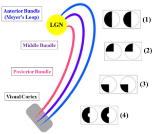
Optic Radiations (ORs) are two white matter fiber bundles allowing direct connection between homolateral Lateral Geniculate Nucleus (LGN) and visual cortex. From the anatomical point of view, ORs are conventionally divided in three different portions, namely anterior, middle and posterior; the Meyer’s loop represents the anterior portion, and it has been shown to be a bundle with a very high variability in human brain.1,2,3 The latter aspect has to be taken into account when studying ORs in clinical and surgical contexts. Indeed, ORs represent eloquent white matter bundles often reconstructed by means of Magnetic Resonance Imaging (MRI) based approaches in order to assess their involvement by pathological conditions as well as to prevent post-surgical damages. Both these conditions can compromise visual function by causing, for instance, visual field deficits.4,5 Schematic representation of ORs’ bundles and common visual field deficits associated with ORs’ lesions is shown in Figure 1.

Figure 1: Schematic representation of ORs, from their origin from LGN (yellow) to visual cortex (grey) and common visual field deficits associated with ORs’ lesions. Each bundle is colored separately. LGN’s damage is followed by homonymous hemianopia (1). Lesions of the anterior bundle (blue) cause homonymous superior quadrantanopia (2) whereas damages of the posterior bundle (pink) are followed by homonymous inferior quadrantanopia (3). Middle bundle is shown in violet. Homonymous hemianopia with macular sparing can be seen after damages at visual cortex level (4).

ROJ-1-e002Fig1

ORs are largely studied by means of diffusion MRI based modelling techniques, such as Diffusion Tensor Imaging (DTI). These methods allow both qualitative evaluation of white matter bundles as well as quantitative analysis of several diffusion parameters, such as Fractional Anisotropy. The noninvasive investigation of ORs by means of diffusion MRI has provided several morphological information regarding ORs’ connectivity. In particular, moving beyond well demonstrated link with V1 and V2,6 direct connections with V3,7 V48 and V58,9 have been recently shown. Functional implications of such connectivity include both primary visual signal elaboration as well as more complex functions; for example, direct connection with extra-striatal areas have been supposed to be implicated in the genesis of complex functional phenomena such as blindsight.10 Moreover, other studies have hypothesized that such extra-striate connectivity might be the anatomical basis for the functional compensation performed after damages of striate visual cortex.11

In the context of tractographic driven investigation of ORs, several issues should be considered, especially if using conventional DTI. Indeed, ORs reconstruction might be potentially affected by well-known DTI limitations, such as partial volume effects.12,13 Furthermore, several bundle-specific issues may compromise ORs tractographic reconstructions, e.g. complexity of ORs anatomical course as well as their relationship with other white matter bundles and brain structures.1,2,8,14,15 From a technical perspective, several voxels with complex geometriesmay be involved, which are known to cause an inaccurate analysis when applying DTI model. These potential pitfalls raised several criticisms regarding reliability of tractographic findings regarding both ORs detection as well as extrastriate connectivity.16 To overcome these limitations, other diffusion models have been tested to improve tractographic reconstructions, such as High Angular Resolution Diffusion-weighted Imaging (HARDI), Q Ball Imaging (QBI) and Diffusion Spectrum Imaging (DSI) algorithms. Although tractographic output provided by these approaches was found to outperform DTI based one, these algorithmsare however difficult to be applied in clinical contexts because they are really scanner demanding and time-consuming or they showed poor angular resolution.17,18 From this point of view, Constrained Spherical Deconvolution (CSD)18 has been found a powerful technique feasible in clinical settings. Recently, Arrigo and colleagues8 have shown how the use of CSD model combined with probabilistic tractography provided robust ORs detection in healthy human brain. They were able to provide a more complete reconstruct of all ORs’ portion, especially in case of Meyer’s loop, justifying such result as being due to the higher angular resolution reached by CSD if compared to other models, as well asits ability to resolve voxels with complex fibers’ configurations.8,13,18 These voxels are indeed known to represent more than 90% of the total number in each brain, thus causing DTI reconstruction troubles for other eloquent bundles of White Matter (WM), such as corticospinal tract and arcuate fasciculus.19 It is worthy to note that CSD has been already used to reconstruct ORs, providing accurate tracking of these white matter bundles and increasing reliability and details of ORs morphological profile.20,21

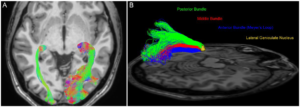
Arrigo et al8 reinforced the hypothesis of ORs’ extrastriate connectivity as well as their involvement in higher order visual functions. Moreover, direct LGN connection with V4 was shown for the first time in humans, reinforcing the findings provided by previous analyses performed in animals.22,23 The comparative view of DTI based and CSD based ORs reconstructions is shown in Figure 2.

Figure 2: OR stractographic reconstruction in healthy human brain. (A) Axial view of DTI based (left side of the image) and CSD based (right side of the image) tractographic reconstructions showed a more complete ORs detection provided by CSD. Each bundle was colored according to its main diffusion direction. (B) Tridimensional sagittal view of CSD based reconstruction of left ORs. The use of CSD based tractography allowed a good detection of each ORs component. Anterior, middle and posterior ORs bundle, starting from LGN (yellow volume), were manually colored respectively in blue, red and green.

ROJ-1-e002Fig2

If benefits provided by CSD model are important in physiological contexts, they become extremely important in pathological ones, for instance during pre-surgical planning. Indeed, ORs are investigated both in patients with brain neoplasms as well as for surgical treatment of drug-resistant epilepsy, the latter consisting in the anterior temporal lobe resection.3,24,25,26,27 Regarding brain neoplasms, many additional reconstruction’s issues have been shown in these patients, which are caused by neoplasm’s effects on diffusion signal; these potential pitfalls may be overcome by using more advanced diffusion signal modelling techniques, such as CSD, as previously reported in other studies.28,29 Regarding pre-surgical planning of ORs, the use of CSD based tractography may increase definition of safety resection margins and it might have a good impact on post-surgical visual deficits.

From a quantitative point of view, the adoption of more advanced diffusion based approaches might have an impact on evaluation of diffusion parameters. Indeed, when performing a tract based quantitative analysis, it should be taken into account that DTI would exclude voxels with complex geometry, since they often show Fractional Anisotropy (FA) values below a relative cut-off used for conventional tracking.13 FA decrements can be found both in voxels with multiple orientations and in voxels involved by a pathological process.30 The result of this DTI limitation is the loss of these voxels from the total count, determining an incomplete profiling of a given pathway. With respect to ORs, this issue may particularly affect Meyer’s loop, which is known to be poorly detected when using DTI tractography; moreover, also other ORs’ portions might be potentially affected due to the presence of crossing fibers.8

This brief description attempted to summarize recent anatomical and physiological advances regarding ORs detection and analysis. Moreover, it showed how advanced diffusion signal modelling techniques may improve ORs’ investigation both in healthy and pathological conditions, describing common as well as tract-specific diffusion issues. Furthermore, this study shows that the choice of diffusion model may have a huge impact both on qualitative and quantitative analysis of ORs.

CONFLICTS OF INTEREST

The authors declare that they have no conflicts of interest.

1. Párraga RG, Ribas GC, Welling LC, et al. Microsurgical anatomyof the optic radiation and related fibers in 3-dimensional images. Neurosurgery. 2012; 71(Suppl 1): 160-171. doi: 10.1227/NEU.0b013e3182556fde

2. Pujari VB, Jimbo H, Dange N, et al. Fiber dissection of the visualpathways: analysis of the relationship of optic radiations to lateral ventricle: a cadaveric study. Neurol India. 2008; 56(2): 133-137. doi: 10.4103/0028-3886.41989

3. Nilsson D, Starck G, Ljungberg M, et al. Intersubjectvariability in the anterior extent of the optic radiation assessed by tractography. Epilepsy Res. 2007; 77(1): 11-16. doi: 10.1016/j.eplepsyres.2007.07.012

4. Longo D, Fauci A, Kasper D, et al. Harrison’s Principles of Internal Medicine. 18th ed. McGraw-Hill Professional, 2011: 1 and 2.

5. Greenberg D, Aminoff M, Simon R. Clinical Neurology 8/E. McGraw-Hill Professional, 2012.

6. Baldwin MK, Kaskan PM, Zhang B, et al. Cortical and subcortical connections of V1 and V2 in early postnatal macaque monkeys. J Comp Neurol. 2012; 520(3): 544-569. doi: 10.1002/cne.22732

7. Alvarez I, Schwarzkopf DS, Clark CA. Extrastriate projections in human opticradiation revealed by fMRI-informed tractography. Brain Struct Funct. 2015; 220(5): 2519-2532. doi: 10.1007/s00429-014-0799-4

8. Arrigo A, Calamuneri A, Mormina EM, et al. New Insights in the optic radiations connectivity in the human brain. Invest Ophthalmol Vis Sci. 2016; 57(1): 1-5. doi: 10.1167/iovs.15-18082

9. Bridge H, Thomas O, Jbabdi S, Cowey A. Changes inconnectivity after visual cortical brain damage underlie alteredvisual function. Brain. 2008; 131: 1433-1444. doi: 10.1093/brain/awn063

10. Schmid MC, Mrowka SW, Turchi J, et al. Blindsight depends on the lateral geniculate nucleus. Nature. 2010; 466(7304): 373- 377. doi: 10.1038/nature09179

11. Bridge H, Hicks SL, Xie J, et al. Visual activation of extra-striatecortex in the absence of V1 activation. Neuropsychologia. 2010; 48: 4148-4154. doi: 10.1016/j.neuropsychologia.2010.10.022

12. Jones DK, Cercignani M. Twenty-five pitfalls in the analysis ofdiffusion MRI data. NMR Biomed. 2010; 23(7): 803-820. doi: 10.1002/nbm.1543

13. Farquharson S, Tournier JD, Calamante F, Fabinyi G, et al. White matter fiber tractography: whywe need to move beyond DTI. J Neurosurg. 2013; 118(6): 1367-1377. doi: 10.3171/2013.2.JNS121294

14. Kier EL, Staib LH, Davis LM, Bronen RA. MR imaging of thetemporal stem: anatomic dissection tractography of the uncinatefasciculus, inferior occipitofrontal fasciculus, and Meyer’s loop of theoptic radiation. AJNR Am J Neuroradiol. 2004; 25(5): 677-691.

15. Peltier J, Verclytte S, Delmaire C, et al. Microsurgical anatomy of the temporal stem: clinical relevance and correlations with diffusion tensor imaging fiber tracking. J Neurosurg. 2010; 112(5): 1033-1038. doi: 10.3171/2009.6.JNS08132

16. Rabbo AF, Koch G, Lefevre C, Seizeur R. Direct geniculo-extrastriate pathways: a review of the literature. Surg Radiol Anat. 2015; 37: 891-899. doi: 10.1007/s00276-015-1450-7

17. Tournier JD, Calamante F, Connelly A. Robust determination of the fibre orientation distribution in diffusion MRI: nonnegativity constrained super-resolved spherical deconvolution. Neuroimage. 2007; 35: 1459-1472. doi: 10.1016/j.neuroimage.2007.02.016

18. Tournier JD, Yeh CH, Calamante F, et al. Resolving crossing fibres using constrained spherical deconvolution: validation using diffusion weighted imaging phantom data. Neuroimage. 2008; 42: 617-625. doi: 10.1016/j.neuroimage.2008.05.002

19. Jeurissen B, Leemans A, Tournier JD, et al. Investigating the prevalence of complex fiber configurationsin white matter tissue with diffusion magnetic resonance imaging. Hum Brain Mapp. 2013; 34: 2747-2766. doi: 10.1002/hbm.22099

20. Lim JC, Phal PM, Desmond PM, et al. Probabilistic MRI tractography of the optic radiation using constrained spherical deconvolution: a feasibility study. PLoS One. 2015; 10(3): e0118948. doi: 10.1371/journal.pone.0118948

21. Martínez-Heras E, Varriano F, Prčkovska V, et al. Improved framework for tractography reconstruction of the optic radiation. PLoS One. 2015; 10(9): e0137064. doi: 10.1371/journal.pone.0137064

22. Ferrera VP, Nealey TA, Maunsell JH. Responses in macaquevisual area V4 following inactivation of the parvocellular and magnocellular LGN pathways. J Neurosci. 1994; 14(4): 2080-2088.

23. Ungerleider LG, Galkin TW, Desimone R, Gattass R. Cortical connections of area V4 in the macaque. Cereb Cortex. 2008; 18: 477-499. doi: 10.1093/cercor/bhm061

24. Duncan JS. Imaging in the surgical treatment of epilepsy. Nat Rev Neurol. 2010; 6: 537-550. doi: 10.1038/nrneurol.2010.131

25. Winston GP, Daga P, Stretton J, et al. Optic radiation tractography and vision inanterior temporal lobe resection. Ann Neurol. 2012; 71(3): 334-341. doi: 10.1002/ana.22619

26. Piper RJ, Yoong MM, Kandasamy J, Chin RF. Application of diffusion tensor imaging and tractography of the optic radiation inanterior temporal lobe resection for epilepsy: a systematic review. Clin Neurol Neurosurg. 2014; 124C: 59-65. doi: 10.1016/j.clineuro.2014.06.013

27. Cho JM, Kim EH, Kim J, et al. Clinical use of diffusion tensor image-merged functional neuronavigationfor brain tumor surgeries: review of preoperative, intraoperative, andpostoperative data for 123 cases. Yonsei Med J. 2014; 55(5): 1303-1309. doi: 10.3349/ymj.2014.55.5.1303

28. Mormina E, Longo M, Arrigo A, et al. MRI tractography of corticospinal tract and arcuate fasciculus in high-grade gliomas performed by constrained spherical deconvolution: qualitative and quantitative analysis. AJNR Am J Neuroradiol. 2015; 36(10): 1853-1858. doi: 10.3174/ajnr.A4368

29. Arrigo A, Mormina E, Calamuneri A. The pre-surgical planning of brainneoplasms: from diffusion tensor imaging to more advanced approaches. Radiol Open Journal. 2015; 1(1): e1-e5.

30. Jellison BJ, Field AS, Medow J, et al. Diffusion tensor imaging of cerebral white matter: a pictorialreview of physics, fiber tract anatomy, and tumor imaging patterns. AJNR Am J Neuroradiol. 2004; 25(3): 356-369.

31. Dai H, Mu KT, Qi JP, et al. Assessment of lateral geniculate nucleus atrophywith 3T MR imaging and correlation with clinical stage of glaucoma. AJNR Am J Neuroradiol. 2011; 32(7): 1347-1353. doi: 10.3174/ajnr.A2486

32. El-Rafei A, Engelhorn T, Wärntges S, et al. Glaucoma classification based on histogram analysis of diffusion tensor imaging measures in the optic radiation. ComputerAnalysis of Images and Patterns Lecture Notes in Computer Science. 2011; 6854: 529-536.doi:10.1007/978-3-642-23672-3_64

33. Liu Y, Duan Y, He Y, et al. A tract-based diffusion study of cerebral white matter inneuromyelitis optica reveals widespread pathological alterations. Mult Scler. 2012; 18(7): 1013-1021. doi: 10.1177/1352458511431731

LATEST ARTICLES

Blood Sample from the Patient

Hypertriglyceridemia-Induced Pancreatitis: A Case Report and Literature Review

Maarten Bulterys, Melvin Willems* and Agnes Meersman

doi.

From Neck Pain to a Life-Threatening Condition: A Case Report

Floris Vandewoude* and Sören Verstraete

doi.

Chest X-ray Showed a Hazy Left Upper Lung Infiltrate

A Noteworthy Case of Myasthenic Crisis Induced by Levofloxacin

Ada Young*, Ramya Ramesh and Milind Awale

doi.

The Right Thigh Anterior Compartment was Swollen, and the Skin was Ulcerated due to the Traditional Cautery

Primary Skeletal Muscle Lymphoma: A Case Report and Literature Review

Solomon Bishaw*, Addisu Alemu and Abel Tefera

doi.

LATEST ARTICLES

Blood Sample from the Patient

Case Report

2024 Jun

Maarten Bulterys, Melvin Willems* and Agnes Meersman

Case Report

2024 Jun

Floris Vandewoude* and Sören Verstraete