INTRODUCTION
Megaesophagus is a condition marked by the generalized dilatation of the esophagus, it is common cause of regurgitation in dogs. Megaesophagus is a disorder in which the esophagus becomes dilated, on account of a decrease in the muscle tone. This condition may arise as a secondary consequence to many ailments such as hypothyroidism or other diseases of unknown etiology.1
Canine endocrinopathy megaesophagus is a fairly rare condition.1,2 Hypothyroidism was quoted as the possible etiology of megaesophagus; however, the exact association between them have not yet been fully understood.3,4
Hypothyroidism is one of the most common canine endocrinopathy with bilaterally symmetrical truncal alopecia. Gastrointestinal symptoms are uncommon in hypothyroidism (hormonal diseases), some dogs with megaesophagus may have experienced hypothyroidism, which has been attributed to hypothyroidism-induced myopathy or neuropathy; treatment of these dogs with thyroxine may have improved their esophageal functions.5
The present work aims to report about megaesophagus in littermates Dogo Argentino accompanied with hypothyroidism for scientific community and veterinary small animal’s practioners.
MATERIALS AND METHODS
Description and Clinical Presentation
Four littermates aged 5 years old belonging to the Dogo Argentino breed (one males and 3 bitches) were taken to a small animal clinic, Faculty of Veterinary Medicine, Cairo University, Giza Governorate, Egypt for medical consultation with a history of frequent regurgitation shortly after feeding. In addition to regurgitation which was not linked with feeding (in empty stomach), with environmental stress and minimal exercise, the regurgitated material was described as frothy. Non-pruritic alopecia in the abdomen and tail, intolerance to exercise, lethargy, and poor body condition was reported by owner. The dogs were quite stressed, restless, and afraid of their surroundings.
Diagnosis Workup
The 4 littermates were subjected to radiographic and endoscopic examinations. The radiographic examination was scheduled for the diagnosis of the regurgitation. Endoscopic examination was performed to investigate the dilated esophageal wall, and and if other factors were involved, endoscopy with injectable general anesthesia using atropine sulphate (0.05 mg/kg bwt), xylazine (dosed at 1 mg/kg bwt), and ketamine (dosed at 10 mg/kg bwt), were undertaken. For examining the skin lesions, skin scrapings were done microscopically to exclude parasitological involvement. T4, T3, and TSH were estimated using serum samples.
Management and Follow-up
For megaesophagus, the owner was advised to feed dogs on an elevated platform in small quantities, and several times daily, allowing them to maintain their position for a few minutes after finishing eating or drinking. A proton pump inhibitor, Controloc® 20 mg pantoprazole tablet per day was prescribed for the long-term management of gastroesophageal reflux and gastric hyperacidity.
For the management of hypothyroidism, and its concurrency with megaesphagus, oral levothyroxine (Forthyron® 0.8 mg pills dosed at 20 μg/kg was prescribed every 12 hours for three months, followed by a periodic estimation of T3, T4 and TSH from the serum sample.
The diagnostic procedures and medicinal management of these cases were employed after the agreement of the Ethical Committee of Faculty of Veterinary Medicine, Cairo University, Giza Governorate, Egypt.
RESULTS
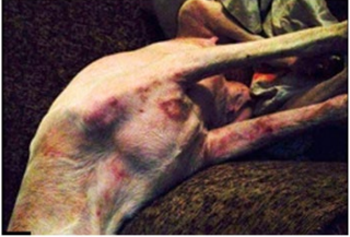
Upon physical examination, a dull coat was reported (Figure 1) along with poor body conditions, and alopecia with increased pigmentation in the tail, and a slightly pale mucous membrane.
Figure 1: Clinical Presentation of Littermate Dogs Showed Bilateral Reddish Patches of Alopecia on Abdomen, Chest, and Medial Aspect of the Fore Limbs.
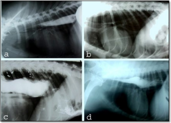
Plain chest and abdomen X-ray analysis did not yield any clinically significant findings. The contrast X-ray examination revealed a diffuse radiopaque esophageal dilation, particularly in the thoracic and abdominal parts of the esophagus as has been represented in Figure 2.
Figure 2: Lateral Views of Positive Contrast Radiographs (a, c & d); Radiopaque Dilated Esophageal Part Dorsal to the Trachea, Extending the Base of Heart, and (b) Plain Radiograph.
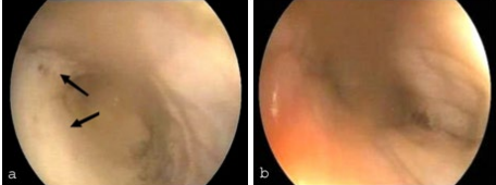
Endoscopic examination revealed a dilated thoracic and abdominal esophagus, with a pale mucosal surface filled with a frothy juice, which moved from the stomach towards the esophagus. The clinical findings indicated gastroesophageal reflux and gastric hyperacidity as presented in Figure 3.
Figure 3: Endoscopic Examination of the Affected Case.
a. Flaccid Dilated Thoracic Esophageal Wall with Frothy Juice from the Stomach (Black Arrows).
b. Flaccid Dilated Abdominal Esophageal Wall.
The gold standard in the diagnosis of hypothyroidism showed a decrease in the T4, T3 levels coupled with an increase in TSH levels. The recorded range for the a T4 level was 0.65±0.012 ng/dL, the T3 level was 2.48±1.006 pcg/ml, and TSH level was 0.08±0.002 uIU/ml relative to the normal range (0.000.60 uIU/ml).
A clinical follow-up of the cases reported showed an improvement in the dermatological signs resulting in a complete return to the bright skin coat, and the regurgitation was significantly improved with continued treatment.
DISCUSSION
Megaesophagus is an important cause of regurgitation in canine population.1 The classical presentation is weight loss, regurgitation shortly after feeding and dehydration. Concurrently, the presenteded cases are exhibited these clinical symptoms as described by German.7
Diagnostic workup depended on combination of history, signs, radiography in addition to endoscopic examination is performed to dismiss obstruction or other causes as mentioned by Moore.8
Many megaesophagus cases in adult dogs can be evolved to be of idiopathic; other postulated causes revolve around toxicity, polyneuro – or polymyopathies and endocrinopathies were described especially hypoadrenocorticism and quite possibly hypothyroidism.8-10 However, hypothyrodism was postulated to play a major role in development of megaesophagus; thyroid dysfunction causes impairment in nerve impulses and neuropathy which consequently may lead to muscle tone reduction.8,11
The dermatologic involvement was assumed to be of endocrine origin, absence of parasitological etiology upon skin scraping and nature of alopecia are main advocate of endocrine role. The bilateral alopecia with non-inflammatory features is quietly indicative of endocrine involvement which described as endocrine alopecia; this is similar to the findings of Nelson and Couto.2 In spite of the clinical presentation of canine hypothyroidism may be vague. Though alopecia and abnormal pigmentation may be indicative but not confirmatory, the confirmation is based on results of serum T3, T4 and TSH estimation.12 Moreover, hyperadrenocorticism and to less extend hypothyroidism are among causes of megaesophagus. However, the natures of signs exhibited by those dogs are more consistent with hypothyroidism; this is concurrent with Ridgway and Graves, Moore.6,8
The usage of levothyroxine for treating hypothyroidism in dogs with megaesophagus is controversial.11 Levothyroxine aided to alleviate megaesophagus after 3-months treatment,12 but other reports showed no improvement.13 Despite that, the presented cases with hypothyroidism and megaesophagus are managed by oral adminstration of levothyroxine.
For those cases hormonal replacement therapy with L-thyroxin has improved the clinical signs dramatically; however some signs may linger but the quality of life is radically improved as mentioned by Huber et al.5
Small multiple meals with high caloric contents accompanied with elevated feeding are offered to those dogs and reported to improve the condition as stated by Moore.8 Gastroesophageal reflux is well documented in megaesophagus and even might be attributed to its evolving as described by Ridgway and Graves,6 proton pump inhibitors as pantoprazole supplementation are well documented in treating acid-related gastric ailments in both human and canine patients.14
CONCLUSION
The authors herein would like to report megaesophagus in dogs accompanied with hypothyroidism, also to state the recommendations of their medicinal management aiming to introduce this for scientific community and veterinarians for solving the problem of such cases.
CONFLICTS OF INTEREST
The authors declare that they have no conflicts of interest.


