Osteology and Rheumatology

Open journal

ISSN 2996-5918

Is Normal Knee Biomechanics Reproduced by Modern Total Knee Arthroplasty Designs? The Role of Fluoroscopy

Pier Francesco Indelli*

Received: March 18th, 2016; Accepted: April 19th, 2016; Published: April 19th, 2016

INTRODUCTION

Total Knee Arthroplasty (TKA) represents a very successful procedure in the treatment of patients affected by severe end-stage tricompartimental knee osteoarthritis. Many patients return to normal daily living activities, including low-impact recreational activities like biking, golfing, swimming and trekking.1 Unfortunately, TKA patients still experience substantial functional impairment compared with their age- and gender-matched peers, especially when doing biomechanically demanding activities.2 Patient satisfaction after TKA reflects these differences: despite recent advances in surgical techniques, implant designs, and patients education, the satisfaction rate after primary TKA between the last decade of the twentieth century and the first decade of the twenty-first century increased only by 3%, leaving 15% of the patients not fully satisfied.3

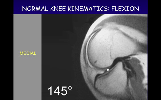
The normal knee motion is quite complex since Giovanni Alfonso Borelli first described the biomechanical patterns of the knee joint.4 In recent times, it has been shown that, as the normal knee goes into flexion, the lateral femoral condyle moves progressively posterior on the tibial plateau (“postero-lateral rollback”), while the medial femoral condyle pivots with a negligible posterior motion.5,6 The authors of the current editorial note quantified this motion in a dynamic MRI study performed in healthy subjects: at 145 degrees of knee flexion, the average lateral condyle rollback was 15 mm while the average medial condyle rollback was 3 mm (Figure 1).7 Such strongly asymmetric “rollback” of the femoral condyles during normal knee flexion is accompanied by a contemporaneous external rotation of the femur with respect to the tibia (“medial pivoting”).8

Figure 1: Dynamic Knee MRI evaluation of the tibiofemoral contact point during deep knee flexion (145°) in healthy subjects.
A) Medial Compartment;

ORHOJ-1-103Fig1a

B) Lateral Compartment. The tibiofemoral contact point is significantly more posterior in the lateral compartment respect to the medial compartment.

ORHOJ-1-103Fig1b

Numerous kinematic studies using static and dynamic fluoroscopy,6,8,9 gait analysis,10 and in vitro techniques11,12 have revealed profound differences between normal knee motion and TKA kinematics. Theoretically, these differences might play a major role in understanding the still high “un-satisfaction” rate among patients following primary TKA.

Video fluoroscopy was first used to define TKA geometry and kinematics in vivo in the early 1990s13: since then, many studies investigated the relationship between TKA designs and joint kinematics.

Currently, TKA systems differ by many characteristics: two of them, the design of the femoral condyle on the sagittal plane (single versus multiple radii) and the level of constraint (Cruciate Retaining-CR, Postero-Stabilized-PS, Medially Congruent-MC, Medial Pivot-MP and Sagittally Stabilized-SS) have been extensively studied with static and, more recently, dynamic fluoroscopy using adapted C-arm units. This editorial note reviews the results of those fluoroscopic studies and compares them with the kinematics of normal knees.

FLUOROSCOPIC ANALYSIS

Komistek et al6 used static fluoroscopy to study the biomechanical proprieties of normal knees: in that report, during deep flexion activities, subjects experienced an average of 12.7 mm of lateral condyle motion, whereas the medial condyle motion was only 2.9 mm. During deep flexion activities, their subjects experienced a significant axial rotation of the tibia relative to the femur (average 13°).

Cruciate-Retaining (CR)-TKA designs have been extensively studied with static fluoroscopy since early 2000s: those designs have shown a lack of posterior femoral rollback and a more anterior tibio-femoral contact point during deep knee flexion14,15 when compared with normal knees. This last phenomenon has been described by many authors as “paradoxical motion”16-19: this unwanted finding has been attributed to the incompetent restraining function of the posterior cruciate ligament.

Because of these biomechanical differences between CR-TKA and the native knee, Posterior-stabilized (PS)-TKA have been introduced in the 1970s as an alternative to CR design in order to reproduce normal knee mechanics. Dennis et al20 fluoroscopically analyzed 811 patients after primary TKA performed using 33 different designs during stance phase of gait or a deep knee bend maneuver: kinematic patterns of fixed versus mobile-bearing designs were similar in which femorotibial contact remained relatively stationary with minimal AP femorotibial translation (“postero-lateral rollback”) respect to the native knee; on the other side, paradoxical anterior femoral translation during deep knee flexion was most commonly observed in the CR-TKA designs. In the same study, the normal medial pivoting kinematic pattern (femoral external rotation/tibial internal rotation) was observed in only 55% of all knees during deep knee flexion.

Shimmin et al,21 in a fluoroscopic study evaluating a medial pivot (MP) TKA design (Saiph, MatOrtho, Leatherhead, UK) characterized by full conformity of themedial articular surface (ball in a socket design) and by partial conformity of the lateral compartment (not flat polyethylene), showed that the medial femoral condyles translated an average of 2 mm posterior to the tibial sulcus while the lateral femoral condyles translated an average of 6 mm posterior to the tibial sulcus during maximum knee flexion. None of the studied knees showed anterior femoral translation (“paradoxical motion”) during flexion.

Recently, Scott et al22 fluoroscopically evaluated a sagitally-stabilized medially spherical TKA (GMK Sphere, Medacta International AG, Castel San Pietro, Switzerland): this TKA design is characterized by a completely spherical femoral condyles and a tibialpolyethylene which is spherical medially and completely flat laterally. In this study, the deep flexion activity in implanted knees showed a mean of 8° tibial internal rotation, 2 mm medial posterior translation and 8 mm posterior translation on the lateral condyle.

The anatomy of the posterior femoral condyles has been extensively studied in the last thirty years: standard radiologic,23 magnetic resonance assisted24 and computer-assisted studies25 have shown that the posterior femoral condyles have single-radius geometry. Historically, TKA manufactures have released two different femoral designs: a single-radius (SR) and a multiple-radii (MR). The SR TKA consists of symmetrical femoral condyles that incorporate a SR in the sagittal plane. The MR-TKA consists of a multiradii femoral design in which the lateral femoral condyle is larger than the medial femoral condyle in the sagittal plane. Recently, Grieco et al9 compared in vivo the kinematic differences in patients implanted with a SR vs. a MR-cruciate retaining (CR) design during deep knee flexion using a novel mobile fluoroscopy system. These authors reported that SR and MR groups experienced statistically similar average AP motion in the lateral condyle: – 0.43 mm (SR) vs. – 1.0 mm (MR); the SR group had significant more AP motion (+ 3.51 mm) compared to the MR group (- 0.42 mm) in the medial condyle. On the other side, they showed that the SR group had a significantly larger amount of physiological axial rotation (average 5.20°) compared to the MR group (average 0.75°).

CONSIDERATIONS

Many methods have been historically used to evaluate normal and following TKA knee kinematics. The great advantage of video fluoroscopy is to allow analysis of multiple “invivo” activities done in weight-bearing conditions. The major limitation of this technique has always been that only on-site activities (i.e. deep knee flexion) were originally tested by many authors. The introduction of mobile fluoroscopy9 allows to study TKA in vivo kinematics during normal walking and walking up and down an inclined ramp.

Classic fluoroscopic studies6,20-22 (Table 1) have shown that normal knee kinematics is not fully reproduced by many TKA designs. Severe kinematic abnormalities, including reduced posterior femoral rollback, paradoxical anterior femoral translation, reduced axial rotation patterns, are commonly present in our TKA patients. The “perfect” TKA should be biomechanically characterized by posterior femoral rollback during deep knee flexion and by an axial rotation in its medial compartment. Recent fluoroscopic studies by Shimmin et al21 on medial pivot (MP) and by Scott et al22 on sagittally-stable (SS) designs showed that “medially-constrained” designs reproduce closer kinematic patterns to normal knees when compared with cruciate-retaining (CR) and posterior-stabilized (PS) designs.

Table 1: Influence of level of constraint on fluoroscopically studied TKA kinematics during deep knee flexion. CR=Cruciate Retaining; PS=Posterior-stabilized; MP=Medial pivot; SS=Sagittally stabilized; *Fluoroscopically assisted measurements performed during deep knee flexion (average distance in mm from the tibio-femoral contact point in full knee extension).

Author

Year TKA Design Medial Condyle AP (mm)*

Lateral Condyle AP (mm)*

Axial Rotation (degrees)*
Komistek et al6

2003

Normal knee – 2.9 mm – 12.7 mm

13°

Dennis et al20

2003

CR + 1 mm (anterior translation) -1.6 mm

NA

Dennis et al20

2003

PS – 1 mm – 3.7 mm

NA

Shimminet al21

2015

MP -2 mm – 6 mm

Scott et al22

2016

SS – 2 mm – 8 mm

Further, long-term clinical investigations on “medially-constrained” TKA designs are needed to understand if their promising fluoroscopic findings correlate with a higher postoperative satisfaction rate in our TKA patients.

1. Chatterji U, Ashworth MJ, Lewis PL, Dobson PJ. Effect of total knee arthroplasty on recreational and sporting activity. ANZ J Surg. 2005; 75: 405-408. doi: 10.1111/j.1445- 2197.2005.03400.x

2. Noble PC, Gordon MJ, Weiss JM, Reddix RN, Conditt MA, Mathis KB. Does total knee replacement restore normal knee function? Clin Orthop Relat Res. 2005; (431): 157-165. doi: 10.1097/01.blo.0000150130.03519.fb

3. Schulze A, Scharf HP. [Satisfaction after total knee arthroplasty. Comparison of 1990-1999 with 2000-2012]. Orthopade. 2013; 42(10): 858-865. doi: 10.1007/s00132-013-2117-x

4. Pope MH. Giovanni Alfonso Borelli: the Father of Biomechanics. Spine. 2005; 30(20): 2350-2355. doi: 10.1097/01.brs.0000182314.49515.d8

5. Dennis DA, Mahfouz MR, Komistek RD, et al. In vivo determination of normal and anterior cruciate ligament-deficient knee kinematics. J Biomech. 2005; 38(2): 241-253. doi: 10.1016/j. jbiomech.2004.02.042

6. Komistek RD, Dennis DA, Mahfouz M. In vivo fluoroscopic analysis of the normal human knee. Clin Orthop Relat Res. 2003; 410: 69-81. doi: 10.1097/01.blo.0000062384.79828.3b

7. Baldini A, Aglietti P, Vena LM, Lup D, Indelli PF. Postoperative recovery and early results: Meniscal Bearing Knee vs Legacy PS. Arthroscopy. 2001; 17(6): Suppl 2(p1): S1-S55.

8. Leszko F, Hovinga KR, Lerner AL, et al. In vivo normal knee kinematics: Is ethnicity or gender an influencing factor? Clin Orthop Relat Res. 2011; 469(1): 95-106. doi: 10.1007/s11999- 010-1517-z

9. Grieco TF, Sharma A, Komistek RD, Cates HE. Single versus multiple-radii cruciate-retaining total knee arthroplasty: An invivo mobile fluoroscopy study. J Arthroplasty. 2016; 31: 694- 701. doi:10.1016/j.arth.2015.10.029

10. Joglekar S, Gioe TJ, Yoon P, Schwartz MH. Gait analysis comparison of cruciate retaining and substituting TKA following PCL sacrifice. The Knee. 2012; 19(4): 279-285. doi: 10.1016/j.knee.2011.05.003

11. Iwaki H, Pinskerova V, Freeman MAR. Tibiofemoral movement 1: the shapes and relative movements of the femur and tibia in the unloaded cadaver knee. J Bone Joint Surg Br. 2000; 82(8):1189. doi: 10.1302/0301-620x.82b8.10717

12. Li G, Zayontz S, DeFrate LE, et al. Kinematics of the knee at high flexion angles: An in vitro investigation. J Orthop Res. 2004; 22(1): 90-95. doi: 10.1016/S0736-0266(03)00118-9

13. Banks SC, Hedge WA. Direct measurement of 3D knee prosthesis kinematics using single plane fluoroscopy. Proceedings of the Orthopaedic Research Society. l993; 18: 428.

14. Bellemans J, Banks S, Victor J, et al. Fluoroscopic analysis of the kinematics of deep flexion in total knee arthroplasty: influence of posterior condylar offset. J Bone Joint Surg Br. 2002; 84(1): 50-53. doi: 10.1302/0301-620X.84B1.12432

15. Victor J, Banks S, Bellemans J. Kinematics of posterior cruciate ligament retaining and -substituting total knee arthroplasty. A prospective randomized outcome study. J Bone Joint Surg Br. 2005; 87(5): 646-655. doi: 10.1302/0301-620X.87B5.15602

16. Stiehl JB, Komistek RD, Dennis DA, Paxson RD, Hoff WA. Fluoroscopic analysis of kinematics after posterior-cruciateretaining knee arthroplasty. J Bone Joint Surg Br. 1995; 77(6): 884-889.

17. Dennis DA, Komistek RD, Hoff WA, et al. In vivo knee kinematics derived using an inverse perspective technique. Clin Orthop Relat Res. 1996; 331: 107-117. doi: 10.1097/00003086-199610000-00015

18. Yoshiya S, Matsui N, Komistek RD, Dennis DA, Mahfouz M, Kurosaka M. In vivo kinematic comparison of posterior cruciate-retaining and posterior stabilized total knee arthroplasties under passive and weight-bearing conditions. J Arthroplasty. 2005; 20(6): 777-783. doi: 10.1016/j.arth.2004.11.012

19. Incavo SJ, Mullins ER, Coughlin KM, Banks S, Banks A, Beynnon BD. Tibiofemoral kinematic analysis of kneeling after total knee arthroplasty. J Arthroplasty. 2004; 19(7): 906-910. doi: 10.1016/j.arth.2004.03.020

20. Dennis DA, Komistek RD, Mahfouz MR, Haas BD, Stiehl JB. Multicenter Determination of In Vivo Kinematics After Total Knee Arthroplasty. Clin Orthop Relat Res. 2003; (416): 37-57. doi: 10.1097/01.blo.0000092986.12414.b5

21. Shimmin A, Martinez-Martos S, Owens J, Iorgulescu AD, Banks SA. Fluoroscopic motion study confirming the stability of a medial pivot design total knee arthroplasty. Knee. 2015; 22(6): 522-526. doi: 10.1016/j.knee.2014.11.011

22. Scott G, Iman MA, Eifert A, et al. Can a total knee arthroplasty be both rotationally unconstrained and anteroposteriorly stabilized? Bone Joint Res. 2016; 5: 80-86. doi: 10.1302/2046- 3758.53.2000621

23. Elias SG, Freeman R, Gokcay EI. A correlative study of the geometry and anatomy of the distal femur. Clin Orthop Relat Res. 1990; 260: 180-186.

24. Hollister AM, Jatana SAK, Sullivan WW, Lupichuk A. The axes of rotation of the knee. Clin Orthop Relat Res. 1993; 290: 259-268.

25. Kurosawa H, Walker PS, Abe S, Garg AT, Hunter T. Geometry and motion of the knee for implant and orthotic design. J Biomech. 1985; 18(7): 487-491. doi: 10.1016/0021-9290(85)90663-3

LATEST ARTICLES

Use of Black Soldier Fly (Hermetia illucens) Prepupae Reared on Organic Waste

review

2024 Mar

Isayas A. Kebede*, Haben F. Gebremeskel and Gelan D. Dahesa,
Figure 11. Risk Map for the Introduction of Ruminant Diseases at Borders

Original Research, peer reviewed

2024 Mar

Yassir Lezaar*, Mehdi Boumalik, Youssef Lhor, Moha El-Ayachi, Abelilah Araba and Mohammed Bouslikhane

Commentary, End-of-Life, Family Dynamics, palliative Care, peer reviewed

2024 Mar

Neil A. Nijhawan*, Rasha Mustafa and Aqeela Sheikh

Retrospective Study

2024 Mar

Shrenik J. Shah*, Abhishek Jha, Chirag Davara, Rushi Mistry and Kapil Kachhadiya

Attention, Disaster, Mindfulness, Original Research, peer reviewed

2024 Mar

Christina Hagen*, Lars Lien, Edvard Hauff and Trond Heir

review

2024 Mar

Isayas A. Kebede* and Gelan D. Dahesa

review

2024 Feb

Bekiyad S. Daro*; Yared A. Deneke, Jiregna G. Negasa and Ishetu N. Dibaba