INTRODUCTION
GAS can cause a wide spectrum of disease ranging from non-invasive infections like pharyngitis and cellulitis, to invasive infections like bacteremia, streptococcal toxic shock syndrome, necrotizing fasciitis and pneumonia. Pneumonia related to GAS is known to be associated with a high rate of occurrence of pleural effusion and empyema, contributing to significant morbidity and mortality that may reach up to 50%, especially in the settings of streptococcal toxic shock syndrome.1,2,3 Pneumonia related to GAS is rarely associated with a rapidly progressing pleural effusion termed “explosive pleurisy” that is usually a complicated exudative parapneumonic effusion that is considered a medical emergency requiring immediate intervention.
CASE PRESENTATION
We report a case of explosive pleurisy that was successfully identified and treated with decortication and drainage through VATS. A 27-year-old male school teacher who has been healthy with no significant past medical or surgical history presented to the hospital 7 days after the development of what was felt to be a simple cold. The patient reported developing sore throat and nasal congestion 2 days after coming back from vacation. His symptoms improved over the next few days but he then developed fever and chills, which were associated with shortness of breath and productive cough. The shortness of breath progressed rapidly and his primary physician diagnosed him with pneumonia and concomitant reactive airway disease. He was started on amoxicillin-clavulanic acid, prednisone and an albuterol inhaler, to be used as needed. His shortness of breath continued to worsen over the next 24 hours and was then associated with upper abdominal pain, nausea, vomiting and diarrhea. He then presented to the Emergency Department (ED) 7 days after developing his initial symptoms. He was unaware of any sick contacts prior to the development of his symptoms. He had a brief history of smoking as a teen but quit more than 10 years prior to this presentation, and he denied any alcohol or illicit drug abuse.
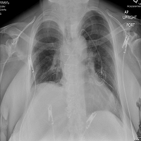
In the ED, the patient appeared to be in distress, with his vitals remarkable for a temperature of 102.4 F, blood pressure of 89/64 mmHg, heart rate of 124, respiratory rate of 40 and an oxygen saturation of 84% on ambient air by pulse oximetry. His exam revealed mottled skin with cold extremities, and dry mucous membranes with pharyngeal erythema with some exudate. His chest exam was remarkable for decreased chest expansion on the right along with diminished breath sounds and dullness to percussion of the right lung field. His abdomen was mildly tender along the right upper quadrant with deep palpation. His blood work showed a leukocytosis with 13,000/mcl WBC (normal 3,500-10,500 cells/mcl) with a left shift with 5,500/mcl bands (normal <1,200 cell/mcl). His lactic acid was 12 mmol/L (normal 0.5-2.2 mmol/L), and his basic metabolic panel was concerning for acute renal failure with a creatinine of 7.7 mg/dL (normal 0.8-1.4 mg/dL) and a Blood Urea Nitrogen (BUN) of 54 mg/dL (normal 7-20 mg/dL). His Chest X-Ray (CXR) in the ED demonstrated a mild hazy opacity within the right hemi-thorax, consistent with early pneumonia with an associated small pleural effusion (Figure 1).
The working diagnosis at that point was considered to be sepsis related to pneumonia or to an unidentified infectious fluid with the possibility of an intra-abdominal infectious source and it was felt that the patient would need further imaging with a computed tomography of the chest and abdomen which was performed 4 hours after the initial presentation. This imaging revealed no evidence of intra-abdominal source, but a dense consolidation of the right upper and lower lobes and a patchy consolidation of the right middle lobe of the lungs were identified, suggestive of extensive right sided pneumonia, along with a moderate-sized right pleural effusion (Figure 2).
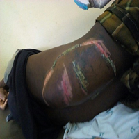
In the ED, the patient received fluid resuscitation with normal saline along with broad-spectrum antibiotic coverage with vancomycin, and piperacillin/tazobactam. He was then admitted to the Medical Intensive Care Unit (MICU) for further management of sepsis secondary to pneumonia. In the MICU, the patient was found to have increased work of breathing with a respiratory rate in the 40s despite being on a 100% oxygen nonrebreather mask. Given the rapid progression of parapneumonic effusion on serial imaging and the clinical deterioration, a decision was made to place a bedside chest tube to drain the effusion that was felt to be contributing to the respiratory distress. The procedure yielded 600 ml of serosanguinous pleural fluid. On the second day of hospitalization, the patient was feeling slightly better but started to develop a non-pruritic, whole-body, diffuse erythematous rash sparing the palms, soles and mucous membranes. His vitals were remarkable for a temperature of 98.5 F, blood pressure of 140/80 mmHg, heart rate of 107, respiratory rate of 36 with oxygen saturation of 95% on 2 L/min of oxygen via nasal cannula. Chest auscultation revealed coarse breath sounds along the right side of the chest with diminished sounds along the right lung base. His labs continued to show leukocytosis but his lactic acid was now improved at 2 mmol/L (normal 0.5-2.2 mmol/L). A gram stain of the pleural fluid showed grampositive cocciin chains and the preliminary fluid culture demonstrated beta hemolytic streptococci. The working diagnosis was invasive GAS infection in the settings of a recent pharyngitis. His clinical picture was also consistent with streptococcal Toxic Shock Syndrome (TSS) in the setting of hypotension on admission, the acute renal failure and the diffuse erythematous “sunburn-like” rash that developed and hence his antibiotics were modified to include vancomycin, ceftriaxone and clindamycin for streptococcal toxin control. The patient was also started on hemodialysis due to worsening renal function with a Cr of 8.9 mg/dL (normal 0.8-1.4 mg/dL).
The patient’s diagnosis was confirmed within the next 2 days of hospitalization as his pleural culture results were finalized as positive for streptococcus pyogenes (GAS) and the antibiotic regimen was further modified to include intravenous penicillin G, 2 million units every 4 hours, along with continuing the clindamycin for his TSS. Follow up CT scan of the chest 2 days after admission demonstrated significant improvement compared to the admission CT, consistent with the improvement in his symptoms (Figure 3).
Unfortunately, despite the initial clinical improvement, over the next 2 days, the patient developed progressive worsening shortness of breath and persistent high grade fevers. On chest exam he was found to have decreased air entry and increased crackles on the right. Labs showed a increasing leukocytosis with a white blood cell count of 36,000/mcl (normal 3,500- 10,500 cells/mcl). A repeat CXR showed worsening pleural effusion. The chest tube, despite what appeared to be appropriate positioning, drained minimal pleural fluid. A repeat CT scan of the chest was performed 6 days after admission which showed significant progressive worsening of the right-sided pleural effusion (Figure 4) and a decision was made then to treat the parapneumonic effusion surgically with decortication and drainage through VATS. The pleural peel biopsy from the surgery showed Gram-positive cocci consistent with the diagnosis of invasive GAS infection.
The patient continued to show progressive clinical improvement in the post-operative period although he required ongoing hemodialysis. The patient received the full course of his antibiotic therapy in the hospital and was then discharged home after 18 days of hospitalization, 12 days post-surgical decortication (Figure 5).
DISCUSSION
Invasive GAS continues to be continues to be associated with considerable morbidity and a mortality rate of upto 50 %.1,2 Invasive GAS infections can cause pneumonia and complicated parapneumonic effusions (empyema) requiring chest tube drainage or surgical decortication along with the appropriate antibiotic therapy to ensure successful outcome.3 Explosive pleurisy has been reported in cases of GAS pneumonia. Explosive pleurisy was first described by Braman and Donat in 1986 when they reported two cases of rapidly progressive pleural effusions attributed to GAS.4 Both of the cases reported were young healthy individuals, with no identifiable risk factors for severe GAS infection, who presented to the hospital with symptoms similar to our patient, notably fever, shortness of breath and pleuritic chest pain, preceded by pharyngitis. Both patients had rapidly progressive pleural effusion within hours of admission to the hospital, and both were initially started on antibiotics but eventually required chest tube drainage. Later in 2001, Johnson and then Sharma reported 2 more cases of explosive pleurisy that required thoracotomy for surgical drainage as chest tube drainage was insufficient for cure.5,6
An interesting feature associated with the above referenced cases with invasive GAS infections is the lack of identifiable risk factors for severe GAS infection as most of the reported cases have been in young and otherwise healthy individuals,7 which is similar to our presented case. The lack of significant comorbidities in our patient likely resulted in delaying the diagnosis of invasive GAS. Once the diagnosis has been established, aggressive supportive care and management is required in the setting of a multi-disciplinary approach.
The management of complicated parapneumonic effusions and explosive pleurisy includes the prompt initiation of antibiotics with GAS coverage including agents such as penicillin, plus the use of clindamycin for toxin control.8 Early thoracentesis should be considered for diagnostic and therapeutic purposes. Gram stain and pleural fluid cultures often assist with rapid diagnosis and antibiotic choice.9 The initial tap should be followed by prompt drainage with a chest tube if purulent or turbid fluid is aspirated, the pleural fluid has a positive gram stain or culture, a pleural fluid pH of less than 7.2 or an effusion that is rapidly progressive requiring drainage for symptomatic relief.10,11 Our patient was treated appropriately with chest tube placement for his rapidly progressive effusion, but his symptoms continued to worsen despite a brief period of initial improvement. Further deterioration of symptoms despite chest tube drainage is an indication for surgical drainage through thoracotomy or VATS for decortication and drainage, which was eventually done with our patient resulting in complete resolution.
CONCLUSION
It is important to maintain a high index of clinical suspicion for invasive GAS infections in young, otherwise healthy patients. Review of the previously reported cases of GAS induced-explosive pleurisy have shown that aggressive management with chest tube drainage and surgical decortication is often needed in addition to antibiotics. The timing of surgery should be based on clinical progression. VATS has demonstrated better outcomes and a higher rate of success compared to antibiotics alone in the management of early parapneumonic effusions.12 Close clinical monitoring, with consideration for early VATS in the setting of explosive pleurisy and parapneumonic effusions is likely to be associated with reduced morbidity and a more rapid resolution of illness.
CONSENT
No consent is required for this article publication.
CONFLICTS OF INTEREST
The Authors declare that there is no conflict of interests regarding the publication of this paper.
IRB POLICY
Our institution does not require an IRB approval for a single case study, as it is an educational activity that does not meet the definition of research.


