INTRODUCTION
Convenience surgery is the definition of a set of surgical interventions in pets that are not justified from a veterinary medical perspective.1 These interventions include tail-docking, earcropping, declawing, and debarking. Animal tails are known to be important for social signaling in some animal species.2 Docking is a surgical removal animal’s tail either for therapeutic reason or prevention of different injuries. Tail docking can be done in either of the two techniques. The first technique involves blocking the blood supply towards the tail with a rubber ligature for a few days until the tail falls off. Whereas, the second one involves the amputation of the tail with surgical scissors or a scalpel. The length to which tails are docked varies by breed and is often specified in the breed standard.3
Tail docking is practiced for centuries and although it could be painful, it is often done without any anesthetic.4 With tail docking, all or part of an animal’s tail is amputated.5 The tail docking has a long history since the beginning of domesticating animals. It is understood to date at least to the Roman Empire. Tail docking is done for several reasons. For instance, in most long-tail dog breeds, it is done for preventing injuries during exercising and fighting whereas, in hunting dogs, it is done to minimize the risk of getting cut up by brushes and thorns as the dog wags its tail. Thus, this is contested by a wide range of groups and is sometimes considered a form of animal cruelty, and this practice is restricted by many European countries.3,6
Historically, tail docking in many dog breeds is an established custom believed to have been introduced some 2000-yearsago.5 In larger breed dogs, docking has been practiced to prevent a different type of injuries and infections as a result of the type of work (guard work) (not to be confused with patrol work where a handler can provide secondary aid) may be docked to prevent their tails from being grabbed in a fight.5,6 The European Convention for the Protection of Pet Animals that is established by the Council of Europe in 1987 restricted the practice of tail docking for nonmedical reasons, however, member countries are free to opt-out of this provision, and almost half of them have done so. Norway completely banned the practice in 1987.7 Other countries where docking is banned include Australia and the United Kingdom.8
Nowadays, tail docking is done for prophylactic (to prevent injury), therapeutic, and cosmetic purposes. In some dogs such as hunting dogs, herding dogs, or terrier dogs, tails can be subject to abrasion or other injuries while moving through dense brush or thickets and this causes pain and infection since it collects burrs and foxtails when the tail wags. Bones in the tail can be broken due to different causes like accidents, injuries, and infections. This, in turn, causes spinal injury to the tail and this needs to remove tail in order to protect the dog from spinal injuries or trauma. However, the largest veterinary professional organization in the United States, the American Veterinary Medical Association, did not accept these since it lacks substantial scientific support.3,6,9
Tail docking is usually performed in puppies at an age of 3-4-days. Dogs, like most carnivores, are born in a much lessdeveloped nervous system than are most herbivores of the same age.5 The most suitable and recommended age of docking in a dog is between the first 3 and 5-days-old. At a later age, it might be expected that, since sensory and perceptive processes are more developed, any pain associated with docking may be intensified in all animal species.10 The American Veterinary Medical Association (AVMA) has expressed a concern that pain during this early development period could cause long-lasting damage and there is evidence in many species that noxious stimuli in the perinatal period may permanently alter the normal development of the central nervous system and have negative long-term consequences.3,8
During the evaluation of the techniques of tail docking, the assessment of acute pain during the procedure, and post-operative pain is the key issue. For this purpose, different parameters such as behavioral and physiological indicators are used to assess the pain. Vocalizations, reluctant to come in contact with a painful stimulus and distress are the behavioral indicators of pain, whereas an increase in plasma cortisol or corticosterone, and increased heart rate are considered as physiological indicators of pain.5 Besides, the behaviors and parameters from neurobiology must be considered for a sufficient understanding of the nature of pain in animals.3,6
Tail amputation should only be performed on those dogs whose tail, or associated structures have been injured or where there is occult pathology of this appendage.11,12 Amputation of the tail is indicated for therapeutic of different conditions such as traumatic lesions, infections, neoplasia, and perinanal fistula. Complications include infection, dehiscence, scarring, fistula recurrence, and anal sphincter and rectal trauma.13 In mature dogs, tail amputation is most commonly performed for the treatment of traumatic skin loss, ischemia, or denervation. Combined with other therapies, tail amputation may also improve outcome.14 In the present case report, a successful cosmetic tail amputation in a dog has been described.
CASE HISTORY AND CLINICAL EXAMINATION FINDINGS
A 2-year-old male dog weighing about 18 kg was presented to Veterinary Teaching Hospital (VTH) with a complaint of tail injured due to biting by another dog during the fighting and the dog was admitted to the hospital a few hours after injuries. Upon presentations, most of the dog’s tail was damaged and injured with bleeding from the injured part of the tail. Also, the dog was aggressive in condition due to pain. Besides, the injured area of the tail was covered with fresh clotted blood and contaminated with debris. The dog was diagnosed with different clinical parameters like heart rate, respiration rate, and the temperature was within physiological limits. Finally, the amputation of the dog’s tail was made above the level of injury.
Animal Handling, Pre-operative Patient Preparation, and Anesthetic Protocol
Pre-operatively, the dog was withheld 12-hours from feed and 6-hours from the water before coming to the clinic based on an appointment. The dog then was properly restrained and premedicated with chemical methods using Ana-Sed® (xylazine HCl, Heartland Vet Supply Ltd, CL, USA at a dose rate of 1.1 mg/kg, intramuscularly). After proper sedation, the dog was positioned in lateral recumbency and the area around the injured tail was washed with water and soap thoroughly after deep sedation. Then, the area around the wound was aseptically prepared by clipping, shaving, scrubbing, washing with water, and Salvon® (cetrimide 3% and Chlorhexidine gluconate 0.5% solution).
Besides, a tourniquet was applied to the base of the tail root and lumbosacral epidural nerve block was done with 2% lidocaine (Lidocaine hydrochloride 2%, Vedco Inc. Saint Joseph Missouri, USA) at a dose rate of 1ml/6 kg body weight to reduce hemorrhage.15 The dog was kept on fluid therapy soon after sedation (5% Dextrose solution plus 0.9% NaCl, 1000 ml stat, IV, Addis Pharmaceutical, Adigrat Ethiopia, at a surgical rate of 10 ml/kg/ hr. and calculated rate 1 drop/sec) to maintain the acid-base balance.
Surgical Management
After proper anesthetic protocol and aseptically preparing the area, the amputation of the tail was carried out according to Tobias.16 A V-shaped skin incision was made 1-2 cm caudal to the joint space at the proposed amputation site, at the base of the tail. After ligating the lateral caudal and medial caudal arteries cranial to the amputation site using chronic catgut 2-0, the coccygeal muscles were transected and the coccygeal vertebrae disarticulated both dorsally and ventrally. The dorsal and ventral skin flaps were pulled cranially to provide a tension-free closure using a scalpel blade. The skin was closed with a cross mattress suture pattern using size 1-0 nylon non-absorbable suture material. Finally, the area was properly cleaned and dressed with a 1% povidone-iodine solution and the dog was admitted home after full anesthetic recovery (Figure 1). was closed with a cross mattress suture pattern using size 1-0 nylon non-absorbable suture material. Finally, the area was properly cleaned and dressed with a 1% povidone-iodine solution and the dog was admitted home after full anesthetic recovery (Figure 1).
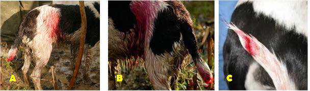
Figure 1. A and B: Clinical Presentation of an Injured Dog after Bite C: The Appearance of the Tail after Aseptic Preparation and Regional Anesthesia of Injured Tail
Post-operative Care and Outcome
Post-operatively, the area was bandaged with gauze having iodine, and oxytetracycline wound spray was applied on the incision site of the tail. Besides, antibiotic Pen Strep (30 mg/kg) and Meloxicam (0.5 mg/kg, IM) were administered IM, SID (once a day) for three days to manage post-operative complications and pain, respectively. The suture was removed on 10th-day post-surgery and the tail stump healed uneventfully after 2-weeks post-operation.
DISCUSSION
In the current case report, the tail was severely injured due to a bite during fighting with another dog. This finding in line with Olatunji-Akioye et al13 who has been reported tail amputation in a single case in association with tail fracture injury sustained through fighting with a cage mate as mainly therapeutic. Besides, this finding also supported by Ural et al17 who employed amputation in severely affected cattle tail.
In the present case report, the tail docking was conducted the recommended procedure for amputating the tail and this was in line with the previous report of Tobias16 and Eyarefe et al,18 that uses the same procedure to amputate tail in various species including dogs. The recommended age for tail docking is in puppies of 2-5-days of age usually performed. However, in this case, report, it was in a dog of 2-years of old that disagrees with the previous report of Eyarefe et al18 and Bennett et al5 that was performed during the first weeks of their age.
The reasons for tail docking include prevention of fecal soiling in lambs which can predispose animals to flystrike19 and tail biting in pigs to prevent serious injury.20 Tail docking in dairy cattle may have originated for two reasons: to control disease transmission and improve the milker’s comfort.2 In Karakkas lambs, deposition of fat in the tail which requires more energy than deposition of lean meat makes tail docking necessary to improve carcass characteristics.21
Nowadays, many countries restricted the practice of cropping and docking because of the effect of the procedure such as pain. European countries that have signed and ratified the European Convention for the Protection of Pet Animals have banned the practice of cropping of ears. However, some countries that ratified the convention made exceptions for tail docking.3,6,8 Thus, the use of cosmetic tail docking remains controversial for acute and chronic pain associated with the procedure.5
In this case, the tail amputation was done to prevent further infection. This work agreed with Olatunji-Akioye et al.13 Complications associated with tail amputation include infection, dehiscence, scarring, fistula recurrence, and anal sphincter and rectal trauma.13 Arguments against tail docking include acute pain, chronic health problems associated with docking, impaired locomotion, and impaired communication by docked dogs.5
CONCLUSION
In this case, successful surgical management of tail amputation was done in a dog. In conclusion, tail amputation remains the last option of treatment for severing traumatic tail if medical treatment is unsuccessful.
DECLARATION
The Mekelle University, College of Veterinary Science ethical review committee had critically reviewed and conclude that there was no ethical misconduct.
The approval of ethical committee was taken for conducting this study and followed all the animal ethics and welfare guidelines.


