Women’s Health

Open journal

ISSN 2380-3940

Cesarean Hysterectomy for Abnormal Placentation Using Balloon Occlusion of the Common Iliac Artery: Case Series

Masafumi Koshiyama*, Shingo Ukita, Masashi Ueda, Kenji Hishikawa, Yoko Furukawa, Yumi Itaya, CNM, Yumiko Watanabe, Natsuko Motooka Meguro, Haruko Horiuchi, and Makiko Oowaki

Masafumi Koshiyama, MD, PhD

Professor, Department of Women’s Health, Graduate School of Human Nursing, The University of Shiga Prefecture Shiga, 522-8533, Japan, Tel. +81-749-28-8664; Fax: +81-749-28-9532; E-mail: koshiyamam@nifty.com

INTRODUCTION

The rates of abnormal placentation, such as placenta accreta, increta, and percreta, have increased.1,2 The condition in which the placenta invades the entire depth of the uterine wall is called “placenta percreta”. Placenta accreta and increta are less severe variants of the same condition.3,4 The major risk factors of abnormal placentation are a placenta previa and previous cesarean section.5,6 The recent increased frequency of cesarean section has resulted in a high incidence of abnormal placentation.

Placenta accreta, increta, and percreta often result in massive peripartum hemorrhaging and are sources of maternal morbidity and mortality in modern obstetrics.7,8,9,10 In particular, uterine rupture and profuse hemorrhaging have been associated with placenta percreta.11 Placenta percreta represents the most difficult management situation of these disorders.

The options for treating abnormal placentation include both conservative and extirpative approaches.12 The generally accepted treatment for the most severe form of abnormal attachment of the placenta is cesarean hysterectomy without attempts to detach the placenta. Recently, the role of interventional radiology in the field of obstetrical hemorrhaging has been widely investigated. Preoperative internal iliac artery balloon occlusion (IIABO) has been widely performed to reduce the blood loss during cesarean hysterectomy.13,14,15,16

We herein report the safe performance and management of cesarean hysterectomy using common iliac artery balloon occlusion (CIABO).

PATIENTS AND METHODS

Four patients who underwent cesarean hysterectomy at 33-36 weeks of gestation using balloon occlusion at Otsu Red Cross Hospital, (Shiga, Japan) from 2009-2013 were recruited for the study. We informed the patients and their families of all of the risks involved in the surgery. All the 4 patients had previously undergone cesarean section. They had placenta previa and myometrial involvement on old operation scars. The myometrial invasion depths of the placenta were determined on ultrasonography (US) and magnetic resonance imaging (MRI). All the 4 patients then underwent cesarean hysterectomy using CIABO (In one case, the balloon was moved from the internal iliac artery to the common iliac artery in order to reduce blood loss). The study protocol was approved by the ethics committee of Otsu Red Cross Hospital, Otsu, Shiga Prefecture, Japan.

Procedure of Cesarean Hysterectomy

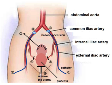
Prior to the surgery, no infiltrations of the placenta into the mucosa of the urinary bladder were shown by cystoscopy, and bilateral ureteral stent-catheters were inserted by urologists. Under general anesthesia, the balloon catheters were inserted via the bilateral femoral arteries and fixed in the common iliac arteries in the operation room by radiologists (Figure 1). During cesarean hysterectomy, these balloons were not inflated. The surgeons started the cesarean section, followed by a vertical incision of the abdominal skin and transverse uterine fundal incision. In case 1 alone, the balloons were first fixed in the internal iliac arteries, but were subsequently moved to the common iliac arteries due to a large amount of blood loss.17


Figure 1: Anatomy in the Pelvis and Cesarean Hysterectomy Procedure. For more Information, see the Text of this.

WHOJ-3-117 Fig 1


As soon as the child was born, the balloons in the bilateral common iliac arteries were inflated. The uterine incision was sutured without detaching the placenta. This was followed by performing a total abdominal hysterectomy as shown in Figure 1. The procedure entailed; ligating and cutting of the uterine arteries at the point of intersection with the ureter. Incision of recto-cervical fold of peritoneum was made to have access to the posterior vaginal fornix followed by a circumferential incision of the vagina at the level of the vaginal fornices. Ligating and cutting the bilateral round ligaments and ligament ovaries were performed. We then incised the left and right connective tissues of the uterus (the sites of ascending and descending branches of uterine arteries) in the upper direction. We were then able to remove the uterus completely. While repairing the incisions, the bilateral catheters were removed.

The success of these procedures was evaluated based on the amount of blood loss and presence of complications.

RESULTS

The clinical data of 4 patients treated with cesarean hysterectomy using balloon occlusion of the common iliac artery are shown in Table 1.


Table 1: The Clinical Data of 4 Patients Treated with Cesarean Hysterectomy using Balloon Occlusion of the Common Iliac Artery.

Patient

Case 117 Case 2 Case 3 Case 4
Age 36 yr 32 yr 35 yr

33 yr

Clinical diagnosis

Preg. 35 w 6 d

previous 4 times c-sec

total placenta previa

s/o placenta increta

Preg. 35 w 5 d

previous 1 time c-sec

total placenta previa

s/o placenta accreta

Preg. 36 w 0 d

previous 1 time c-sec

total placenta previa

s/o placenta increta

Preg. 33 w 5 d

previous 1 time c-sec

total placenta previa

s/o placenta accreta

Balloon position  internal iliac a. =>common iliac common iliac a. common iliac a.

common iliac a.

Occluded time

1 hr 15 min 56 min 50 min 55 min
Blood loss 7720 ml
(including amniotic fluid)
3781 ml
(including amniotic fluid)
3851 ml
(including amniotic fluid)

1361 ml
(including amniotic fluid)

Blood transfusion

Autologous blood 1200 ml

MAP 10 iu, FFP 10 iu

Autologous blood 1050 ml Autologous blood 1750 ml Autologous blood 1200 ml
Complication None None None

None

Surgical time

2 hr 49 min 3 hr 43 min 4 hr 25 min 2 hr 48 min
Pathologic diagnosis Placenta percreta Placenta increta Placenta percreta

Placenta accreta


The patients’ ages ranged from 32-36 years old (mean 34 years old). Two were ultimately diagnosed with placenta percreta, 1 with placenta increta and one with placenta accreta. There was only a case where the clinical diagnosis before surgery corresponded to the final diagnosis after surgery. The actual invasive depth of the placenta was deeper than that estimated before surgery in three cases. On comparing the diagnoses before and after surgery, the final diagnoses were worse after surgery in three cases. All of the placentas were attached to the old operation scars. However, the myometrial invasion depths of the placentas were found to have not been accurately determined on US and MRI.

As we previously reported, we were unable to control the massive bleeding with IIABO.17 We therefore, immediately moved the balloon from the internal iliac artery to the common iliac artery. Because of our experience with case 1, we placed the balloon in the common iliac arteries from the start of the operation in cases 2-4.

Regarding the outcomes of cesarean hysterectomy, the total volume of blood lost was 7720 ml in case 1, 3781 ml in case 2, 3851 ml in case 3 and 1361 ml in case 4. The volume of blood lost in the 3 patients whose balloons were placed in the common iliac artery from the start of the surgery ranged from 1361-3851 ml (including amniotic fluid and fewer amounts of bleeding than these), and these patients received only autologous blood transfusion (1050 ml, 1200 ml and 1750 ml; mean 1333 ml). The time of occlusion of the common iliac arteries ranged from 50 min to 1h 15 min with intervals of 10 min for deflating the balloons.

Complications did not occur in any of the 4 patients treated with these methods and management strategies. Our staff (midwives and doctors) visited the patients many times and also supported their mental healing. The 4 patients were discharged from our hospital on the 7th or 8th post-operative day. Post-operative evaluation of the patients 30 days after the surgery did not reveal any complications with all mothers and children being in good health.

DISCUSSION

Although, the impact of morbidly abnormal placentation on pregnancy outcomes has been well-described, no randomized trials and few studies have examined the management of pregnancies complicated by this disorder. As a result, the recommendations for its management are based on case series and reports, personal experience and expert opinion.

The options for treating abnormal placentation include both conservative and extirpative approaches. O’Brien et al12 reported that conservative treatment was preferred in hemodynamically stable patients. However, the patients treated with such conservative therapy are at continued risk of massive hemorrhaging and require prolonged intensive observation. Postpartum hemorrhage necessitating hysterectomy has been observed from 3 hours to 7 weeks after delivery in women who were initially managed conservatively.18,19 In contrast, the extirpative approach is completed within a certain period and usually requires an adjuvant hemostatic technique, such as pre-operative placement of balloon occlusion.13,14,15,16 In our hospital, we have safely performed cesarean hysterectomies using arterial balloon occlusion. The size of their balloons should be optimally tailored to the anatomically targeted vessels being occluded using pre-operative MRI, because a poorly occluded vessel will yield incomplete hemostasis and increased hemorrhage.20

As we previously reported in case 1,17 we attempted to perform IIABO to treat patients with placenta percreta. However, massive vaginal bleeding suddenly occurred due to detachment of the placenta following uterine contraction during cesarean hysterectomy. A quick shift from IIABO to CIABO was very effective in reducing the volume of blood loss. The use of IIABO in patients has reported no improved surgical outcomes compared with patients without the treatment.21,22 Failure of IIABO can be explained by the presence of extensive anastomosis bridging the internal and external iliac arteries.23 We therefore, subsequently performed CIABO during cesarean hysterectomy (cases 2-4). Theoretically, proximal balloon occlusion of the large vessels such as the aorta and common iliac arteries is more effective for hemostasis. However, it is possible to cause the potential risk of ischemia to the extremities if prolonged occlusion is required in these cases.20

In cases 2-4, we were able to control the hemorrhage using CIABO during cesarean hysterectomy (range of blood loss: 1361-3851 ml including amniotic fluid). Regarding blood transfusion, we were able to use autologous blood alone in cases 2-4. In addition, all of the patients were discharged from our hospital without any complications, such as thrombus, embolism, coagulopathy, severe anemia, hematoma, infection or organ disorders. Balloon occlusion has been thought not to stop blood flow completely.24

While a few other reports have also described the superiority of CIABO to other methods,23,25,26,27,28 rare cases complicated by thrombosis have been reported after CIABO, necessitating conservative managements with a heparin drip.28,29,30 Therefore, the occlusion time should be as short as possible.

Following cesarean hysterectomy, the patients are at risk for complications, such as persistent coagulopathy, anemia, thromboembolism and other organ disorders, and death. As such, the patients may experience anxiety or even post-traumatic stress disorder. They may also experience pain, uterine loss, numbness, and issues in communicating or bonding with their baby. These patients therefore, require close monitoring, sometimes in an intensive-care setting.31 Psychiatrists, special midwives and social workers should focus on the mental health care of these patients. We were unable to follow the patients for more than a month after surgery because we could not set up a system of the further examination. Understanding patients’ experiences with emergency peripartum hysterectomy can help practitioners not only address patients’ initial complications but also provide needed long-term support.32

CONCLUSIONS

CIABO during cesarean hysterectomy was effective in controlling blood loss in 4 cases and can be conducted safely. Given that only 4 cases were managed using this technique in this study, further evaluation of the efficacy and outcomes of CIABO is needed. Patients treated with cesarean hysterectomy should be managed by close monitoring in an intensive-care setting with ample support for their mental healthcare.

CONFLICTS OF INTEREST

The authors declare that they have no conflicts of interest.

1. Weckstein LM, Masserman JSH, Gaite TJ. Placenta accrete: A problem of increasing clinical significance. Obstet Gynecol. 1986; 69: 480-482. Web site. https://www.ncbi.nlm.nih.gov/pubmed/3808530. Accessed December 15, 2016

2. Khan M, Sachdeva P, Arora R, Bhasin S. Conservative management of morbidity adherent placenta- A case report and review literature. Placenta. 2013; 34: 963-966. doi: 10.1016/j.placenta.2013.04.016

3. Morison JE. Placenta accrete. A clinicopathologic review of 67 cases. Obstet Gynecol Annu. 1978; 7: 107-123. Web site. https://www.ncbi.nlm.nih.gov/pubmed/662213. Accessed December 15, 2016.

4. Miller D, Cholletr JA, Goodwin TM. Clinical risk factors for placenta previa-placenta accrete. Am J Obtet Gynecol. 1997; 177: 210-214. doi: 10.1016/S0002-9378(97)70463-0

5. Kistner RW, Hertig AT, Reid DE. Simultaneously occurring placenta previa and placenta accrete. Surg Gynecol Obstet. 1952; 94: 141-151. Web site. https://www.ncbi.nlm.nih.gov/pubmed/14901248. Accessed December 15, 2016.

6. Clark SL, Koonings PP, Phelan JP. Placenta previa accrete and prior cesarean section. Obstet Gynecol. 1984; 66: 89-92. Web site. https://www.ncbi.nlm.nih.gov/pubmed/4011075. Accessed December 15, 2016.

7. Rossi AC, Lee RH, Chmait RH. Emergency postpartum hysterectomy for uncontrolled postpartum bleeding: A systematic review. Obstet Gynecol. 2010; 115: 637-644. doi: 10.1097/AOG.0b013e3181cfc007

8. Wright JD, Pri-Paz S, Herzog TJ, et al. Predictors of massive blood loss in women with placenta accrete. Am J Obstet Gynecol. 2011; 205: 38e1-6. doi: 10.1016/j.ajog.2011.01.040

9. Bateman BT, Mhyre JM, Callaghan WM, Kukina EV. Peripartum hysterectomy in the United States: Nationwide 14 year experience. Am J Obstet Gynecol. 2012; 206: 63 e1-8. doi: 10.1016/j.ajog.2011.07.030

10. Eshkoli T, Weintraub Ay, Sergienko R, Sheiner E. Placenta accrete: Risk factors, perinatal outcomes, and consequences for subsequent births. Am J Obstet Gynecol. 2013; 208: 219.e1-e7. doi: 10.1016/j.ajog.2012.12.037

11. Lloyd-Jones R, Winterton WR. Spontaneous rupture of the uterus due to placenta percreta: A case report. J Obstet Gynecol Br Common. 1961; 68: 273-276. Web site. https://www.ncbi.nlm.nih.gov/pubmed/8697340. Accessed December 15, 2016.

12. O’Brien JM, Barton JR, Donaldson ES. The management of placenta percreta: Conservative and operative strategies. Am J Obstet Gynecol. 1996; 175: 1632-1638. doi: 10.1016/S0002-9378(96)70117-5

13. Dubois J, Garel L, Grignon A, Lemay M, Leduc L. Placenta percreta: Balloon occlusion and embolization of the internal iliac arteries to reduce intraoperative blood losses. Am J Obstet Gynecol. 1997; 176: 723-726. Web site. https://www.ncbi.nlm.nih.gov/pubmed/9077641. Accessed December 15, 2016.

14. Ojala K, Perälä J, Kariniemi J, Ranta P, Raudaskoski T, Terkay A. Arterial embolization and prophylactic catheterization for the treatment for severe obstetric hemorrhage. Acta Obstet Gynecol Scand. 2005; 84: 1075-1080. doi: 10.1111/j.0001-6349.2005.00727.x

15. Tan CH, Tay KH, Steah K, et al. Perioperative endovascular internal iliac artery occlusion balloon placement in management of placenta accreta. AJR. 2007; 189: 1158-1163. doi: 10.2214/AJR.07.2417

16. Carnevale FC, Kondo MM, de Oliveira Sousa W Jr, et al. Perioperative temporary occlusion of the internal iliac arteries as prophylaxis in cesarean section at risk of hemorrhage in placenta accreta. Cardiovasc Intervent Radiol. 2011; 34: 758-764. doi: 10.1007/s00270-011-0166-2

17. Hishikawa K, Koshiyama M, Ueda M, et al. Exchange of intraoperative balloon occlusion of the internal iliac artery for the common iliac artery during cesarean hysterectomy in a patient with placenta percreta. Am J Case Rep. 2013; 14: 409-411. doi: 10.12659/AJCR.889449

18. Jaffe R, DuBeesheter B, Sherer DM, Thompson EA, Woods JR Jr. Failure of methotrexate for the term placenta percreta. Am J Obstet Gynecol. 1994; 171: 558-559.doi: 10.1016/0002-9378(94)90300-X

19. Veenstra M, Spinder T, Dekker GA, van Geijin HP. Post partum intra-abdominal hemorrhage due to placenta percreta. Eur J Onstet Gynecol. 1995; 62: 253-256. doi: 10.1016/0301-2115(95)02183-8

20. Knuttinen MG, Jani A, Gaba RC, Bui JT, Carrillo TC. Balloon occlusion of the hypogastric arteries in the management of placenta accrete: A case report and review of the literature. Semin Intervent Radiol. 2012; 29: 161-168. doi: 10.1055/s-0032-1326924

21. Levine AB, Kuhlman K, Bonn J. Placenta accreta: Comparison of cases managed with and without pelvic artery balloon catheters. J Matern Fetal Med. 1999; 8: 173-176. doi: 10.1002/(SICI)1520-6661(199907/08)8:4<173::AID-MFM7
>3.0.CO;2-V

22. Bodner LJ, Nosher JL, Gribbin C, Siegel RL, Beale S, Scorza W. Balloon-assisted occlusion of the internal iliac arteries in patients with placenta accreta/percreta. Cardiovasc Intervent Radiol. 2006; 29: 354-361. doi: 10.1007/s00270-005-0023-2

23. Shih JC, Liu KL, Shyu MK. Temporary balloon occlusion of the common iliac artery: New approach to bleeding control during cesarean hysterectomy for placenta percreta. Am J Obstet Gynecol. 2005; 193: 1756-1758. doi: 10.1016/j.ajog.2005.08.033

24. Matsubara S, Nonaka H, Kobayashi M, Kuwata T, Fujii H. Intrauterine balloon occlusion during cesarean hysterectomy for placenta previaaccreta: The internal or common iliac artery? Acta Obstet Gynecol Scand. 2014; 93: 122-123. doi: 10.1111/aogs.12273

25. Clausen C, Stensballe J, Albrechtsen CK, Hansen MA, Lonn L, Langhoff-Roos J. Balloon occlusion of the internal iliac arteries in the multidisciplinary management of placenta percreta. Acta Obstet Gynecol Scand. 2013; 92: 386-391. doi: 10.1111/j.1600-0412.2012.01451.x

26. Nishida Y, Hirakawa T, Utsunomiya Y, Tsusue H, Narahara H. Transfundal uterine incision performed with prophylactic common iliac artery balloon occlusion for patient with placenta previatotalis. Clin Med Insights Case Rep. 2012; 5: 93-97. doi: 10.4137/CCRep.S9777

27. Minas V, Gul N, Shaw E, Mwenenchanya S. Prophylactic balloon occlusion of the common iliac arteries for the management of suspected placenta accreta/percreta: Conclusions from a short case series. Arch Gynecol Obstet. 2015; 291: 461-465. doi: 10.1007/s00404-014-3436-9

28. Chou MM, Kung HF, Hwang JI, Chen WC, Tseng JJ. Temporary prophylactic intravascular balloon occlusion of the common iliac arteries before cesarean hysterectomy for controlling operative blood loss in abnormal placentation.Taiwan J Obstet Gynecol. 2015; 54: 493-498. doi: 10.1016/j.tjog.2014.03.013

29. Sewell MF, Rosenblum D, Ehrenberg H. Arterial embolus during common iliac balloon catheterization at cesarean hysterectomy. Obstet Gynecol. 2006; 108: 746-748. doi: 10.1097
/01.AOG.0000201992.80130.2c

30. Matsueda S, Hidaka N, Kondo Y, Fujiwara A, Fukushima K, Kato K. External iliac artery thrombosis after common iliac artery balloon occlusion during cesarean hysterectomy for placenta accreta in cervico-isthmic pregnancy. J Obstet Gynecol Res. 2015; 41: 1826-1830. doi: 10.1111/jog.12777

31. Brigham and Women’s Hospital. Caring for the placenta accreta patient. Web site. http://www.brighamandwomens.org/Departments_and_Services/obgyn/Medical_Professionals/caring-for-placenta-accerta-patient.aspx#pageheadline. Accessed June 30, 2016.

32. de la Cruz CZ, Coulter ML, O’Rourke K, Alio PA, Daley EM, Mahan CS. Women’s experiences, emotional responses, and perceptions of care after emergency peripartum hysterectomy: A qualitative survey of women from 6 months to 3 years postpartum. Birth. 2013; 49: 256-263. doi: 10.1111/birt.12070

LATEST ARTICLES

Chest X-ray Showed a Hazy Left Upper Lung Infiltrate

A Noteworthy Case of Myasthenic Crisis Induced by Levofloxacin

Ada Young*, Ramya Ramesh and Milind Awale

doi.

The Right Thigh Anterior Compartment was Swollen, and the Skin was Ulcerated due to the Traditional Cautery

Primary Skeletal Muscle Lymphoma: A Case Report and Literature Review

Solomon Bishaw*, Addisu Alemu and Abel Tefera

doi.

An Unusual Presentation of Encephalitis in a Patient with Lyme Neuroborreliosis

Maithily Patel*, Jazmin Jatana, Ramya Ramesh and Milind Awale

doi.

Practical Pointers for Drug Development and Medical Affairs

Gerald L. Klein*, Roger E. Morgan, Shabnam Vaezzadeh, Burak Pakkal and Pavle Vukojevic

doi.

10.17140/CTPOJ-7-125

Prevalence and Risk Factors of Subclinical Mastitis of Goats in Banadir Region, Somalia

Omar M. Salah*, Yasin H. Sh-Hassan, Moktar O. S. Mohamed, Mohamed A. Yusuf and Abas S. A. Jimale

doi.10.17140/VMOJ-9-184

LATEST ARTICLES