OVERVIEW OF THE ILLUSTRATION
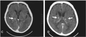
A 68-year old woman was referred to our hospital because of dry cough with persistent numbness in the extremities. Chest roentgenogram showed right hilar tumor with mediastinal lymphnode and brain magnetic resonance imaging (MRI) revealed multiple tiny nodules. After performing the transbronchial needle aspiration obtained from subcarinal lymphnode, she was diagnosed with small cell lung cancer (SCLC) accompanied by multiple brain metastasis (clinical stage IV: T4N3M1). After completion of four cycles of chemotherapy, the size of lung lesion was regressed together with complete resolution of brain lesions, and she discharged uneventfully. However, her numbness was remained. Four months later, she was hospitalized again for severe numbness in her trunk and extremities. Brain MRI (contrast-enhanced T1-weighted image) demonstrated new symmetric metastatic lesions in bilateral thalamus (Figure A and B) as well as left putamen. Another two months later, numbness exacerbated and spread from the top to toe with the growing of primary and bilateral thalamic metastatic lesions on computed tomography (CT) (Figure C and D). In spite of whole-brain irradiation, she had suffered from severe numbness, and died of obstructive pneumonia. To our knowledge, this is the first case of bilateral symmetric thalamic metastasis in SCLC with refractory numbness over the whole body.
Figure A and B. Brain MRI (contrast-enhanced T1-weighted image) Demonstrated New
Symmetric Metastatic Lesions in Bilateral Thalamus (arrow heads) as well as Left Putamen.

Figure C and D. Head CT with Contrast-media Depicted that Bilateral Thalamic Metastatic Lesions are getting Larger in Size.


