Cancer Studies and Molecular Medicine

Open journal

ISSN 2377-1518

Application of Radionuclides and Antibody -Drug Conjugates to Target Cancer

Sean L. Kitson*

Sean L. Kitson, PhD

Department of Biocatalysis and Isotope Chemistry, Almac, 20 Seagoe Industrial Estate, Craigavon, BT63 5QD, UK E-mail: sean.kitson@almacgroup.com

INTRODUCTION

A century ago, the bacteriologist Paul Ehrlich (1908 Noble Prize) a pioneer of chemotherapy and haematology, first postulated the concept of targeted therapy towards the treatment of disease causing agents.1 This concept was to create an ideal therapeutic agent termed the ‘magic bullet’ which went directly to specific cellular targets in order to attack the disease. Currently, Ehrlich’s vision is now being realized in the treatment of cancer with the development of targeted therapies, mainly based on monoclonal antibodies.2

A major breakthrough was made in 1975, by the Nobel Prize winners Milstein and Köhler, in the development of hybridoma technology. This technology platform revolutionised the production of antibodies by having a single specificity towards the cognate antigen, in the development of targeted therapies.3 Moreover, this approach is being exploited by several biopharmaceutical companies to develop strategies for delivering radionuclides to image and destroy a variety of cancers including adequate cytotoxic drug payloads.4

These cytotoxic drug payloads are utilised in the development of Antibody-Drug Conjugates (ADCs) and include the anti-neoplastic agents: Mono Methyl Auristatin E (MMAE) and mertansine (DM1) to target the microtubules in cancerous cells. These other payloads include the DNA damaging agents calicheamicins and duocarmycins extending to the topoisomerase II inhibitors doxorubicins and camptothecins.5 All of these cytotoxic drugs have demonstrated in vitro potency against several tumour cell lines, down to the picomolar level. This compares to first generation ADCs using nanomolar amounts of doxorubicin.6

Currently, other emerging drug payloads such as the sequence selective DNA alkylating agents called Pyrrolo Benzo Diazepines (PBDs) will form the basis of the next generation of ADCs.7 These PBDs have shown to be ten thousand times more potent than systemic chemotherapeutics and nearly a thousand times more potent than other cytotoxins used in ADCs.

The ideal treatment plan for a patient is first to locate the cancerous site(s) by using a radionuclide antibody capable of imaging the tumour volume. The following imaging modalities can be applied: planar imaging; Single Photon Emission Computed Tomography (SPECT) and Positron Emission Tomography (PET). These techniques can be extended to state-of-the-art clinical hybrid imaging systems which combine SPECT (or PET) with Computed Tomography (CT) and more recently PET scanners with functional magnetic resonance imaging (fMRI) instruments.8,9

However, if the cancer site is shown to retain an appropriate level of the antibody – through the application of these imaging techniques – based on gamma or positron emitters: it would be reasonable for the patient to receive a therapeutic dose of the same antibody – labelled with a radionuclide emitting alpha or beta radiation – which ever proves more capable of killing the cancer cells.

The limitation of utilising murine antibodies has been circumvented by the use of chimeric, humanized, or fully human monoclonal antibodies.10 The challenge of targeting solid tumours with alpha and beta radiation is the dilemma of inducing a sufficient response on the cancerous mass without producing lethal toxic side effects. Therefore, it is paramount to analyse and control the radiation dose being delivered to tumour site(s) and compare the effects of this dose on the surrounding healthy cells.

Consequently, it is important to calculate the risk to other normal and/or non-neoplastic sites, capable of concentrating radioactivity, especially in the excretory organs (e.g. kidneys).11 To help to define the risk, it is important to obtain pharmacokinetic data about the therapeutic radionuclide. This will enable a calculation of the percentage of injected dose per gram tissue therefore limiting normal tissue damage.12-14

The majority of precedents set by Radio-Immuno Therapy (RIT) were made by antibodies labelled with beta emitters (e.g. iodine-131). Today, after extensive research and clinical trials, RIT therapy against various cancers has now been accepted.15 A continuation of research in the application of alpha emitters to treat cancer has been proposed for radiolabelling of many molecules, transported by various bio-vectors such as monoclonal antibodies.16

The main emphasis is the utilisation of radiolabelled antibodies as agents for Radio-Immuno Therapy (RIT). Following labelling with alpha emitters, Radionuclide Antibody-Conjugates (RACs) became the prototype for Targeted Alpha Therapy (TAT) using other targets and bullets, as in the case of peptides17 and somatostatin receptors.18

Ongoing clinical trials have shown that somatostatin receptor peptides labelled with the beta emitters yttrium-90 and lutetium-77 have been effective in the treatment of neuroendocrine tumours.19

RADIONUCLIDES TARGETING CANCER

Currently, there are around 100 radionuclides that emit alpha radiation; the majority of them produced in nuclear reactors. Only a few are considered useful as therapeutics agents. These include bismuth-213 (generator produced),20 astatin211 (cyclotron produced),21 actinium-225 (generator produced)22 and thorium-227 (generator produced).23 These radiolabelled therapeutic agents transported by bio-vectors such as monoclonal antibodies can be utilized in the treatment of a variety of cancers such as lymphomas, leukaemia and melanomas.24 This is demonstrated further on by the ability of Xofigo® to form complexes within the area of bone metastases.25 This is due to theactive moiety radium-223, in the form of radium-223 dichloride, to mimic calcium and the ability to complex with the bone mineral hydroxyapatite.26

A clinical precedent, on the practice of using alpha therapy towards bone metastases, with radium-223 dichloride (half-life=11.4 days) marketed as Alpharadin® became a firstin-class therapeutic.27 In May 2013, Alpharadin® now called Xofigo®, was given FDA approval to treat patients with castration-resistant prostate cancer, symptomatic bone metastases with no known visceral metastatic disease.28

Xofigo® is the first and only alpha particle-emitting radioactive therapeutic agent approved by the FDA that has demonstrated improvement in overall survival rates.29 To date, the most promising advance in cancer therapy is connected to the evolvement of using Radiolabelled Antibody-Conjugates (RACs) to deliver alpha particles.30

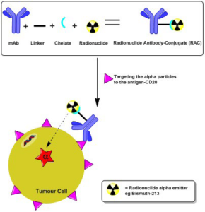
The basic principle of the TAT technique relies on the emission of alpha particles in which the radionuclide (e.g. actinium-225, bismuth-213, astatine-211) is held in a crown shaped chelate (e.g. derivatives of DTPA, DOTA), connected preferably to a low molecular weight drug (Figure 1). Alternatively, it may be more frequently linked to a monoclonal antibody, antibody fragments or peptide via a linker-chelate.31,32 Therefore, it is paramount to get the right combination of radionuclide, linker-chelate and/or peptide, drug substrate or antibody for a particular cancer.33-35 This is to ensure that an adequate amount of radiation is delivered to the cancer site by targeting the specific antigen (e.g. CD20) to annihilate it.

Figure 1: TAT targetingCD20 antigens on tumour cells

CSMMOJ-1-102Fig1

Several TAT research groups have shown that the ideal radionuclide for this approach to be effective must have the following basic parameters.36-38:

• The radionuclide must emit an energy lower than 40 keV;

• Alpha particles have a short pathlength (50-80 microm) and high linear energy transfer of approximately 100 keV/mi crom;

• The radionuclide should have an ideal half-life of 30 min utes to 10 days to allow for logistics and treatment plan for the patient;

• For the generation of ‘medical’ radionuclides, the daugter radionuclide must be stable with a half-life greater than 60 days;

• The radiopharmaceutical in the form of kits and/or synthsis must be able to incorporate the radioactive label into carrier substrates as rapidly as possible for patient use.

Numerous clinical trials have utilised a wide range of bio-vectors to target cancer and include: HuM195 for acute myelogenous leukaemia;39 Astatinated MX35-F(ab’)2 monoclonal antibodies for ovarian cancer;40 Radium-223 dichloride for bone metastases;41 Murine 9.2.27 to target the Melanomaassociated Chondroitin Sulfate Proteoglycan (MCSP) antigen on melanoma;42 CD20 antigen for lymphoma43 and human IgG2/mouse chimeric anti-tenascin 81C6 forglioblastoma multiforme.44

Currently several preclinical trails include the biovectors: Monoclonal antibody C595 labelled with bismuth-213 to target MUC1 gene expressed by the prostate;45 PA12 human recombinant protein to target urokinase-type Plasminogen Activator (uPA) system which is expressed in several types of cancer (e.g. breast cancer);46 Monoclonal antibody J591 to target the Prostate Specific Membrane Antigen (PSMA)47 and Bevacizumab (Avastin®) in the treatment of recurrent glioblastoma.48

Currently, other approaches to target cancer include the following FDA approved radiopharmaceuticals:

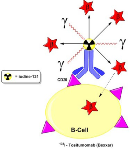
This targeted approach continues with Bexxar®, which contains the antibody tositumomab, radiolabelled with iodine-131 to target the CD20 antigen.49 The patient first receives tositumomab, followed by the infusion of tositumomab radiolabelled with iodine-131. This is the same antibody covalently bound to the radionuclide iodine-131. The iodine-131 emits both beta and gamma radiation and decays with a half-life of 8 days (Figure 2). A successful clinical study involving 40 patients led to the approval in 2003 of Bexxar® for the treatment of rituximabrefractory, low-grade, follicular non-Hodgkin’s lymphoma.50

Figure 2: Bexxar® firing beta and gamma radiation to kill B-cells (Adaptation from Kitson et al51)

CSMMOJ-1-102Fig2

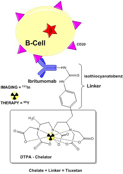
The unique feature of Zevalin® is that it can be used to target the CD20 antigen on B-cell non-Hodgkin’s lymphoma to allow imaging and in therapy to destroy it. The radiopharmaceutical is first labelled with the radiometal indium-111, using tiuxetan chelation. This gamma-emitter is transported to the lymphoma sites by the monoclonal antibody ibritumomab which detects B-cells. SPECT imaging then can be used to verify that the antibody is properly distributed within the body.52 The indium-111 is swapped with radionuclide yttrium-90, to transport beta particles to kill B-cells (Figure 3).

Figure 3: Zevalin® firing beta-particles at B-cells (Adaptation from Kitson et al.51 )

CSMMOJ-1-102Fig3

Zevalin® therapy has useful indications for relapsed or refractory, low grade or follicular, B-cell non-Hodgkin’s lymphoma.53 In 2002, the FDA gave approval for Zevalin® to be used in treatment of relapsed or refractory low-grade follicular or transformed B-cell non-Hodgkin’s lymphoma; including patients with rituximab refractory follicular non-Hodgkin’s lymphoma. In 2008, Zevalin® was approved as the first-line consideration for follicular lymphoma in the European Union.54

ANTIBODY-DRUG CONJUGATES (ADCS) TARGETING CANCER

These immunotherapeutic agents called Antibody Drug Conjugates (ADCs), target specific antigens particularly on cancer B-cellssuch as CD19, CD20, CD21, CD22, CD40, CD72, CD79b and CD180.55 In 2011, the FDA gave approval to Adcetris® to treat Hodgkin’s lymphoma and systematic anaplastic large-cell lymphoma.56 The continued success of this therapeutic agent arrived in February 2013, when the FDA announced the approval of Kadcyla®, for the treatment of metastatic breast cancer.57

Adcetris® consists of the bio-vector brentuximab (IgG1 cAC10), which is a chimeric monoclonal antibody, to target the human CD30 antigen on B-cells.58 The antibody is attached to a combination linker, via the Cysteine Sulfhydryl (Cys-SH) groups. These are generated from the mild reduction of the inter-chain hinge disulfide bonds of the antibody.59 This linker combination is made up of a thiol-reactive maleimidocaproyl (mc) spacer, the dipeptide Valine-Citrulline (Val-Cit) linker and a 4-aminobenzylcarbamate (PABC) self-immolative spacer.60 This set-up facilitates the conjugation of on average four drug Molecules of Monomethyl Auristatin E (MMAE) on the antibody (Figure 4).61 MMAE is so toxic to healthy cells that it cannot be used as a stand-alone chemotherapeutic.62

Figure 4: Structure of Adcetris®

CSMMOJ-1-102Fig4

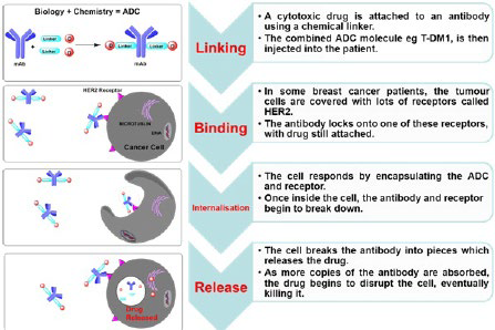
In the mechamism of action ADC binds to the antigen on the B-cell to from an ADC-antigen complex (Figure 5). The ADC-antigen complex in the case of Adcetris® is internalized by clathrin-mediated endocytosis and transported to the intracellular lysosome compartment. The ADC-antigen complex fuses with the lysosome and the action of cathepsin-B proteases initiates a spontaneous intramolecular [1,6]-elimination of PABC to release the free-drug MMAE (picomolar potency) into the cytoplasm.63 This drug then inhibits microtubule assembly, causing depolymerization, leading to cell cycle arrest.

Figure 5: Mechanism of Action of ADCs

CSMMOJ-1-102Fig5

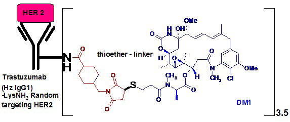
Kadcyla® consists of three components (Figure 6): the humanized MAb (IgG1) trastuzumab (Herceptin®) to target HER2 tumour antigens; the microtubule polymerization inhibitormaytansinoid DM1 drug and the (N-Maleimidomethyl) Cyclohexane-1-Carboxylate (MCC) non-cleavable thioether linker. Once the ADC is internalised into the cancer cell it undergoes catabolic metabolism releasing the cytotoxic drug DM1 from the antibody.65

Figure 6: Structure of Kadcyla®

CSMMOJ-1-102Fig6

The majority of ADCs contain a number of the same drug attached to the monoclonal antibody, thereby producing heterogeneous mixtures. Kadcyla®, exists in such a heterogeneous form, ranging from 0-9 DM1 drug-molecules on each monoclonal antibody, with an average of 3.5 DM1 molecules per monoclonal antibody.66 The tumour killing action of DM1, is in the inhibition of cell division, by binding tubulin, arresting the target cell in the G2/M stage of the cell cycle which results in apoptosis.67

Currently, strategies are being developed to produce ADCs with a greater degree of homogeneity. This is particularly directed to the Drug to Antibody Ratio (DAR), to circumvent regulatory issues.68 The majority of ADCs typically contain a binomial distribution of cytotoxic drugs per monoclonal antibody, typically varying from 0-8 drugs moieties per ADC molecule. These emerging technologies are able to influence and aid the homogeneity of the DAR ratio.69

FUTURE PROSPECTS

Targeting cancer cells with specific monoclonal antibodies which carry cytotoxic drugs and radionuclide payloads is now a reality. This was first envisaged by Paul Ehrlich over 100 years ago. The main aim of this approach is to limit the damage to surrounding healthy cells in the vicinity of tumour cells. Currently, over 130 patients have received this experimental cancer treatment which is called Targeted Alpha Therapy (TAT). Information gathered from these first clinical trials will contribute to future safety profiles for the administration of alpha emitters in future patients.

The real successes have come from Bexxar® used in the treatment of non-Hodgkin’s lymphoma, by delivering beta and gamma radiation from iodine-131. Conversely, the radiopharmaceutical Zevalin® is used both as an SPECT imaging agent and also as a therapeutic bullet. The destruction of the cancer is achieved by the usage of the beta emitter yttrium-90 to target and destroy B-cell non-Hodgkin’s lymphoma.

At present, the biopharmaceutical industry is excited by the FDA approvals of Adcetris® to treat Hodgkin’s lymphoma and Kadcyla® for the treatment of metastatic breast cancer. The only alpha particle emitting radioactive therapeutic agent approved by the FDA is Xofigo®, for the treatment of castrationresistant prostate cancer. This advancement of medical imaging techniques will deliver greater success to the targeted therapy approach in the management and treatment of oncological disease states.

1. Strebhardt K, Ullrich A. Paul Ehrlich’s magic bullet concept: 100 years of progress. Nat Rev Cancer. 2008; 8(6): 473-480. doi: 10.1038/nrc2394

2. Kitson SL, Quinn DJ, Moody TS, Speed D, Watters W, Rozzell D. Antibody-drug conjugates (ADCs) – A new generation of biotherapeutic bullets. Chim Oggi. 2013; 31(4): 30-36.

3. Kohler G, Milstein C. Continuous cultures of fused cells secreting antibody of predefined specificity. Nature. 1975; 256(5517): 495-497. doi: 10.1038/256495a0

4. Steiner M, Neri D. Antibody-radioniclide conjugates for cancer therapy: historical consideration and new trends. Clin Cancer Res. 2011; 17(20): 6406-6416. doi: 10.1158/1078-0432.CCR11-0483

5. Flygare JA, Pillow TH, Aristoff P. Antibody-drug conjugates for the treatment of cancer. Chem Biol Drug Des. 2013; 81(1): 113-121. doi: 10.1111/cbdd.12085

6. Sievers EL, Senter PD. Antibody-drug conjugates in cancer therapy. Annu Rev Med. 2013; 64: 15-29. doi: 10.1146/ annurev-med-050311-201823

7. Rahman KM, James CH, Thurston DE. Effect of base sequence on the DNA cross-linking properties of pyrrolobenzodiazepine (PBD) dimers. Nucleic acid Res. 2011; 39(13): 5800-5812. doi: 10.1093/nar/gkr122

8. Kitson SL, Cuccurullo V, Ciarmiello A, Salvo D, Mansi L. Clinical applications of positron emission tomography (PET) imaging in medicine: oncology, brain diseases and cardiology. Curr Radiopharm. 2009; 2: 224-253. doi: 10.2174/1874471010902040224

9. Mansi L, Ciarmiello A, Cuccurullo V. PET/MRI and the revolution of the third eye. Eur J Nucl Med Mol Imaging. 2012; 39(10): 1519-1524. doi: 10.1007/s00259-012-2185-x

10. Winter G, Harris WJ. Humanized antibodies. Trends Pharmacol Sci. 1993; 14(5): 139-143. doi: 10.1016/0165-6147(93)90197-r

11. Vegt E, Jong de M, Wetzels JMF, et al. Renal Toxicity of Radiolabeled Peptides and Antibody Fragments: Mechanisms, Impact on Radionuclide Therapy, and Strategies for Prevention. J Nucl Med. 2010; 51(7): 1049-1058. doi: 10.2967/ jnumed.110.075101

12. Paganelli G, Bartolomei M, Ferrari M, et al. Pre-targeted locoregional radioimmunotherapy with 90Y biotin in glioma patients: phase I study and preliminary therapeutic results. Cancer Biother Radiopharm. 2001; 16(3): 227-235. doi: 10.1089/10849780152389410

13. Palm S, Elgqvist J, Jacobsson L. Patient-specific alphaparticle dosimetry. Curr Radiopharm. 2011; 4(4): 329-335. doi: 10.2174/1874471011104040329

14. Sgouros G, Hobbs RF, Song H. Modelling and dosimetry for alpha-particle therapy. Curr Radiopharm. 2011; 4(3): 261- 265. doi: 10.2174/1874471011104030261

15. Brans B, Linden O, Giammarile F, Tennvall J, Punt C. Clinical applications of newer radionuclide therapies. Eur J Cancer. 2006; 42(8): 994-1003. doi: http://dx.doi.org/10.1016/j. ejca.2005.12.020

16. Lindegren S, Frost SH. Pretargeted radioimmunotherapy with α-particle emitting radionuclides. Curr Radiopharm. 2011; 4(3): 248-260. doi: 10.2174/1874471011104030248

17. Miao Y, Hylarides M, Fisher DR, et al. Melanoma therapy via peptide-targeted (alpha)-radiation. Clin Cancer Res. 2005; 11(15): 5616-5621. doi: 10.1158/1078-0432.CCR-05-0619

18. Maecke HR, Reubi JC. Somatostatin receptors as targets for nuclear medicine imaging and radionuclide treatment. J Nucl Med. 2011; 52(6): 841-844. doi: 10.2967/jnumed.110.084236

19. Wang L, Tang K, Zhang Qi, et al. Somatostatin receptor-based molecular imaging and therapy for neuroendocrine tumors. BioMed Research International. 2013. doi: 10.2174/1568009053202054

20. Morgenstern A, Bruchertseifer F, Apostolidis C. Targeted alpha therapy with 213Bi. Curr Radiopharm. 2011; 4(4): 295-305. doi: 10.2174/1874471011104040295

21. Zalutsky MR, Vaidyanathan G. Astatine-211-labeled radiotherapeutics: an emerging approach to targeted alpha-particle radiotherapy. Curr Pharm Design. 2000; 6(14): 1433-1455. doi: 10.2174/1381612003399275

22. Scheinberg DA, McDevitt MR. Actinium-225 in targeted alpha-particle therapeutic applications. Curr Radiopharm. 2011; 4(4): 306-320. doi: 10.2174/1874471011104040306

23. Ogawa K, Washiyama K. Bone target radiotracers for palliative therapy of bone metastases. Curr Med Chem. 2012; 19(20): 3290-3300. doi: 10.2174/092986712801215865

24. Salvatori M, Indovina L, Mansi L. Targeted α-particle therapy: a clinical overview. Curr Radiopharm. 2008; 1(3): 251-253. doi: 10.2174/1874471010801030251

25. El-Amm J, Freeman A, Patel N, Aragon-Ching JB. BoneTargeted therapies in metastatic castration-resistant prostate cancer: Evolving Paradigms. Prostate Cancer. 2013. doi: 10.1155/2013/210686

26. Jadvar H, Quinn DI. Targeted α-particle therapy of bone metastases in prostate cancer. Clin Nucl Med. 2013; 38(12): 966- 971. doi: 10.1097/RLU.0000000000000290

27. Liepe K. Alpharadin, a 223Ra-based alpha-particle-emitting pharmaceutical for the treatment of bone metastases in patients with cancer. Curr Opin Investig Drugs. 2009; 10(12): 1346- 1358.

28. U.S. Food and Drug AdministrationPress Release. FDA approves new drug for advanced prostate cancer. Website: http:// www.fda.gov/newsevents/newsroom/pressannouncements/ ucm352363.htm 2013; Accessed March 15, 2014.

29. Parker C, Nilsson S, Heinrich D, et al. Alpha emitter radium223 and survival in metastatic prostate cancer. N Engl J Med. 2013; 369(3): 213-223. doi: 10.1056/NEJMoa1213755

30. Sartor O, Maalouf BN, Hauck CR, Macklis RM. Targeted use of Alpha Particles: Current Status in Cancer Therapeutics. J Nucl Med Radiat Ther. 2012; 3: 136. doi: 10.4172/2155- 9619.1000136

31. Carroll V, Demoin DW, Hoffman TJ, Jurisson SS. Inorganic chemistry in nuclear imaging and radiotherapy: current and future directions. Radiochimica Acta. 2012; 100: 653-667. doi: 10.1524/ract.2012.1964

32. Fisher DR. Commercial availability of alpha-emitting radio nuclides for medicine. Curr Radiopharm. 2008; 1: 127-134. doi: 10.2174/1874471010801030127

33. Kassis AI, Adelstein SJ. Radiobiologic principles in radionuclide Therapy. J Nucl Med. 2005; 46(Suppl): 4S-12S.

34. Unak P. Targeted tumour radiotherapy. Brazilian Archives of Biology and Technology. 2002; 45: 97-110. doi: http://dx.doi. org/10.1590/S1516-89132002000500014

35. Regaud C, Lacassagne A. La radiosensibilite cellulaire envisage dans ses manifestations generalis. In- Radiophysiologie et radiotherapie, Paris. Archieves de L’Institut du Radium de L’Universite de Paris and La Foundation Curie. 1927; 95-116.

36. Dahle J, Abbas N, Bruland ØS, Larsen RH. Toxicity and relative biological effectiveness of alpha emitting radioimmumoconjugates. Curr Radiopharm. 2011; 4(4): 321-328. doi: 10.2174/1874471011104040321

37. Cascini GL, Cuccurullo V, Tamburrini O, Rotondo A, Mansi L. Peptide imaging with somatostatin analogues: more than cancer probes. Curr Radiopharm. 2013; 6(1): 36-40. doi: 10.2174/1874471011306010006

38. Cuccurullo V, Mansi L. Toward tailored medicine (and beyond): the phaeochromocytoma and paraganglioma model. Eur J Nucl Med Mol Imaging. 2012; 39(8): 1262-1265. doi: 10.1007/ s00259-012-2156-2

39. Jurcic GJ, Larson SM, Sgouros G, et al. Targeted alpha particle immunotherapy for myeloid leukaemia. Blood. 2002; 100(4): 1233-1239.

40. Andersson H, Cederkrantz E, Bäck T, et al. Intraperitoneal alpha-particle radioimmunotherapy of ovarian cancer patients; pharmacokinetics and dosimetry of 211At-MX35 F(ab’)2 -a phase I study. J Nucl Med. 2009; 50(7): 1153-1160.

41. Harrison MR, Wong TZ, Armstrong AJ, George DJ. Radium223 chloride: a potential new treatment for castration-resistant prostate cancer patients with metastatic bone disease. Cancer Manag Res. 2013; 5: 1-14. doi: 10.2147/CMAR.S25537

42. de Bruyn M, Rybczynska AA, Wei Y, et al. Melanoma-associated Chondroitin Sulfate Proteoglycan (MCSP)-targeted delivery of soluble TRAIL potently inhibits melanoma outgrowth in vitro and in vivo. Mol Cancer. 2010; 9: 301. doi: 10.1186/1476-4598-9-301

43. Maloney DG. Anti-CD20 antibody therapy for B-cell lymphomas. N Engl J Med. 2012; 366(21): 2008-2016. doi: 10.1056/ NEJMct1114348

44. Zalutsky MR, Reardon DA, Akabani G, et al. Clinical experience with α-particle-emitting 211 At: treatment of recurrent brain tumor patients with 211At-labeled chimeric antitenascin monoclonal antibody 81C6. J Nucl Med. 2008; 49(1): 30-38. doi: 10.2967/jnumed.107.046938

45. Wang L, Chen H, Pourgholami MH, Beretov J, Hao J, et al. (2011) Anti-MUC1 monoclonal antibody (C595) and doce taxel markedly reduce tumor burden and ascites, and prolong survival in an in vivo Ovarian Cancer Model. PLoS ONE. 6(9): e24405. doi: 10.1371/journal.pone.0024405

46. Allen BJ, Tian Z, Rizvi SMA, Li Y, Ranson M. Preclinical stiudies of targeted alpha therapy for breast cancer using 213Bilabelled-plasminogen activator inhibitor type 2. Brit J Cancer. 2003; 88(6): 944-950. doi: 10.1038/sj.bjc.6600838

47. Smith-Jones PM, Vallabhajosula, S, Navarro, V, Bastidas, D, Goldsmith, SJ, Bander NH. Radiolabeled monoclonal antibodies specific to the Extracellular domain of prostate-specific membrane antigen: preclinical studies in nude mice bearing LNCaP Human Prostate Tumor. J Nucl Med. 2003; 44(4): 610-617.

48. Cohen MH, Shen Y Li, Keegan P, Pazdur R. FDA drug approval summary: bevacizumab (Avastin) as treatment of recurrent glioblastoma multiforme. Oncologist. 2009; 14(11): 1131- 1138. doi: 10.1634/theoncologist.2009-0121

49. Mayes S, Brown N, Illidge TM. New antibody drug treatments for lymphoma. Expert Opin Biol Ther. 2011; 11(5): 623- 640. doi: 10.1517/14712598.2011.560569

50. Lin FI, Iagaru A. Current concepts and future directions in radioimmunotherapy. Curr Drug Discov Technol. 2010; 7(4): 253-262. doi: 10.2174/157016310793360684

51. Kitson SL, Cuccurullo V, Moody TS, Mansi L. Radionuclide antibody-conjugates, a targeted therapy towards cancer. Curr Radiopharm. 2013; 6(2): 57-71. doi: 10.2174/1874471011306020001

52. Otte A. Diagnostic imaging prior to 90Y-ibritumomab tiuxetan (Zevalin) treatment in follicular non-Hodgkin’s lymphoma. Hell J Nucl Med. 2008; 11(1): 12-15.

53. Cicone F, Baldini R, Cox MC, et al. Radioimmunotherapy of heavily pre-treated, non-Hodgkin’s lymphoma patients: efficacy and safety in a routine setting. Anticancer Res. 2009; 29(11): 4771-4778.

54. Mace JR. Radioimmunotherapy in follicular lymphoma: an update. Clin Ad. Hematol Oncol. 2012; 10(6): 394-396.

55. Teicher BA, Chari RVJ. Antibody conjugate therapeutics: challenges and potential. Clin Cancer Res. 2011; 17(20): 6389- 6397. doi: 10.1158/1078-0432.CCR-11-1417

56. U.S. Food and Drug AdministrationPress Release. FDA approves Adcetris to treat two types of lymphoma. Website: http:// www.fda.gov/NewsEvents/Newsroom/PressAnnouncements/ ucm268781.htm 2011; Accessed March 15, 2014.

57. FDA approves new treatment for late-stage breast cancer: http://www.fda.gov/NewsEvents/Newsroom/PressAnnouncements/ucm340704.htm 2013; Accessed March 15, 2014.

58. Bhatt S, Ashlock BM, Natkunam Y, et al. CD30 targeting with brentuximab vedotin: a novel therapeutic approach to primary effusion lymphoma. Blood. 2013; 122(7): 1233-1242. doi: 10.1182/blood-2013-01-481713

59. Senter PD, Sievers EL. The discovery and development of brentuximab vedotin for use in relapsed Hodgkin lymphoma and systemic anaplastic large cell lymphoma. Nat Biotechnol. 2012; 30(7): 631-637. doi: 10.1038/nbt.2289

60. Nolting B. Linker technologies for antibody-drug conjugates. Methods Mol Biol. 2013; 1045: 71-100. doi: 10.1007/978-1- 62703-541-5_5

61. Beck A, Lambert J, Sun M, Lin K. Fourth world antibodydrug conjugate summit: February 29-March 1, 2012, Frankfurt, Germany. MAbs. 2012; 4(6): 637-647. doi: 10.4161/ mabs.21697

62. Dosio F, Brusa P, Cattel L. Immunotoxins and anticancer drug conjugate assemblies: The role of the linkage between components.Toxins (Basel). 2011; 3(7): 848 883. doi: 10.3390/toxins3070848

63. Ducry L, Stump B. Antibody-drug conjugates: linking cytotoxic payloads to monoclonal antibodies. Bioconjugate Chem. 2010; 21(1): 5-13. doi: 10.1021/bc9002019

64. Doronina SO, Toki BE, Torgov MY, et al. Development of potent monoclonal antibody auristatin conjugates for cancer therapy. Nat Biotechnol. 2003; 21(7): 778-784. doi: 10.1038/ nbt832

65. LoRusso PM, Weiss D, Guardino E, Girish S, Sliwkowski MX. Trastuzumab emtansine: a unique antibody-drug conjugate in development for human epidermal growth factor receptor 2-positive cancer. Clin Cancer Res. 2011; 17(20): 6437-6447. doi: 10.1158/1078-0432.CCR-11-0762

66. Kozak KR, Tsai SP, Fourie-O’Donohue A, et al. Total antibody quantification for MMAE-conjugated antibody-drug conjugates: impact of assay format and reagents. Bioconjug Chem. 2013; 24(5): 772-779. doi: 10.1021/bc300491k

67. Tijink BM, Buter J, De Bree R, et al. A Phase I dose escalation study with anti-CD44v6 bivatuzumab mertansine in patients with incurable squamous cell carcinoma of the head and neck or esophagus. Clin Cancer Res. 2006; 12(20): 6064-6072. doi: 10.1158/1078-0432.CCR-06-0910

68. Sassoon I, Blanc V. Antibody-drug conjugate (ADC) clinical pipeline review. Methods Mol Biol. 2013; 1045: 1-27. doi: 10.1007/978-1-62703-541-5_1

69. Ouyang J. Drug-to-antibody ratio (DAR) and drug load distribution by hydrophobic interaction chromatography and reversed phase high-performance liquid chromatography. Methods Mol Biol. 2013; 1045: 275-283. doi: 10.1007/978-1-62703- 541-5_17

LATEST ARTICLES

Chest X-ray Showed a Hazy Left Upper Lung Infiltrate

A Noteworthy Case of Myasthenic Crisis Induced by Levofloxacin

Ada Young*, Ramya Ramesh and Milind Awale

doi.

The Right Thigh Anterior Compartment was Swollen, and the Skin was Ulcerated due to the Traditional Cautery

Primary Skeletal Muscle Lymphoma: A Case Report and Literature Review

Solomon Bishaw*, Addisu Alemu and Abel Tefera

doi.

An Unusual Presentation of Encephalitis in a Patient with Lyme Neuroborreliosis

Maithily Patel*, Jazmin Jatana, Ramya Ramesh and Milind Awale

doi.

Practical Pointers for Drug Development and Medical Affairs

Gerald L. Klein*, Roger E. Morgan, Shabnam Vaezzadeh, Burak Pakkal and Pavle Vukojevic

doi.

10.17140/CTPOJ-7-125

Prevalence and Risk Factors of Subclinical Mastitis of Goats in Banadir Region, Somalia

Omar M. Salah*, Yasin H. Sh-Hassan, Moktar O. S. Mohamed, Mohamed A. Yusuf and Abas S. A. Jimale

doi.10.17140/VMOJ-9-184

LATEST ARTICLES