Radiology

Open journal

ISSN 2473-0947

Will Plain Abdominal Radiographs become Obsolete?

Habib Mohamed Bin Ismail* and Akif Malik

Habib Mohamed Bin Ismail, MBBS

Buckinghamshire Health Trust, Amersham HP7 0JD, UK; Tel. 07510306339; E-mail: habib.mohamed91@gmail.com

INTRODUCTION

The Royal College of Radiologists (RCR) recommend plain abdominal radiographs in the evaluation of suspected bowel obstruction or perforation, inflammatory bowel disease flare ups, acute and chronic pancreatitis, foreign bodies and blunt or penetrating abdominal injuries.1 Other possible indications include suspected ureteric colic, constipation and palpable abdominal masses.

Plain abdominal radiographs are the initial radiological investigation performed in most patients who present to hospital with abdominal pain. These can be performed supine and erect, with the addition of an erect chest radiograph if perforation is suspected. Plain abdominal radiographs can be categorised as normal, abnormal or non-specific. Kellow et al2 conducted a large retrospective analysis of interpretation of plain abdominal radiographs in 874 non-trauma patients. Forty-six percent of abdominal radiographs were interpreted as non-specific whilst 72% of patients with normal abdominal radiographs, who went on to have further imaging, had subsequent abnormalities. Other studies have replicated similar results outlining interpretation of plain abdominal radiographs as inaccurate and non-specific.3,4,5 Computed tomography (CT), as expected, has been shown to be more sensitive (96%) compared to plain abdominal radiographs (30%).6 Despite this, plain abdominal radiographs are sensitive in selected patients – those with bowel obstruction, viscus perforation, foreign bodies and ureteric calculi.6,7,8 Thus, one might argue that CT should be used first line, in certain instances if available, given its greater sensitivity in picking up pathology; however, one must remember that theradiation dose of CT should be considered as it is approximately ten times that of a plain abdominal film.

THE PROBLEM WITH IONISING RADIATION

While we rely heavily on ionising radiation for both diagnosis and intervention, it is not without risk. It can damage tissues and promote carcinogenesis. Stochastic and deterministic effects are two effects of ionising radiation. Stochastic effect quantifies the probability of carcinogenesis occurrence and is proportional to the dose. Deterministic effects are effects that could potentially cause functional impairment of the organ or tissue. However, this only occurs above a certain threshold. Some examples of deterministic effects include skin erythema or necrosis, infertility and cataract formation.9

CT scanning was first used on a patient in 1971 when a CT of the Head was performed for a suspected frontal lobe tumour; the first CT scan of the body was performed in 1974.10

CT requires a large dose of radiation and patient dose quantification is important. CT Dose Index (CTDI) is the radiation output of a CT scanner. This allows the comparison of safety and effectiveness of different CT scanners. Dose length product (DLP) is the dose of a complete CT examination (i.e., all series in a scan) and this is related to the stochastic risk. DLP is derived from CTDI Volume which quantifies helical scanning.9,11,12,13

Radiation dose is quantified by effective dose or absorbed dose. Effective dose is an estimate of the overall harm to the patient by radiation. It is difficult to quantify accurately as the dose for each radiosensitive organ will need to be estimated. It reflects the relative risk from exposure to ionising radiation and is therefore not individual-dependent but provides an estimate for all individuals. It is measured in Sieverts (Sv) or rem (roentgen equivalent man). Absorbed dose is energy absorbed per unit mass/ organ. It quantifies risk of organ damage from radiation. It is measured in Grays (Gy).12,14 Reducing effective dose is challenging as it is dependent on patient size.15

Putting things into perspective, the UK average annual radiation dose is about 3 millisieverts (mSv) a year, most of it occurring from natural radioactivity.16 Abdominal and pelvis radiographs require approximately 0.7 mSv each.17 The average dose for CT Abdomen/ Pelvis is between 10 and 24 mSV.18,19 However, a recent study showed that the median effective dose for CT scan of a multiphase abdomen and pelvis is 31 mSV.20

ABDOMINAL RADIOGRAPHS IN THE EVALUATION OF SMALL BOWEL OBSTRUCTION

Abdominal radiographs are generally over-requested.2 The commonest reasons for request are for the investigation of obstruction, perforation and foreign body ingestion.21,22

The most common cause of small bowel obstruction (SBO) is postoperative adhesions. Other causes include incarceration secondary to hernias, neoplasm and Crohn’s disease.23,24 The sensitivity and specificity of abdominal radiographs in the evaluation of mechanical small-bowel obstruction is poor, with failure to confirm diagnosis in a third of cases.25,26,27,28 Thus, in these patients, further imaging is inevitably required.25 These often include ultrasound or CT with contrast. CT has been shown to be superior in specificity and sensitivity compared to plain abdominal radiograph in determining the presence and cause of obstruction.26,28 If abdominal radiography is suggestive of SBO, it is frequently followed-up with a CT scan.27,29 Dilatation of small bowel is a common finding on plain abdominal radiograph which could suggest small bowel obstruction, paralytic ileus or intra-abdominal disorder.30 Such patients will eventually require a CT scan to identify the cause of the blockage. Thus, if a strong clinical suspicion of small bowel obstruction exists, is it necessary to perform a plain abdominal radiograph first? Plain abdominal radiographs can either be false negative, non-specific or positive. Most of the above scenarios will result in the request of a CT scan, thus resulting in even more radiation than if a CT scan were to be performed as the first radiological investigation on its own.

Having said this, in adhesional SBO, the cause is not always readily identified by a CT scan.31 In such patients, small bowel obstruction is conservatively managed (and, indeed, in most patients with SBO). Thus, plain abdominal radiographs may have a specific role in managing these patients out-of-hours where the availability of CT scans is limited.


Figure 1: Small Bowel Obstruction on a Plain Abdominal Radiograph.49

ROJ-2-116 Fig 1


ABDOMINAL RADIOGRAPHS IN THE EVALUATION OF SIGNIFICANT BOWEL OBSTRUCTION

Large bowel obstruction (LBO) is a surgical emergency with the commonest cause being colorectal cancer.32 Other causes include caecal volvulus, sigmoid volvulus, diverticulitis, hernias, foreign bodies, medications (including opiod-based illicit drugs), inflammatory bowel disease, external compression, adhesions and intussusception.33 Radiological findings of LBO include dilatation of the large bowel proximal to the occlusion in the colon. Air-fluid levels can be seen on supine abdominal radiographs which suggests acute obstruction.33 Common sites of obstruction include the caecum, the hepatic and splenic flexures, and the recto-sigmoid colon. Similar to patients with SBO, abdominal radiograph is the first imaging modality used in patients suspected of having LBO, with the sensitivity and specificity of abdominal radiographs in LBO being 84% and 72% respectively.33 Abdominal ultrasound can also aid in the diagnosis of LBO.34 Patients with suggestion of LBO on the abdominal radiograph will go on to have an urgent CT scan to identify the cause of LBO – intraluminal, mural and extraluminal. Furthermore, CT can also aid in the detection of metastases in LBO secondary to malignancy. The sensitivity and specificity of CT scans in LBO is 96% and 93% respectively.35,36 As with SBO, should patients with suspected LBO have an initial plain abdominal radiograph if they will ultimately require a CT scan, especially when the commonest cause (colorectal cancer) is unlikely to be managed “conservatively”?


Figure 2: demonstrates large bowel obstruction on an abdominal X-ray.

ROJ-2-116 Fig 2


GASTROINTESTINAL PERFORATION

Gastrointestinal perforation is also a surgical emergency, with peptic ulcer disease being reported as the most common cause of perforation.37 It is usually identified as pneumoperitonem on an erect chest and/or abdominal radiograph.38 Specific signs include Rigler sign, football sign and triangle sign.31,39 Plain abdominal radiography is not accurate for pneumoperitoneum with studies on specificity ranging from 53% to 89.2%.39,40,41,42 False positive results can result in unnecessary laparotomy and needless exposure to general anaesthesia.6,39 Undoubtedly, patients with pneumoperitoneum should urgently be taken to theatre. However, given advances in imaging and the availability of CT scanners, most patients with pneumoperitoneum on abdominal radiograph will have further imaging. CT scans are extremely sensitive (92%) and specific (94%) for GI perforation.43,44 The signs suggestive of gastrointestinal perforation on CT include the ligamentum teres sign and falciform ligament sign (indicating free gas).45 Other symptoms include the presence of free intraperitoneal fluid and leaking of contrast agent through the bowel wall.46 Furthermore, CT is also sensitive for identifying the site of perforation (90%).47

Solis et al38 showed in a small retrospective study that CT delays surgery in patients with pneumoperitoneum on abdominal radiograph. Thus, given the risk of unnecessary laparotomy and unnecessary delays in surgery where it is required, patients with strong clinical suspicion of GI perforation should ideally have a CT instead of Abdominal X-ray (AXR) as first line investigation (Figure 3).


Figure 3: Gastrointestinal Perforation on a Plain Abdominal Radiograph.51

ROJ-2-116 Fig 3


LIMITATIONS

There are various restrictions proposed to replacing plain abdominal radiographs with CT-scans, as a first line modality. Firstly, CT scans are less readily available compared to plain film imaging, given that they require increased resources in terms of time and overall expense. Secondly, many of the previous studies may well be limited by limited by recall bias in which the overall outcome of the CT scan or AXR may not always include the initial clinical suspicion. Finally, if CT examinations replaced plain abdominal radiographs as an initial modality in certain instances, it would significantly impact on the workload of Radiologists. The National Health Service (NHS) produced its annual report on NHS imaging and radiodiagnostic activity and reported that the average growth per year in the last 10 years for CT requests has been 10.3%.48 The RCR state that the UK has 48 radiologists per million population, with most Western countries having double this number. The suggestion that we move CT imaging to a first-line modality before abdominal radiographs may not be pragmatic.

CONCLUSION

AXR are known to have a low sensitivity and specificity compared to CT scans in the assessment of patients presenting with acute abdominal pain. Despite this, it is the view of the authors that the role of AXRs will not become obsolete given aforementioned pragmatic difficulties in getting a CT scan CT scan as a first-line image and the risk of the much greater radiation dose that CT carries. However, if it is almost certain that a patient will go on to have a CT scan based on clinical findings alone, there is scope for bringing this into practice to avoid the combined radiation dose of AXR and CT together versus CT alone. Otherwise, the iRefer guidelines developed by the RCR should be adhered to, where an abdominal X-ray may be useful under certain circumstances.

ACKNOWLEDGEMENT

We thank Radiopaedia for providing permission to reuse their images and these are appropriately cited.

CONFLICTS OF INTEREST

The authors declare that they have no conflicts of interest.

1. Royal College of Radiologists (Great Britain). iRefer: Making the best Use of Clinical Radiology. London, UK. The Royal College of Radiologists; 2016: 207.

2. Kellow ZS, Macinnes M, Kurzencwyg D, et al. The role of abdominal radiography in the evaluation of the nontrauma emergency patient. Radiology. 2008; 248(3): 887-893. doi: 10.1148/radiol.2483071772

3. Anyanwu AC, Moalypour SM. Are abdominal radiographs still overutilized in the assessment of acute abdominal pain? A district general hospital audit. J R Coll Surg Edinb. 1998; 43(4): 267-270.

4. Field S, Guy PJ, Upsdell SM, Scourfield AE. The erect abdominal radiograph in the acute abdomen: should its routine use be abandoned? Br Med J (Clin Res Ed). 1985; 290(6486): 1934-1936. doi: 10.1136/bmj.290.6486.1934

5. Morris-Stiff G, Stiff RE, Morris-Stiff H. Abdominal radiograph requesting in the setting of acute abdominal pain: temporal trends and appropriateness of requesting. Ann R Coll Surg Engl. 2006; 88(3): 270-274. doi: 10.1308/003588406X98586

6. Mackersie AB, Lane MJ, Gerhardt RT, et al. Nontraumatic acute abdominal pain: Unenhanced helical CT compared with three-view acute abdominal series. Radiology. 2005; 237: 114-122. doi: 10.1148/radiol.2371040066

7. Gupta K, Bhandari R, Chander R. Comparative study of plain abdomen and ultrasound in non-traumatic acute abdomen. Indian J Radiol Imaging. 2005; 15(1): 109-115. doi: 10.4103/0971-3026.28760

8. Ahn SH, Mayo-smith WW, Murphy BL, Reinert SE, Cronan JJ. Radiology abdominal pain in adult patients: Abdominal radiography compared with CT evaluation 1. Radiology. 2002; 225: 159-164.

9. Tsalafoutas IA, Koukourakis GV. Patient dose considerations in computed tomography examinations. World J Radiol. 2010; 2(7): 262-268. doi: 10.4329/wjr.v2.i7.262

10. Beckmann EC. CT scanning the early days. Br J Radiol. 2006; 79(937): 5-8. doi: 10.1259/bjr/29444122

11. Christner J, Kofler J, McCollough C. Estimating effective dose for CT using dose–length product compared with using Organ doses: Consequences of adopting international commission on radiological protection publication 103 or Dual-Energy Scanning. Med Phys Informatics. 2010; 194: 881-889. doi: 10.2214/AJR.09.3462

12. Yeh D, Tsai H, Tyan Y, Chang Y. The population effective dose of medical computed tomography examinations in Taiwan for 2013. PLoS One. 2016; 11(10): e0165526. doi: 10.1371/journal.pone.0165526

13. Huda W, Ogden KM, Khorasani MR. Converting dose-length product to effective dose at CT. Radiology. 2008; 248(3): 995-1003. doi: 10.1148/radiol.2483071964

14. Ware DE, Huda W, Mergo PJ, Litwiller AL. Radiation effective doses to patients undergoing abdominal CT examinations 1. Med Phys. 1999; 210(8): 645-650. doi: 10.1148/radiology.210.3.r99mr05645

15. Tamm EP, Rong XJ, Cody DD, Ernst RD, Fitzgerald NE, Kundra V. Quality initiatives: CT radiation dose reduction: How to implement change without sacrificing diagnostic quality. Radio Graphics. 2011; 31(7): 1823-1832. doi: 10.1148/rg.317115027

16. Lin EC. Radiation risk from medical imaging. Mayo Clin Proc. 2010; 85(12): 1142-1146; quiz 1146. doi: 10.4065/mcp.2010.0260

17. Gans SL, Stoker J, Boermeester MA. Plain abdominal radiography in acute abdominal pain; past, present, and future. Int J Gen Med. 2012; 5: 525-533. doi: 10.2147/IJGM.S17410

18. Osei EK, Darko J. A survey of organ equivalent and effective doses from diagnostic radiology procedures. ISRN Radiol. 2013; 2013. doi: 10.5402/2013/204346

19. Van Unnik JG, Broerse JJ, Geleijns J, Jansen JT, Zoetelief J, Zweers D. Survey of CT techniques and absorbed dose in various Dutch hospitals. Br J Radiol. 1997; 70(832): 367-371. doi: 10.1259/bjr.70.832.9166072

20. Smith-Bindman R, Lipson J, Marcus R, et al. Radiation dose associated with common computed tomography examinations and the associated lifetime attributable risk of cancer. Arch Intern Med. 2009; 169(22): 2078-2086. doi: 10.1001/archinternmed.2009.427

21. Karkhanis S, Medcalf J. Plain abdomen radiographs: The right view? Eur J Emerg Med. 2009; 16(5): 267-270. doi: 10.1097/MEJ.0b013e328323d6e5

22. Smith JE, Hall EJ. The use of plain abdominal x rays in the emergency department. Emerg Med J. 2009; 26(3): 160-163. doi: 10.1136/emj.2008.059113

23. Miller G, Boman J, Shrier I, Gordon PH. Etiology of small bowel obstruction. Am J Surg. 2000; 180(1): 33-36. doi: 10.1016/S0002-9610(00)00407-4

24. Maglinte DD, Balthazar EJ, Kelvin FM, Megibow AJ. The role of radiology in the diagnosis of small-bowel obstruction. AJR Am J Roentgenol. 1997; 168(5): 1171-1180. doi: 10.1016/S0002-9610(00)00407-4

25. Shrake PD, Rex DK, Lappas JC, Maglinte DD. Radiographic evaluation of suspected small bowel obstruction. Am J Gastroenterol. 1991; 86(2): 175-178.

26. Maglinte DD, Reyes BL, Harmon BH, et al. Reliability and role of plain film radiography and CT in the diagnosis of small-bowel obstruction. AJR Am J Roentgenol. 1996; 167(6): 1451-1455. doi: 10.2214/ajr.167.6.8956576

27. Frager D, Medwid SW, Baer JW, Mollinelli B, Friedman M. CT of small-bowel obstruction: value in establishing the diagnosis and determining the degree and cause. AJR Am J Roentgenol. 1994; 162(1): 37-41. doi: 10.2214/ajr.162.1.8273686

28. Suri S, Gupta S, Sudhakar PJ, Venkataramu NK, Sood B, Wig JD. Comparative evaluation of plain films, ultrasound and CT in the diagnosis of intestinal obstruction. Acta Radiol. 1999; 40(4): 422-428. doi: 10.3109/02841859909177758

29. Gazelle GS, Goldberg MA, Wittenberg J, Halpern EF, Pinkney L, Mueller PR. Efficacy of CT in distinguishing small-bowel obstruction from other causes of small-bowel dilatation. AJR Am J Roentgenol. 1994; 162(1): 43-47. doi: 10.2214/ajr.162.1.8273687

30. Mullan CP, Siewert B, Eisenberg RL. Small Bowel Obstruction. AJR Am J Roentgenol. 2012; 198(2): W105-W117. doi: 10.2214/AJR.10.4998

31. Stoker J, van Randen A, Laméris W, Boermeester MA. Imaging patients with acute abdominal pain. Radiology. 2009; 253(1): 31-46. doi: 10.1148/radiol.2531090302

32. Hayakawa K, Tanikake M, Yoshida S, Urata Y, Yamamoto E, Morimoto T. Radiological diagnosis of large-bowel obstruction: neoplastic etiology. Emerg Radiol. 2013; 20(1): 69-76. doi: 10.1007/s10140-012-1088-2

33. Jaffe T, Thompson WM. Large-bowel obstruction in the adult: classic radiographic and CT findings, etiology, and mimics. Radiology. 2015; 275(3): 651-663. doi: 10.1148/radiol.2015140916

34. Ogata M, Imai S, Hosotani R, Aoyama H, Hayashi M, Ishikawa T. Abdominal sonography for the diagnosis of large bowel obstruction. Surg Today. 1994; 24(9): 791-794. doi: 10.1007/bf01636308

35. Frager D, Rovno HD, Baer JW, Bashist B, Friedman M. Prospective evaluation of colonic obstruction with computed tomography. Abdom Imaging. 23(2): 141-146. doi: 10.1007/s002619900307

36. Beattie GC, Peters RT, Guy S, Mendelson RM. Computed tomography in the assessment of suspected large bowel obstruction. ANZ J Surg. 2007; 77(3): 160-165. doi: 10.1111/j.1445-2197.2006.03998.x

37. Guniganti P, Bradenham CH, Raptis C, Menias CO, Mellnick VM. CT of gastric emergencies. Radio Graphics. 2015; 35(7): 1909-1921. doi: 10.1148/rg.2015150062

38. Solis C V, Chang Y, De Moya MA, Velmahos GC, Fagenholz PJ. Free air on plain film: Do we need a computed tomography too? J Emerg Trauma Shock. 2014; 7(1): 3-8. doi: 10.4103/0974-2700.125631

39. Levine MS, Scheiner JD, Rubesin SE, Laufer I, Herlinger H. Diagnosis of pneumoperitoneum on supine abdominal radiographs. AJR Am J Roentgenol. 1991; 156(4): 731-735. doi: 10.2214/ajr.156.4.2003436

40. Chen SC, Yen ZS, Wang HP, Lin FY, Hsu CY, Chen WJ. Ultrasonography is superior to plain radiography in the diagnosis of pneumoperitoneum. Br J Surg. 2002; 89(3): 351-354. doi: 10.1046/j.0007-1323.2001.02013.x

41. Cho KC, Baker SR. Extraluminal air. Diagnosis and significance. Radiol Clin North Am. 1994; 32(5): 829-844.

42. Braccini G, Lamacchia M, Boraschi P, et al. Ultrasound versus plain film in the detection of pneumoperitoneum. Abdom Imaging. 1996; 21(5): 404-412. doi: 10.1007/s002619900092

43. Maniatis V, Chryssikopoulos H, Roussakis A, et al. Perforation of the alimentary tract: Evaluation with computed tomography. Abdom Imaging. 2000; 25(4): 373-379. doi: 10.1007/s002610000022

44. Sherck J, Shatney C, Sensaki K, Selivanov V. The accuracy of computed tomography in the diagnosis of blunt small-bowel perforation. Am J Surg. 1994; 168(6): 670-675. doi: 10.1016/S0002-9610(05)80142-4

45. Singh JP, Steward MJ, Booth TC, Mukhtar H, Murray D. Evolution of imaging for abdominal perforation. Ann R Coll Surg Engl. 2010; 92(3): 182-188. doi: 10.1308/003588410X12664192075251

46. Hainaux B, Agneessens E, Bertinotti R, et al. Accuracy of MDCT in predicting site of gastrointestinal tract perforation. AJR Am J Roentgenol. 2006; 187(5): 1179-1183. doi: 10.2214/AJR.05.1179

47. Imuta M, Awai K, Nakayama Y, et al. Multidetector CT findings suggesting a perforation site in the gastrointestinal tract: Analysis in surgically confirmed 155 patients. Radiat Med. 2007; 25(3): 113-118. doi: 10.1007/s11604-006-0112-4

48. National Health Service. NHS Imaging and Radiodiagnostic activity in England. 2013. Web site. http://www.england.nhs.uk/statistics/wp-content/uploads/sites/2/2013/04/KH12-release-2012-13.pdf. Accessed June 20. 2017

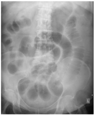
49. Bickle I. Small bowel obstruction. Radiopaedia.org; rID 33226. Web site. https://radiopaedia.org/cases/small-bowel-obstruction-12. Accessed June 20. 2017.

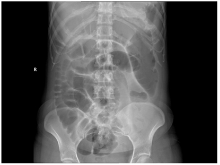
50. Babu V. Large bowel obstruction. Radiopaedia.org; rID 18015. Web site. https://radiopaedia.org/cases/large-bowel-obstruction. Accessed June 20. 2017.

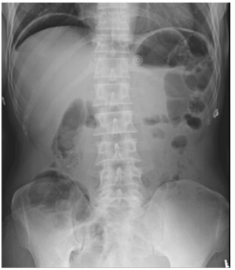
51. Kulkarni R. Bowel perforation. Radiopaedia.org; rID 21444. Web site. https://radiopaedia.org/cases/bowel-perforation. Accessed June 20. 2017.

LATEST ARTICLES

Unraveling the Mysteries of Type-A Aortic Dissection Using POCUS/Echocardiography

Syeda Rukh*, Sathyanarayana Machani and Milind Awale

doi.

Blood Sample from the Patient

Hypertriglyceridemia-Induced Pancreatitis: A Case Report and Literature Review

Maarten Bulterys, Melvin Willems* and Agnes Meersman

doi.

From Neck Pain to a Life-Threatening Condition: A Case Report

Floris Vandewoude* and Sören Verstraete

doi.

LATEST ARTICLES