Advances in Food Technology and Nutritional Sciences

Open journal

ISSN 2377-8350

Treatment of Acute Antibody-Mediated Rejection in Children Post-Kidney Transplantation: A Single Center’s Experience

Issa Alhamoud, Ei Khin, Rong Huang, Lesli McConnell and Mouin G. Seikaly*

Received: November 21st, 2020; Revised: January 24th, 2021; Accepted: January 27th, 2020; Published: February 8th, 2021

INTRODUCTION

Kidney transplantation has significantly changed the quality of life of patients with advanced chronic kidney disease (CKD). Unfortunately, renal allografts have a finite longevity; this is despite significant advancements in post-renal transplant management. Long-term graft survival is often impacted by events such as rejection,recurrence of original disease, immunosuppression toxicity, non-adherence to antirejection therapy, and opportunistic infections. The incidence of acute antibody-mediated rejection (aAMR) in pediatric renal transplant recipients (pRTRs) has not been well defined.1 This form of rejection is responsible for approximately 35.6% of renal allograft loss.2,3,4 Histopathologic diagnostic criteria for aAMR have been recently redefined.5,6

Currently, there has not been a collective approach to the management of patients with aAMR. Previously reported therapies to treat aAMR included: escalation of maintenance immunosuppressive agents, pulse methylprednisolone, intravenous gamma globulin (IVIG) to neutralize pathogenic circulating antibodies, rituximab to deplete B-lymphocytes, rabbit antithymoglobulin (rATG) to deplete T-cells, bortezomib to destroy mature plasma cells, apheresis to temporarily remove circulating antibodies, and recently eculizumab to block terminal complement pathway.7 Those treatment options have been used individually or in combination with variable outcomes.8

METHODS

Study Design

A retrospective analysis was conducted on all pRTRs younger than 21-years-old who were diagnosed with aAMR between January 2014 and November 2015. Kidney biopsy (index biopsy) was performed for graft dysfunction (elevation of serum creatinine> 20% of baseline)using 2013 Banff criteria for AMR diagnosis.5,6

Primary endpoints included estimated glomerular filtration rate (eGFR) as well as graft survival at one year after aAMR. eGFR was estimated using bedside Schwartz equation.9 Secondary endpoints involved iDSAs responseand treatment complications. Serum creatinineand serum iDSAs levels were monitored periodically to determine the response to treatment. The occurrence of opportunistic infections, malignancy, and bone marrow suppression parameters were monitored as markers of tolerance to therapy.

Baseline serum creatinine level to calculate eGFR was arbitrarily defined as the lowest serum creatinine level during thelast three months before the aAMR event. Baseline eGFR used the aforementioned baseline serum creatinine was calculated using bedside Schwartz equation.9 As per our practice guidelines to manage aAMR, Immuno-dominant donor-specific anti-HLA antibody titers (iDSAs) titers were initially monitored every two weeks. The monitoring interval was then individualized based on the clinical and laboratory parameters.

Immunosuppression Regimen

Induction treatment for cadaveric kidney transplant recipients included rabbit anti-thymoglobulin (rATG) 1.5 mg/kg (total not to exceed 6 mg/kg) every other day till tacrolimus level is in therapeutic range (12-15 ng/mL), and methylprednisolone 10 mg/kg initially then tapered gradually. Living-donor kidney recipients receive basiliximab as an induction dose of 10 mg for weight <30 kg and 20 mg for weight >30 kg on day 0 and 4 after renal transplantation in addition to methylprednisolone as aforementioned. Once patient’s serum creatinine level dropped to 50% of pre-transplant level, we started tacrolimus as a maintenance immunosuppressive therapy (initial dose 0.1 mg/kg/dose twice daily). Target level of tacrolimus had been maintained at 12-15 ng/mL during the first 4-weeks, 10- 12 ng/mL in 5-12-weeks, 7-10 ng/mL in 13-16-weeks, 5-7 ng/mL in 17-24-weeks, and 3-5 ng/mL after 6-months of transplantation. Mycophenolate mofetil (MMF) 600 mg/m2 /day divided in two doses, and prednisone 15 mg daily between 1-3-months, 10 mg daily between 3-6-months, then gradually tapered till off over one year.

Treatment of aAMR

All aAMR subjects were treated with our institutional protocol. This protocol consisted of methylprednisolone 30 mg/kg for 5 doses (Max 1 gram/dose), IVIG 1 g/kg for 2 doses, rituximab 375 mg/m2 for 2 doses (Max 1 gram/dose), and bortezomib 1.3 mg/ m2 for 4 doses (Max 2.5 mg/dose). The protocol was modified as deemed necessary by the primary nephrologist based on the clinical and laboratory parameters of recipients. One unresponsive patient received Eculizumab at the dose of 900 mg weekly for four-weeks then 1200 mg biweekly for two doses. Table 2 summarized our institutional protocol.

Table 2. aAMR Protocol

Medication/Dose Day 0 Day 1 Day 2 Day 3 Day 4 Day 5 Day 6 Day 7 Day 8 Day 9 Day 10
Methylprednisolone
30 mg/kg/dose (max 1 g/dose)

X

X

X

X

X

IVIG
1 g/kg/dose (max 70 g/dose)

X

X

Bortezomib
1.3 mg/m2/dose (max 2.5 mg/dose)

X

X

X

X

Rituximab
375 mg/m2/dose (max 1000 mg/dose)

X

X

Maintenance immunosuppressive therapy was also intensified where tacrolimus level has been increased to be above the target level where rejection occurred, and mycophenolate mofetil dose has also been increased from 600 mg/m2 /day to 1200 mg/m2 /day divided in two doses. In some non-adherent recipients, tacrolimus was switched to intravascular. Belatacept infusion biweekly then monthly.

This study was approved by the Institutional Review Board (IRB  #  STU  112016081) at the University of Texas Southwestern Medical Center in Dallas, Texas, United States.

Statistical Methods

Descriptive analyses of the continuous and categorical data were performed using means, standard deviations, proportions and frequencies. Statistical testing of sub-groups included Chi-square test, Fisher’s Exact test, two-sample t-test, and Wilcoxon Rank Sums test, as appropriate to the variable’s level of measurement and distribution. Linear regression model was used to investigate relationship between single or multiple independent variables and outcome variable. The statistical analyses were performed with SAS 9.4.

RESULTS

Patient’s Demographics

Over the study period, 10 of 105 pRTRs were diagnosed with aAMR (9.5%). All subjects were diagnosed at a median graft age of 43-months (range; 10-74 months). aAMRwas found predominantly in females (70%). The median patient’s age was 15-years (range; 11-18-years). Therapeutic tacrolimus concentration was found in 6 patients (60%) with aAMR. Only one aAMR subject had high panel reactive antibodies (PRAs) prior to the second kidney transplantation and patient was desensitized using rituximab, bortezomib, and IVIG as per protocol. Demographic and clinical data of patients are summarized in Table 1.

Table 1. Patient Demographics and Clinical Information

aAMR Subjects N=10

Patientage at Rejection (year) Median, range

15 (11-18)

Graftage at rejection date (months) Median, range

43 (10-74)

Female %, n

30% (3)

ESRD Cause
Time on dialysis prior to transplant

70% (7)

Glomerular disease %, (n)

10% (1)

Others %, (n)

10% (1)

Time on Dialysis Prior to Transplant
Pre-emptive %, (n)

0% (0)

≤12-months %, (n)

20% (2)

>12-months %, (n)

80% (8)

Dialysis Modality

Pre-emptive %, (n)

0% (0)

Peritoneal dialysis %, (n)

80% (8)

Hemodialysis %, (n)

20% (2)

Transplant Type

Deceased-Donor %, (n)

90% (9)

Living-Donor %, (n)

10% (1)

Prior transplant %, (n)

10% (1)

Pre-transplant PRA >20%

Class I PRA %,(n)

10% (1)

Class II PRA %, (n)

10% (1)

Prior rejection %, (n)

10% (1)

Mismatch score/10 (mean, std)

8.2 (1)

Positive cross match %, (n)

0%, (0)

Pre-rejection Tacrolimus Level

Therapeutic level %, (n)

60%, (6)

Non-therapeutic level %, (n)

40% (4)

1-year graft survival %, (n)

100% (10)

1-year patient survival %, (n)

100% (10)

ESRD: End Stage Renal Disease. CAKUT: Congenital Anomalies of Kidney and Urinary Tract

Acute cellular rejection (ACR) was concurrently diagnosed with aAMR in 6 patients out of 10 pRTRs as shown in Table 3. A C4d immunohistochemical stain was negative in 2 patients, focal in 5 patients (<50%), and diffuse in 3 patients (>50%). Allograft glomerulopathy was present in 2 grafts. The histopathological features of aAMR cases arepresented in Table 4.

Table 3. Banff Classification of aAMR among Our pRTRs

Case 1 Case 2 Case 3 Case 4 Case 5 Case 6 Case 7 Case 8 Case 9 Case 10
Banff Score aAMR ACR IA aAMR ACR IB aAMR ACR IIA aAMR ACR IA aAMR aAMR aAMR aAMR ACR IB aAMR aAMR ACR IB


Table 4. Histopathological Features of pRTRs Diagnosed with aAMR

Case 1 Case 2 Case 3 Case 4 Case 5 Case 6 Case 7 Case 8 Case 9 Case 10
Tubulitis (t)

1

3

3

2

1

0

0

3

0

3

Interstitial inflammation (i)

1

2

3

2

0

0

0

3

0

3

Glomerulitis (g)

1

2

0

2

1

0

2

1

0

1

Arterial hyaline thickening (ah)

0

0

0

1

0

0

1

0

0

0

Intimal arteritis (v)

0

0

1

0

0

0

0

0

0

0

C4d

1

2

0

3

2

0

3

1

3

0

Peritubular capillaritis (ptc)

2

2

3

2

1

2

2

2

2

2

Allograft glomerulopathy (cg)

0

Ia

0

1a

0

0

0

0

0

0

Interstitial fibrosis (ci)

1

1

2

1

0

2

1

0

0

1

Tubular atrophy (ct)

1

1

2

1

0

2

1

0

1

1

Fibrous intimal thickening (cv)

0

0

2

0

0

0

0

0

0

1

Mesangial matrix increase (mm)

0

0

0

1

0

0

0

0

0

0

6 cases had concurrent AAMR and ACR (IA in 2 cases, IB in 3 cases, and IIA in one case). t: tubulitis. i: interstitial inflammation. g: glomerulitis. ah: arterial hyaline thickening. v: intimal arteritis. ptc: peritubular capillaritis. cg: allograft glomerulopathy. ci: interstitial fibrosis. ct: tubular atrophy. cv: fibrous intimal thickening. mm: mesangial matrix increase.


Outcomes of aAMR Treatment on Renal Allograft Function and Survival

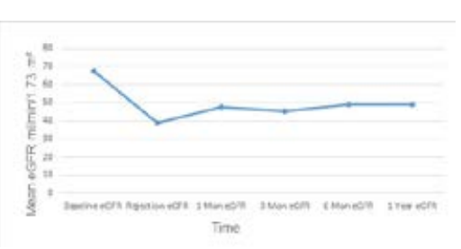
Patient and graft survival was 100% at one-year of the observation period. At the time ofindexbiopsy, none of the patients needed dialysis. aAMR caused a decline in mean eGFR of 42% of baseline mean (mean baseline eGFR=67.2±19.5 mL/min/1.73 m2 versus mean eGFR at rejection=38.9±14.2 mL/min/1.73 m2; p=0.002).

Four-weeks post-treatment, eGFR increased by 22% in comparison to baseline eGFR (mean eGFR=47.4±8.9 mL/ min/1.73 m2 vs 38.9±14.2 mL/min/1.73 m2. p=0.02).Despite not returning to baseline eGFR, 1-year eGFR had been stabilized and increased by 26% as compared to the rejection eGFR (mean eGFR=49.0±13.2 ml/min/1.73 m2; p=0.001) as shown in Figure 1.

Figure 1. Estimated GFR Change in pRTRs Treated for aAMR Over Time

NPOJ-6-125 Fig 1
Mean eGFR dropped significantly at the index biopsy 38.9 ml/min/1.73 m2 vs baseline mean eGFR 67.2 ml/min/1.73 m2; +p=0.002. However, mean eGFR increased significantly at 4-weeks (47.4 ml/min/1.73 m2; *p=0.02), and had stayed stable (49.0 ml/min/1.73 m2; *p=0.01) over a 1-year of observation period.


A functional response (FR)of eGFR was determined as the ratio of a difference between eGFR at 1-month post-treatment and nadir eGFR level at the rejection time to the difference between the baseline eGFR and eGFR level at the rejection time.

Functional graft response wass tratified as a complete response (CR) when the ratio had a value greater than 70%, an incomplete response (IR) when this ratio was between 30% and 70%, and no functional response if the ratio was less than 30%.10

As a functional trend, none of aAMR cases had the complete response. However, the incomplete response (IR) at 4-weeks post-treatment was accomplished in 7 patients with aAMR. Three functional allografts were considered unresponsive to therapy.

Outcomes of treatment on iDSAs

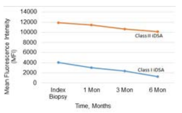
iDSAs were detected using the LabScreen Single Antigen Assay (Thermofisher, MA, USA). The method for calculating bead threshold has previously been published by Sullivan et al.11 We considered the rise in iDSAs, quantified as mean fluorescence intensity (MFI), was significant if the level was above 1500 MFI. Donor-specific antibodies (DSAs) were detected in 9 patients, where 5 patients had both class I and II DSAs and 4 patients had only class II DSAs as illustrated in Table 5. Immuno-dominant DSAs class II were 3 folds higher than iDSAs class I. iDSA was mainly HLA-A for class I and HLA-DQ for class II.

Table 5. Donor-Specific Antibodies in aAMR Cases at Index Biopsy

Case 1 Case 2 Case 3 Case 4 Case 5 Case 6 Case 7 Case 8 Case 9 Case 10
DSA (MFI)

C4 (1435)

A29 (1053)

A1 (2475)

A29 (6342)

DQ2 (21697)

Neg

DQ4 (14878)

DRB5 (DR51-1671)

DQ2 (14489)

A31 (2889)

Interstitial inflammation (i)

DQ7 (5699)

B44 (1248)

A2 (2150)

B44 (3346)

DQ4 (21856)

DQ7 (13922)

DQ6 (6916)

DQ4 (15920)

A2 (1807)

Glomerulitis (g)

B50 (7258)

B13 (829)

B45 (5001)

DQ2/DQA2 (854)

B60 (437)

Arterial hyaline thickening (ah)

DQ5 (13125)

B51 (758)

C16 (2642)

DQ2 (8779)

Intimal arteritis (v)

DR7 (5251)

DR7 (2594)

DR7 (812)

C4d

DR-B4 (5028)

DR17 (7029)

Peritubular capillaritis (ptc)

DQ (7446)

DQ2 (21531)

Allograft glomerulopathy (cg) DR-B3 ((DR52-1860)
Interstitial fibrosis (ci) DP0201 (2036)


                                                         eGFR@1 month post therapy-eGFR@aAMR
Functional Response = ——————————————

                                         Baseline eGFR-eGFR@aAMR


Monthly follow-up iDSAs data were available on 9 aAMR patients. Immuno-dominant DSA- class I titers had declined by 26% (3003±3745 MFI vs 4080±2560 MFI at rejection time) at 1 month, by 42% (3003±3745 MFI vs 4080±2560 MFI at rejection time) at 3-months, and by 69% (1265±1947 MFI vs 4080±2560 MFI at rejection time; p=0.04) at 6-months of follow-up. iDSAs class II had decreased by 4% (11468±6065 MFI vs 11930±5609 MFI) at 1-month and 10% (10629±6440 MFI vs 11930±5609 MFI) at 3-months, and by 15% (10130±7355 MFI vs 11930±5609 MFI: p>0.05) at 6-months of follow up period as shown in Figure 2. IDSA class I became negative in 3 patients and iDSA class II became negative in one receipient.

Figure 2. Mean Change Inimmuno-dominant DSA-HLA Class I and II Titers in Response to treatment of aAMR

NPOJ-6-125 Fig 2
MOf the 10 aAMR cases, 9 patients with functional graft had available DSA data over 6-12 months. One patient had aAMR with negative DSA. A significant decrease in iSDA-HLA class I occurred late 3-6 months post treatment (*paired t-test; p=0.04). Treatment effects on iDSA-HLA class II were minimal and did not reach statistical significance. No follow-up data of DSA were available for case 4.


Safety Profile

Overall, the protocol was well-tolerated. No serious bacterial infections nor malignancywere reported in our subjects. Adverse effects on bone marrow parameters included lymphopenia (n=8), thrombocytopenia (n=3), and anemia (n=6).

Four patients developed opportunistic infectionswithin 4- months following the administration of our treatment protocol. These infections include: BK viremia (n=2), EBV viremia (n=1), CMV viremia (n=1), and herpes zoster infection (Shingles) (n=1). All infections were successfully treated without sequelae. Adverse effects occurring during drug administration included: Rituximab-related fever/chills (n=1), methylprednisoloneinduced hypertension (n=2), IVIG-induced hypertension (n=1), Bortezomib-related paresthesia (n=1), and posterior reversible encephalopathy syndrome (PRES) (n=1).

DISCUSSION

Despite a considerable progress in the post-renal transplant management, aAMR remains an important event that threatens the short-and long-term outcomes of kidney transplantation. In the present case series, we investigated the outcomes of treatment of pRTRs who developed aAMR post-transplantation. Out of 10 cases, pure aAMR was diagnosed in 4 pRTRs, and 6 cases were combined aAMR and ACR. The incidence of aAMR without concurrent ACR was 3.8% of total pRTRs within our observation period, which is comparable to our previous case series as well as other reports in the literature.1,12 In our cohort, the overall incidence of aAMR with ACR was up to 9.5%, which is also similar to some other reports.13,14 aAMR in our population occurred late, beyond 1-year post-transplantation. Previous reports suggested that late aAMR heralds a poor response to conventional treatment and carries a high risk of graft loss.15,16,17,18,19

There is no consensus as to the treatment of aAMR in pRTRs.7,8 Most published data were derived from case reports or series, with variable approaches and inconsistent outcomes. Most regimens include inhibition of the B-Cells with monoclonal antibodies against cluster designation (CD20) with rituximab, destruction of antibody-secreting plasma cell (CD138) with bortezomib, antibodies removal and immunomodulation (plasma exchange, intravenous immunoglobulin, andcorticosteroids), and inactivation of antibody-mediated terminal complement activation (eculizumab). Despite the current plethora in the immunosuppressive agents, limited data are available to support any single agent or combined agents that offer clear advantages over others.20

Over the past few years, the use of bortezomib in pRTRs who developed aAMR has been rising with mixed outcomes.1,21,22 Bortezomib, proteasome s26 inhibitor, causes apoptosis of mature plasma cells, B-cells and activated T-cells through complex series of interactions.23,24,25,26,27 There are promising data of bortezomib in the treatment of aAMR in multiple case reports. Everly et al22 reported that bortezomib therapy alone provides an effective treatment of AMR and ACR with minimal toxicity and provides a sustained reduction in iDSAs levels. However, the recent published randomized clinical trial failed to show that bortezomib alone prevents GFR loss, improves histologic features, or reduces DSA, with a potential for significant toxicity.28

In our cohort, the study subjects had a partial improvement in graft function (30-70% of baseline eGFR) in 7 patients with no response (<30% of baseline eGFR) in 3 patients at 4-week spost-treatment. None of the pRTRs among the study subjects fully regained baseline renal function. However, all aAMR><30%of baseline eGFR) in 3 patients at 4-week spost-treatment. None of the pRTRs among the study subjects fully regained baseline renal function.

However, all aAMRgrafts had remained dialysis-free during the observation period. Our observation is in agreement with other studies that highlighted a role for bortezomib as a stabilizer of renal allografts function in conventional unresponsive late-aAMR.13 The role of bortezomibcontaining protocols should be further evaluated in randomized trials. Such trials should address strategies with regards to dosing, frequency, and duration of treatment. One limitation of our study is the use of combination therapyto treat aAMR. This does not allow us to define the role of the individual immunosuppressive agent.

Another putative predictor in aAMR prognosis is iDSAs. In our cohort, the decline in iDSAs class I (A) titer was more significant than iDSAs HLA class II (DQ) titer. As we mentioned above, regaining of renal function was independent of iDSAs response. Renal function in all grafts had stabilized without the need for renal replacement therapy. Furthermore, there had been no association between improvement in the graft function and the decrease in iDSAs titers. There was no correlation between the severity of aAMR and the decline in the allograft’s eGFR nor the titers of iDSAs.

B-lymphocytes may also play a role in the development of aAMR since they have the potential to develop to mature antibody-secreting plasma cells, it seems to be prudent to target this type of cells in aAMR. B-cell depleting agents such as rituximab have been used off label in renal transplantation field including aAMR treatment. Several studies in pRTRs have demonstrated the beneficial effects of rituximab on graft function, graft survival, and histopathological reversibility in renal allografts with aAMR,29-34 However, there was no consensus on the dosage and frequency of doses.30,31 Furthermore, its utilization was limited due to a high risk of infectious complications.35

Other antitumoral therapies such as plasmapheresis and immunoadsorption have a limited role in the treatment of aAMR.36,37 Plasmapheres is helps to remove preformed-abnormal circulating antibodies and enhances the effect of bortezomib on plasma cells and B-lymphocytes.38,39

IVIG has been used widely in aAMR treatment despite a limited-evidence of benefit.40,41 Dual treatment with IVIG and plasmapheresis administration assists to substitute lost immunoglobulins and minimizes the risk of infections.42,43,44,45 All aAMR cases received IVIG as per protocol. However, one receipient was given monthly IVIG 1 g/kg for six-months. iDSA class I and II in this subject had decreased substantially compared to other aAMR subjects.

It is common to diagnose ACR concurrently with aAMR. In our cohort, 60% of cases were mixed aAMR and ACR. A combined ACR and aAMR needs a multi-agent therapy as a single therapy is often not optimal. The role of T-cell depleting agents in pure aAMR treatment is not clear. Bortezomib therapy is reported to treat refractory ACR through a complex interactions on T-cell functions.22

In summary, our current study suggests that bortezomibbased protocol during a 1-year follow-up period resulted in 1) 100% graft survival with 26% increase eGFR from time of rejection 2) iDSAs class I antibodies declined more significantly than iDSAs class II antibodies and 3) our protocol was well-tolerated with no significant life-threatening infections nor malignancy were reported.

CONCLUSION

In conclusion, our study suggests a beneficial role for the combined therapy in the management of pRTRs who develop aAMR. However, confirmatory results should come from larger scale, longitudinal, multicenter study, which would allow for better understanding of efficacy and safety of our protocol.

CONFLICTS OF INTEREST

The authors declare that they have no conflicts of interest.

1. Twombley K, Thach L, Ribeiro A, Joseph C, Seikaly M. Acute antibody-mediated rejection in pediatric kidney transplants: A single center experience. Pediatr Transplant. 2013; 17: E149-E155. doi: 10.1111/petr.12129

2. North American Pediatric Renal Trials and Collaborative Studies. 2010 Annual Transplant Report 2010.

3. Wiebe C, Gibson IW, Blydt-Hansen TD, Pochinco D, Birk PE, Ho J, et al. Rates and determinants of progression to graft failure in kidney allograft recipients with de novo donor-specific antibody. Am J Transplant. 2015; 15: 2921-2930. doi: 10.1111/ajt.13347

4. Kim JJ, Balasubramanian R, Michaelides G, Wittenhagen P, Sebire NJ, Mamode N, et al. The clinical spectrum of de novo donor-specific antibodies in pediatric renal transplant recipients. Am J Transplant. 2014; 14: 2350-2358. doi: 10.1111/ajt.12859

5. Solez K, Colvin RB, Racusen LC, Haas M, Sis B, Mengel M, et al. Banff 07 classification of renal allograft pathology: Updates and future directions. Am J Transplant. 2008; 8: 753-760. doi: 10.1111/j.1600-6143.2008.02159.x

6. Haas M, Sis B, Racusen LC, Solez K, Glotz D, Colvin RB, et al. Banff 2013 meeting report: Inclusion of c4d-negative antibodymediated rejection and antibody-associated arterial lesions. Am J Transplant. 2014; 14: 272-283. doi: 10.1111/ajt.12590

7. Kasiske BL, Zeier MG, Chapman JR, Craig JC, Ekberg H, Garvey CA, et al. KDIGO clinical practice guideline for the care of kidney transplant recipients: A summary. Kidney Int. 2010; 77: 299-311. doi: 10.1038/ki.2009.377

8. Roberts DM, Jiang SH, Chadban SJ. The treatment of acute antibody-mediatedrejection in kidney transplant recipients-a systematic review. Transplantation. 2012; 94: 775-783. doi: 10.1097/TP.0b013e31825d1587

9. Schwartz GJ, Work DF. Measurement and estimation of GFR in children and adolescents. J Am Soc Nephrol. 2009; 4: 1832-1843. doi: 10.2215/CJN.01640309

10. Minervini MI, Torbenson M, Scantlebury V, Vivas C, Jordan M, Shapiro R, et al. Acute renal allograft rejection with severe tubulitis (Banff 1997 grade IB). Am J Surg Pathol. 2000; 24: 553-558. doi: 10.1097/00000478-200004000-00009

11. Sullivan D, Ahn C, Gao A, Lacelle C, Torres F, Bollineni S, et al. Evaluation of current strategies for surveillance and management of donor-specific antibodies: Single-center study. Clin Transplant. 2018; 32 : e13285. doi: 10.1111/ctr.13285

12. Mauiyyedi S, Colvin RB. Humoral rejection in kidney transplantation: New concepts in diagnosis and treatment. Curr Opin Nephrol Hypertens. 2002; 11: 609-618. doi: 10.1097/00041552-200211000-00007

13. Pearl MH, Nayak AB, Ettenger RB, Puliyanda D, Palma Diaz MF, Zhang Q, et al. Bortezomib may stabilize pediatric renal transplant recipients with antibody-mediated rejection. Pediatr Nephrol. 2016; 31(8): 1341-1348. doi: 10.1007/s00467-016-3319-3

14. Gulleroglu K, Baskin E, Bayrakci US, Turan M, Ozdemir BH, Moray G, et al. Antibody-mediated rejection and treatment in pediatric patients: One center’s experience. Exp Clin Transplant. 2013; 11: 404-407. doi: 10.6002/ect.2012.0242

15. Dorje C, Midtvedt K, Holdaas H, Naper C, Strom EH, Oyen O, et al. Early versus late acute antibody-mediated rejection in renal transplant recipients. Transplantation. 2013; 96: 79-84. doi: 10.1097/TP.0b013e31829434d4

16. Pefaur J, Diaz P, Panace R, Salinas P, Fiabane A, Quinteros N, et al. Early and late humoral rejection: A clinicopathologic entity in two times. Transplant Proc. 2008; 40: 3229-3236 doi: 10.1016/j.transproceed.2008.03.123

17. Sun Q, Liu ZH, Ji S, Chen J, Tang Z, Zeng C, et al. Late and early C4d-positive acute rejection: Different clinical histopathological subentities in renal transplantation. Kidney Int. 2006; 70: 377-383. doi: 10.1038/sj.ki.5001552

18. Gupta G, Abu Jawdeh BG, Racusen LC, Bhasin B, Arend LJ, Trollinger B, et al. Late antibody-mediated rejection in renal allografts: Outcome after conventional and novel therapies. Transplantation. 2014; 97: 1240-1246. doi: 10.1097/01.TP.0000442503.85766.91

19. Banasik M, Koscielska-Kasprzak K, Myszka M, Bartoszek D, Zabinska M, Boratynska M, et al. A significant role for antihuman leukocyte antigen antibodies and antibody-mediated rejection in the biopsy-for-cause population. Transplant Proc. 2014; 46: 2613-2617. doi: 10.1016/j.transproceed.2014.09.028

20. KDIGO clinical practice guideline for the care of kidney transplant recipients. Am J Transplant. 2009; 9 (Suppl 3): S1-S155. doi: 10.1111/j.1600-6143.2009.02834.x

21. Waiser J, Budde K, Schutz M, Liefeldt L, Rudolph B, Schonemann C, et al. Comparison between bortezomib and rituximab in the treatment of antibody-mediated renal allograft rejection. Nephrol Dial Transplant. 2012; 27: 1246-1251. doi: 10.1093/ndt/gfr465

22. Everly MJ, Everly JJ, Susskind B, Brailey P, Arend LJ, Alloway RR, et al. Bortezomib provides effective therapy for antibody- and cell-mediated acute rejection. Transplantation. 2008; 86: 1754-1761. doi: 10.1097/TP.0b013e318190af83

23. Hideshima T, Richardson P, Chauhan D, Palombella VJ, Elliott PJ, Adams J, et al. The proteasome inhibitor PS-341 inhibits growth, induces apoptosis, and overcomes drug resistance in human multiple myeloma cells. Cancer Res. 2001; 61: 3071-3076.

24. Chromik J, Schnürer E, Meyer RG, Wehler T, Tütingb T, Wölfel T, et al. Proteasome-inhibited dendritic cells demonstrate improved presentation of exogenous synthetic and natural HLAclass I peptide epitopes. J Immunol. Methods. 2006; 308: 77-89. doi: 10.1016/j.jim.2005.09.021

25. Nencioni A, Garuti A, Schwarzenberg K, Cirmena G, Dal Bello G, Rocco I, et al. Proteasome inhibitor induced apoptosis in human monocyte-derived dendritic cells. Eur J Immunol. 2006a; 36: 681-689. doi: 10.1002/eji.200535298

26. Nencioni A, Schwarzenberg K, Brauer KM, Schmidt SM, Ballestrero A, Grünebach F, et al. Proteasome inhibitor bortezomib modulates TLR4- induced dendritic cell activation. Blood. 2006b; 108: 551-558. doi: 10.1182/blood-2005-08-3494

27. Vanderlugt CL, Rahbe SM, Elliott PJ, Dal Canto MC, Miller SD. Treatment of established relapsing experimental autoimmune encephalomyelitis with the proteasome inhibitor PS-519. J Autoimmun. 2000; 14: 205-211. doi: 10.1006/jaut.2000.0370

28. Eskandary F, Regele H, Baumann L, Bond G, Kozakowski N, Wahrmann M, et al. A randomized trial of bortezomib in late antibody-mediated kidney transplant rejection. J Am Soc Nephrol. 2018; 29: 591-605. doi: 10.1681/ASN.2017070818

29. Becker YT, Becker BN, Pirsch JD, Sollinger HW. Rituximab as treatment for refractory kidney transplant rejection. Am J Transpl. 2004; 4: 996-1001. doi: 10.1111/j.1600-6143.2004.00454.x

30. Gomes AM, Pedroso S, Martins LS, Malheiro J, Viscayno JR, Santos J, et al. Diagnosis and treatment of acute humoral kidney allograft rejection. Transplant Proc. 2009; 41 :855–858. doi: 10.1016/j.transproceed.2009.01.062

31. Faguer S, Kamar N, Guilbeaud-Frugier C, Fort M, Modesto A, Mari A, et al. Rituximab therapy for acute humoral rejection after kidney transplantation. Transplantation. 2007; 83: 1277-1280. doi: 10.1097/01.tp.0000261113.30757.d1

32. Zarkhin V, Li L, Kambham N, Sigdel T, Salvatierra O, Sarwal MM. A randomized, prospective trial of rituximab for acute rejection in pediatric renal transplantation. Am J Transplant. 2008; 8: 2607-2617. doi: 10.1111/j.1600-6143.2008.02411.x

33. Kaposztas Z, Podder H, Mauiyyedi S, Illoh O, Kerman R, Reyes M, et al. Impact of rituximab therapy for treatment of acute humoral rejection. Clin Transplant. 2009; 23: 63-73. doi: 10.1111/j.1399-0012.2008.00902.x

34. Billing H, Rieger S, Ovens J, Süsal C, Melk A, Waldherr R, et al. Successful treatment of chronic antibody mediated rejection with IVIG and rituximab in pediatric renal transplant recipients. Transplantation. 2008; 86: 1214-1221. doi: 10.1097/TP.0b013e3181880b35

35. Kamar N, Milioto O, Puissant-Lubrano B, Esposito L, Pierre MC, Ould Mohamed A, et al. Incidence and predictive factors for infectious disease after rituximab therapy in kidney-transplant patients. Am J Transplant. 2010: 10: 89-98. doi: 10.1111/j.1600- 6143.2009.02785.x

36. Puttarajappa C1, Shapiro R, Tan HP. Antibody-mediated rejection in kidney transplantation: A review. J Transplant. 2012; 2012: 193724. doi: 10.1155/2012/193724

37. Okafor C, Ward DM, Mokrzycki MH, Weinstein R, Clark P, Balogun RA. Introduction and overview of therapeutic apheresis. J Clin Apher. 2010; 25: 240-249. doi: 10.1002/jca.20247

38. Stegall MD and Gloor JM. Deciphering antibody mediated rejection: New insights into mechanisms and treatment. Curr Opin Organ Transplant. 2010; 15: 8-10. doi: 10.1097/MOT.0b013e3283342712

39. Walsh RC, Alloway RR, Girnita AL, Woodle ES. Proteasome inhibitor-based therapy for antibody-mediated rejection. Kidney Int. 2012; 81: 1067-1074. doi: 10.1038/ki.2011.502

40. Jordan SC, Quartel AW, Czer LS, Admon D, Chen G, Fishbein MC, et al. Posttransplant therapy using high-dose immunoglobulin (intravenous gammaglobulin) to control acute humoral rejection in renal and cardiac allograft recipients and potential mechanism of action. Transplantation. 1998; 66: 800-805. doi: 10.1097/00007890-199809270-00017

41. Luke PP, Scantlebury VP, Jordan ML, Vivas CA, Hakala TR, Jain A, et al. Reversal of steroid- and antilymphocyte antibodyresistant rejection using intravenous immunoglobulin (IVIG) in renal transplant recipients. Transplantation. 2001; 72: 419-422. doi: 10.1097/00007890-200108150-00010

42. Dwyer JM. Manipulating the immune system with immune globulin. N Engl J Med. 1992; 326: 107-116. doi: 10.1056/NEJM199201093260206

43. Debre M, Bonnet MC, Fridman WH, Carosella E, Philippe N, Reinert P, et al. Infusion of Fc gamma fragments for treatment of children with acute immune thrombocytopenic purpura. Lancet. 1993; 342: 945-949. doi: 10.1016/0140-6736(93)92000-j

44. Tyan DB, Li VA, Czer L, Trento A, Jordan SC. Intravenous immunoglobulin suppression of HLAalloantibody in highly sensitized transplant candidates and transplantationwith a histoincompatible organ. Transplantation. 1994; 57: 553-562. doi: 10.1097/00007890-199402270-00014

45. Negi VS, Elluru S, Siberil S, Graff-Dubois S, Mouthon L, Kazatchkine MD, et al. Intravenous immunoglobulin: An update onthe clinical use and mechanisms of action. J Clin Immunol. 2007; 27: 233-245. doi: 10.1007/s10875-007-9088-9

LATEST ARTICLES

Use of Black Soldier Fly (Hermetia illucens) Prepupae Reared on Organic Waste

review

2024 Mar

Isayas A. Kebede*, Haben F. Gebremeskel and Gelan D. Dahesa,
Figure 11. Risk Map for the Introduction of Ruminant Diseases at Borders

Original Research, peer reviewed

2024 Mar

Yassir Lezaar*, Mehdi Boumalik, Youssef Lhor, Moha El-Ayachi, Abelilah Araba and Mohammed Bouslikhane

Commentary, End-of-Life, Family Dynamics, palliative Care, peer reviewed

2024 Mar

Neil A. Nijhawan*, Rasha Mustafa and Aqeela Sheikh

Retrospective Study

2024 Mar

Shrenik J. Shah*, Abhishek Jha, Chirag Davara, Rushi Mistry and Kapil Kachhadiya

Attention, Disaster, Mindfulness, Original Research, peer reviewed

2024 Mar

Christina Hagen*, Lars Lien, Edvard Hauff and Trond Heir

review

2024 Mar

Isayas A. Kebede* and Gelan D. Dahesa

review

2024 Feb

Bekiyad S. Daro*; Yared A. Deneke, Jiregna G. Negasa and Ishetu N. Dibaba