A new multidisciplinary project funded by the City of London Archaeological Trust (CoLAT) Rosemary Green grant will be using the latest imaging techniques on a large sample of archaeological skeletal assemblages from several post-medieval (18th and 19th century) and medieval sites across London as well as other non-metropolitan areas of England for comparison. This project aims to establish for the 1st time a synthetic overview from the physical evidence of how health patterns have changed up to the present day in London and the role that industrialization has played in determining the factors critical to the health of its population, past and present.
One of the most influential changes in our environment, both physical and social, over the past 1000 years has been industrialization, bringing revolutionary wide-spread changes in our material culture, technology, medicine and environment that continue to affect us today. All of these changes have impacted on the health and lifestyles of the modern British population. The industrial period is a pivotal age marking a shift towards increasing longevity and the chronic illnesses associated with aging, as well as a seeming rise in ‘man-made’ diseases such as cancer. To what extent, then, are these illnesses a product of the industrialized environment we have created? Have these diseases always been present or are they arising as a consequence of our modern, mechanized lifestyles?
This project seeks to identify under-recorded diseases in historic skeletal assemblages (such as metastatic cancers, hyperostosis frontalis interna (HFI) and osteoporosis) using direct radiography (DR) and up-to-date imaging modalities on a large sample of archaeological human skeletal remains, in order to enhance our understanding of the wider impact of industrialization on the health of Londoners in the context of the wider country up to the modern day.
AIMS AND OBJECTIVES
The project will examine the skeletal remains of c. 2,500 individuals to include 1000 adult men and women from industrial period London with the post-medieval population consisting of individuals of all social status. To gauge the impact of industrialization on London itself, the results will be compared to data based on the analysis of a further 500 individuals who lived in the medieval city. The process of industrialization throughout the country was not uniform and comparing the London data to that from non-metropolitan settlements will best capture the effect of living within the highly urbanized environment of the city.
In order to achieve this, the remains of 500 post-medieval individuals in addition to 500 medieval individuals from small towns and cathedral cities will be examined to identify cases of the selected diseases. Archaeological and historical evidence for the nature of the physical surroundings for each site will also be gathered to place the skeletal data into its appropriate environmental context.
The project will enable the much needed research of comparative data on post-medieval assemblages in London, allowing a large scale synthesis that has yet to be produced of how our health has changed as a result of industrialisation in the UK. This will simultaneously address a hiatus in our wider palaeopathological and clinical knowledge, currently restricted by a lack of the application of modern imaging techniques to skeletal assemblages.
The project will produce an exciting new resource of digital radiographs, scans and 3D models and will lead to the creation of a large digital archive of human skeletal remains, including virtual 3D models.
METHODS STATEMENT
A total of 18 sites have been identified for radiographic analysis comprising a sample of 1000 post-medieval and 500 medieval London individuals in addition to the remains of 500 medieval and 500 post-medieval non-metropolitan individuals.
We have selected 7 diseases that are often associated with industrialization, urbanization, enriched lifestyles and old age to assess their prevalence over time:
- HFI: Detected using radiography and computed tomography (CT).
- Osteoporosis: Detected using radiography.
- Joint Disease: Detected macroscopically and radiographically.
- Trauma: Detected macroscopically.
- Neoplastic disease (cancer): Detected using radiography and CT.
- Infectious disease (rib lesions): Detected macroscopically.
- DISH: Detected macroscopically.
Radiographic analysis will be undertaken on strategically selected areas of the skeleton that would allow maximum data to be recorded in a timely and effective manner.
- Crania
- Vertebrae (lumbar)
- Pelvis
- Femora
- 2nd Metacarpal(s)
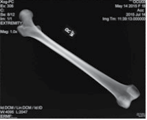
Radiography of the elements is carried out using a portable direct digital radiographic kit using a Canon CDXI Direct Digital X-ray plate system and Sedecal 4 kW generator (Figures 1 and 2). The process of the radiographed images will follow the guidelines of digital imaging and communications in medicine (DICOM). Recording of radiographic lesions will be undertaken in accordance with Burgener et al1 and with the interpretative assistance of a radiologist. The crania, vertebrae, femora and hands will undergo radiography in order to assess HFI, osteoporosis and neoplastic disease.
Figure 1: OCU00_18 Positioning of right femur anterior view.
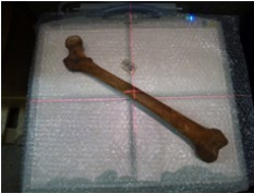
Figure 2: OCU00_18 DICOM image of right femur.
The prevalence of macroscopically observed diseases will be calculated using the Wellcome Osteological Research Database (WORD) and published data. The demographic profile of each assemblage will also be reconstructed to establish the lifespan of men and women. Social status, age, sex and parturition will be taken into account. The data will then be compared to modern clinical data in order to place it in its relevant present day context.
Clinical information and data will be researched via online resources, such as the World Health Organisation (WHO) website. Data would be published in an anonymous format to respect the privacy of the deceased and any living relatives. The archaeological evidence for the physical and social environment of each site will be established through desk-top research, so that the skeletal data can be placed into its relevant context. A series of maps illustrating the expansion of settlement and industry across the city will also be consulted.
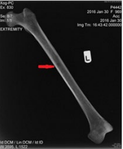
The project is in the initial stages of radiographing individuals from different sites and once completed will then go through all of the radiographic images for analysis and interpretation. The DICOM image below (Figure 3) is of a femur of a female (P4442, SK [969]) from an excavation at Holy Trinity Church, Stratford who died in 1854. The biographical details present for this individual allowed us to obtain a death certificate, which stated the cause of death as ‘carcinoma mammae certified’. From the osteological analysis and recording of her skeletal remains there was no macroscopic disease recorded but in the radiograph there appears to be an isolated possible osteolytic lesion.
Figure 3: P4442_969 DICOM image of femur with possible osteolytic lesion.
Collation of the radiographs investigating lesions not visible from macroscopic analysis will be key to the investigations in to the prevalence of diseases that may be present but that are not always visible from gross bony lesions. The advantages of the associated documentary sources for the post-medieval period is having the opportunity of researching parish cause of death and those certified with a death certificate from 1837 onwards in comparison with the osteological analysis. Research projects engaging the use of modern imaging techniques such as DR with archaeological assemblages are proving very fruitful, expanding our knowledge of the diseases present and further substantiating the curated skeletal collections being on-going valuable research assets.
ACKNOWLEDGEMENTS
Thanks are due to Reveal Imaging Ltd. for their radiography services.
DISCLOSURE
This article is based on the CoLAT funded project “The Impact of Industrialization on London Health”.
CONFLICTS OF INTEREST
The authors declare that they have no conflicts of interest.


