Sports and Exercise Medicine

Open journal

ISSN 2379-6391

The Gluteus Medius Activation in Female Indoor Track Runners is Asymmetrical and may be Related to Injury Risk

Stephanie E. Nevison , Youngmin Jun and James P. Dickey*

James P. Dickey, PhD

Associate Professor, School of Kinesiology, Faculty of Health Sciences, University of Western Ontario, London, ON N6A 5B9, Canada; E-mail: jdickey@uwo.ca

INTRODUCTION

Running is a common recreational and competitive activity consisting of alternating stance and swing phases, and including an airborne phase.1,2 The intermittent application of force on contact with the ground produces a peak vertical impact force equal to about 2.5 times a person’s body weight,3 Achilles tendon forces between 6.1 and 8.2 times body weight, and patellar tendon forces between 4.7 and 6.9 times body weight.4 As a result, runners are prone to “over-use” injuries such as Achilles tendinopathy, chronic knee lesions (patellar cartilage lesions), iliotibial band syndrome and stress fractures.5 International athletic competitions have relatively high rate of injury (81.1±4.2 injuries per 1000 athlete registrations),6 and mixed findings about the injury rates in males and females. For example, a recent review of injuries in major track and field competitions concluded that males have a higher risk of injury than females,5 a retrospective review of injuries sustained by 95 track and field athletes in a 12 month period observed similar injury rates in male and female athletes,7 a recent systematic review that included a total of 4,671 pooled participants observed that most studies did not find that gender was associated with running injuries,8 and a systematic review of injuries in recreational and competitive runners revealed a positive association between overall lower extremity running injuries and the female sex.9 In addition, weak hip abductors and external rotators are associated with the development of general overuse injuries of the foot and ankle, patellofemoral pain, iliotibial band syndrome and anterior cruciate ligament injury.10 Interestingly, decreased hip abductor concentric strength predicts the development of exertional medial tibial pain in females, but not males.11

Competitive track runners exclusively run around the curves of the track in the counter-clockwise direction. Compared to running in a straight path with prominent vertical ground reaction forces, running around curves requires larger lateral ground reaction forces12 and different muscle activations.13 While both legs of a runner generate lateral ground reaction forces when running around a curve, the resultant ground reaction force is significantly greater for the outside leg.12 This asymmetry is also reflected in increased hind foot evertor strength of the right leg and hind foot inverter strength of the left leg of collegiate sprinters and middle distance runners after ten weeks of track training.14 Right foot evertors and left foot invertors, in conjunction with hip abductors and hip adductors, are thought to produce the forces necessary for accelerating around the curves of the track.14 While many studies have evaluated the kinematics15,16 and ground reaction forces during running around curves,12,17 fewer studies have evaluated the muscular patterns during running around curves and related cutting maneuvers.13 These data have been interpreted in light of the maximum forces that limbs can generate and have led to the conclusion that the inside leg is the limiting factor during running around curves.12 For example, the inside leg has smaller propulsive and lateral forces compared to the outside leg.12 However, these papers specifically evaluate the force components along the leg and assume that the lateral component of the ground reaction force is produced by leaning into the corner such that the limb extensor force includes a lateral component.17 If this were the case, then we would expect that the gluteus medius muscle activation would be similar in the straightaways and while running around curves. However, the hip abductor muscles on the outside leg can effectively produce an abduction moment during stance that would result in lateral ground reaction forces. If the hip abductors are contributing to the lateral ground reaction force then we would expect that there would be greater gluteus medius activation on the outside leg while running around curves compared to the straightaways. Given that running around curves requires laterally-directed components of the ground reaction force12 and may place higher demands on the hip abductors,14 and that females have weaker abductors than males,18 females may show differential muscle activations and may be at a higher risk of injury while running on curved tracks.

The purpose of this study was to investigate the asymmetry of gluteus medius muscle activation patterns in female runners as they run around the curves of an unbanked 200 m track. Given the difference in function of the inside and outside legs while running around curves, we hypothesized specific directional hypotheses. We hypothesized that the right (outside leg) gluteus medius activation on curves would be significantly greater than straightaways, which is consistent with these muscles creating abduction moments to contribute to the lateral forces during curves. We hypothesized that the left (inside leg) gluteus medius activation on curves would be significantly less than straightaways, since abduction moments for the inside limb take away from centripetal forces during curves.

METHOD

Participants

Eight middle- to long-distance female runners of Western University’s 2013-2014 Track and Field and Cross Country teams were recruited to participate. Volunteers signed a written consent letter and completed a Training and Medical History Form assessing their eligibility. All participants were between 18 and 27 years of age and currently not suffering from an injury resulting in abnormal running form. The participants’ mean (± SD) age was 20±1.67 years, mean height (± SD) was 167.63±12.04 cm, mean (± SD) leg length was 102.75±6.39 cm and mean weight (± SD) was 57.50±6.46 kg. Each participant ran an average (± SD) of 7.44±1.24 hours per week and had been running for an average (± SD) of 8.50±2.83 years. During the indoor track season, an average (± SD) of 3.31±1.00 hours per week of training time consisted of running timed intervals in the counter-clockwise direction around the 200 m indoor track at Thomson Arena at Western University. The 10 mm polyurethane rubber track was unbanked and included 45 m straightaways and 55 m curves.14 This study was approved by the UWO Ethics Review Board and Women’s Track and Field Head Coaches, and all participants provided informed consent.

Participant Preparation

Wireless surface electromyography electrodes (TrignoTM Wireless Systems, DELSYS, Boston, MA, USA) with 10 mm inter-electrode distance were adhered midway between the crista iliaca and the greater trochanter to capture gluteus medius muscle activation.19 Both lower extremities were instrumented. The skin was wiped with alcohol swabs to reduce impedance and to improve electrode adherence. The electrodes were arranged parallel to the muscle fibers and were secured to the skin using the manufacturer’s double-sided tape interfaces, and additional strips of tape, to prevent electrode movement.

Maximal Voluntary Contractions and Strength Tests

Maximal hip abductor strength was tested three times per leg for each participant. Guidelines for manual resistance testing of the gluteus medius1921 were followed while providing resistance with a Mark-10 Series 5 digital force gauge (Mark-10 Corporation, Copiague, NY, USA), similarly to other researchers.22 The examiner’s standard command was “go-go-go-pushpush” as the participant attempted to abduct her leg against manual resistance while laterally recumbent.22 The participant held the contraction for four seconds and had two minutes of rest between contractions. Maximal strength was defined as the peak force recorded by the digital force gauge.

The digital force gauge was also used to measure hip flexor and adductor strengths. For hip flexor strength, the participant was in a seated position and maintained their thigh 8 inches from the surface of the chair.22,23 With the digital force gauge in hand, force was manually applied onto the thigh to break the muscle contraction.23 The peak force was recorded. For hip adductor strength, the participant was laterally recumbent with her knees extended.24 The participant attempted to adduct her bottom leg without internal/external rotation, flexion or extension of the hip and a breaking force was manually applied below the knee.24

Testing Protocol

To warm-up prior to testing, the participants jogged at a warm-up pace for five minutes. Testing for each participant consisted of three 250 m trials around the innermost lane of the track, from the start of a straightaway around the track and to the end of that straightaway. Participants ran at a target pace of 5 m/s, the approximate race pace for a 3000 m race for the first two trials and walked at a self-selected pace for the third trial. Fifty meters into each trial the electromyography (EMG) system and manual stopwatch were started. At the completion of one lap of the track, the EMG system and stopwatch were stopped. Running trials exceeding ±0.5 m/s of the target speed were repeated. In between tests, participants rested for five minutes. Participants were videotaped from a viewing platform above the track using a Sony HDR-XR550V digital camera mounted on a tripod. The camera was panned to follow the participants as they walked and ran to determine when they were running on curves versus straightaways and times of foot contact.

DATA ANALYSIS

A two-way repeated measures ANOVA was used to compare peak strength measurements for participants hip abductors, hip adductors, hip extensors and hip flexors for their bodies’ right and left sides.

The raw EMG signals were sampled at 2000 Hz, fullwave rectified and low-pass filtered with a 2nd order Butterworth (Fc=20 Hz). The videos were played frame-by-frame using VLC media player (version 2.1.4, Free Software Foundation, Boston, MA, USA) to record foot contact timing. The videotape recording was also used to code the strides as whether they occurred in the straightaway or curve. Individual strides for both the right and left sides were partitioned out of the data record using the timing of foot contacts extracted from the videotape recording. EMG data were normalized to walking on the straightaways to yield percent of walking values similarly to other researchers,25,26 and the mean amplitudes were extracted to represent the magnitude of muscle activation. One-tailed paired t-tests were used to evaluate the statistical significance of differences in average gluteus medius EMG amplitudes between the straightaways and curves for right (outside) and left (inside) legs using Graph Pad Prism (version 6, Graph Pad Software, San Diego, CA, USA). Statistical significance was set at p<0.05.

RESULTS

Peak Strength

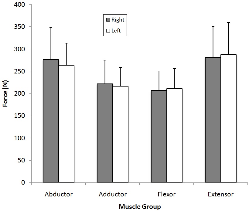
There was no significant interaction between sides and muscle group’s strength (p=0.924), and no significant main effect of side (p=0.928). There was a statistically significant main effect of muscle group (Figure 1; p<0.0001). Post hoc testing revealed that the abductors were significantly stronger than the adductors (p=0.049), and the extensors were significantly stronger than the flexors (p=0.007) while no other comparisons were significantly different.

Figure 1: Maximal Hip Strengths for the Right and Left Sides.

Maximal Hip Strengths for the Right and Left Sides

The differences between the right and left sides were not statistically significant.

Muscle Activation

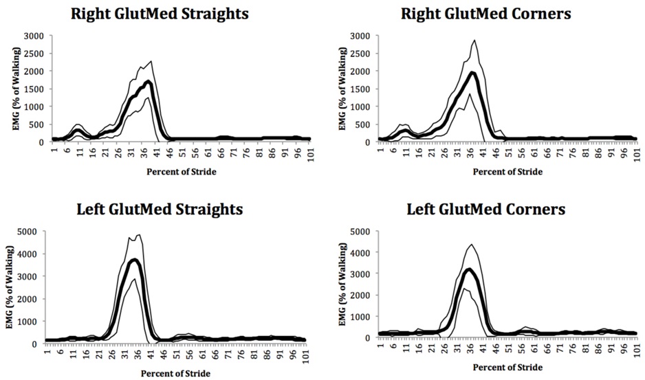
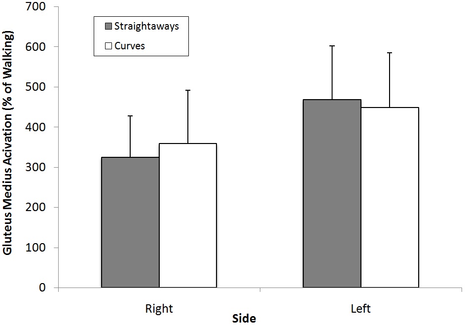
We observed that the gluteus medius showed a burst of activity during stance (Figure 2). In general the gluteus medius was activated between three and five times greater during running than walking (Figure 3). Interestingly we observed that the right gluteus medius muscle appeared to be activated less than the left for both straightaways and curves. When we compared between curves and straightaways for each side, we observed significantly greater activation for the right (outside leg) gluteus medius in curves compared to straightaways (p=0.015), and a trend towards less activation of the left (inside leg) gluteus medius on the curves compared to the straightaways (p=0.065).

Figure 2: Ensemble average patterns of gluteus medius muscle activation for the right and left sides, and for straightaways and curves for a representative participant.

Ensemble average patterns of gluteus medius muscle activation

The mean values are shown with the thick line and the narrow lines represent plus and minus one standard deviation from the mean. The running stride extends from foot contact (0 percent of stride) to subsequent foot contact (100 percent of stride).

Figure 3: Mean muscle activation (±SD) of the left (inside) and right (outside) gluteus medius for runners on curves versus straightaways.

Mean muscle activation (±SD) of the left (inside) and right (outside) gluteus medius

There was a significant increase in gluteus medius activation on the right side in curves compared to straightaways. There was a trend towards decreases in gluteus medius activation on the left side in curves compared to straightaways.

DISCUSSION

The purpose of this study was to determine if the gluteus medius contracts asymmetrically when female runners run around the curves of a 200 m unbanked track. Given the necessity for increased lateral ground reaction forces,12,27 we hypothesized that the magnitude of gluteus medius activation of female runners would be different between straightaways and curves for the right (outside) and left (inside) legs. In specific, we hypothesized increased activation of the right (outside) gluteus medius in curves, and decreased activation of the left (inside) gluteus medius in curves. The results of the study supported our hypotheses: gluteus medius activation in curves was significantly greater for the right (outside) leg, and we observed a trend for decreased gluteus medius activation of the left (inside) leg in curves compared to straightaways when female runners ran on an unbanked 200 m track. These data suggest that the biomechanical and muscular demands of curvilinear running differed from those of straight-path running. Previous studies have thought that extension of the leg with the body leaning into the corner produces the required lateral forces,28,29 and that the inside leg limits curve sprinting performance.12 Although these ideas may have some merit, we have determined that the hip abductor muscles, particularly on the right (outside) leg, play a significant role in generating the lateral forces necessary for turning. These demands may place female track runners at increased risk for injury.

Our findings are consistent with studies that have determined that the outside leg generated greater peak lateral GRFs12,30 and larger lateral impulses27,30 compared to the inside leg. Our findings are also consistent with a study that observed that the trunk lean angle increases while the outside foot is on the ground and decreases when the inside foot is on the ground.31 Similarly, our findings are consistent with a study that observed increases in right side gluteus medius activation during left turns in open cutting maneuvers, though this difference was not statistically significant.13 They observed that the bursts of raw EMG occurred in swing, which they interpret as performing medial rotation of the femur in preparation for foot contact. However, the delay between the EMG signal and the production of force32 means that this activation would actually result in forces during stance. Other researchers have also noted that the gluteus medius is active during stance.33 Accordingly the gluteus medius activation was abducting the hip while the foot is planted, leading to laterally directed ground reaction forces. These lateral forces during stance, primarily from the outside limb,30 provide the centripetal force necessary to change the direction of the runner, allowing them to run around the curve.12 Some authors have presented contrasting understandings of the roles of the inner and outer legs in running. For example, Alt et al.15 concluded that the inside leg provided stability in the frontal plane (eversion adduction strategy) while the outside leg provided motion in the horizontal plane (rotation strategy), although their rationale was based upon stance durations and joint kinematics rather than kinetics or EMG.

Previous studies have determined the gluteus medius stabilizes the hip before and after stance phase during straight path running.34 The present study extends these findings by documenting gluteus medius’ secondary role in hip abduction.35

The uniformity in hip abductor, hip adductor, hip extensor and hip flexor strengths for participants left and right sides indicated that participants did not have pre-existing strength asymmetries. It is important to note that we attempted to carefully control body position, as research has shown that variations, such as side-lying hip abduction and internal rotation angles, influence amount of muscle activation,36 and presumably force. Although all of the participants in this study were highly trained track runners (members of the Varsity Track and Field team), it is possible that increased muscle activation of the outside (right) gluteus medius in curvilinear running may lead to muscular asymmetry, as observed in hindfoot invertor and evertor muscles.14 It is important to note that the role of hindfoot muscle groups is similar to the hip abductors/adductors during curvilinear running;14 both are responsible for producing forces that allow a runner to accelerate around a curve.

The greater demands on the right (outside) leg gluteus medius during curves may place female track runners at increased risk for injury. Repeated asymmetrical muscle activation with existing muscle weakness may result in “over-use” injuries of the lower extremities.37 In fact, there appears to be a relationship between hip abductor strength and iliotibial band syndrome (ITBS); long distance runners with ITBS have weaker hip abduction strength in the affected leg compared to their unaffected leg, and also compared to unaffected long-distance runners.38 They also observed that symptom relief with a successful return to the preinjury training program paralleled improvement in hip abductor strength.38 Thus, to reduce the risk of injury, coaches may wish to supplement track running with strength training targeting hip abductors.38,39 Other studies have shown asymmetries in strength,38 and that large increases in hip muscle strength can be achieved in a two month duration unsupervised exercise program.40 This issue may be more relevant for female track runners as they are at greater risk than male track runners of developing iliotibial band syndrome and other hip-related injuries.41,42

Although it was not related to our hypotheses, and therefore was not evaluated statistically, the gluteus medius muscles on the right side appeared to be activated less than the left side. While many studies only measure EMG from muscles on one side of the body and assume bilateral symmetry (for example43,44), previous studies of walking found bilateral asymmetries in participants for six of seven leg muscles.45 Asymmetry has also been emphasized in gait46 and running.47 Given that we normalized our running EMG trials to the walking trials, the asymmetry that we observed might have been caused by either the walking and/or the running trials, but was not caused by bilateral differences in maximum strength.

LIMITATIONS

The present study analyzed muscle activity when runners ran at one speed around an unbanked track. Other researchers have identified that running speed influences the ground reaction forces30 and the muscle activation levels,44,48 so our findings only reflect running at a steady 3000 m race pace (5 m/s). Accordingly our findings may not be applicable to running at other speeds, at other track radii, while accelerating, or on banked tracks. Our EMG measurements were limited to the gluteus medius muscle, and accordingly we do not have insight into the roles of other abductors49,50 or other lower extremity muscles. Fine-wire EMG may be useful for evaluating the functionally distinct regions of the gluteus medius.33 Our participants were all highly trained members of the female Varsity Track and Field team, and accordingly our findings may not be applicable to other groups. Further research should expand the one-dimensional models of running on a curved path, such as the maximum leg extension force model, to consider the contribution of the hip abductors.

CONCLUSION

In summary, the present study has demonstrated that the gluteus medius contracts asymmetrically in female runners as they run around the curves of an unbanked, 200 m indoor track. This activation in participants’ outside (right) leg likely contributes to lateral forces and centripetal accelerations necessary to run around the curves. This hip abductor mechanism may be a performance constraint for curved running. Furthermore, this asymmetrical muscle activation may lead to injury in track runners, with females at even greater risk than males. Coaches may want to consider injury prevention strategies such as training around both directions of the track, and complementing running with strength training to prevent the development of muscle strength asymmetry, or to correct it if it is present.

ACKNOWLEDGEMENTS

Special thanks to Andrew Dragunas, Peter Wegscheider, Ryan Frayne and Western University’s Varsity Cross Country Team. Finally, thank you to the Scholar’s Electives Program and Western University that provided Stephanie Nevison with this research opportunity.

CONFLICTS OF INTEREST

None.

1. Novacheck TF. The biomechanics of running. Gait Posture. 1998; 7: 77-95. doi: 10.1016/S0966-6362(97)00038-6

2. Nicola TL, Jewison DJ. The anatomy and biomechanics of running. Clin Sports Med. 2012; 31: 187-201. doi: 10.1016/j.csm.2011.10.001

3. Robbins SE, Hanna AM. Running-related injury prevention through barefoot adaptations. Med Sci Sports Exerc. 1987; 19: 148-156. doi: 10.1249/00005768-198704000-00014

4. Scott SH, Winter DA. Internal forces of chronic running injury sites. Med Sci Sports Exerc. 1990; 22: 357-369.

5. Edouard P, Alonso J-M. Epidemiology of Track and Field Injuries. New Studies in Athletics. 2013; 28: 85-92.

6. Feddermann-Demont N, Junge A, Edouard P, Branco P, Alonso J-M. Injuries in 13 international Athletics championships between 2007-2012. Br J Sports Med. 2014; 48: 513-522. doi: 10.1136/bjsports-2013-093087

7. Bennell KL, Crossley K. Musculoskeletal injuries in track and field: incidence, distribution and risk factors. Aust J Sci Med Sport. 1996; 28: 69-75.

8. Saragiotto BT, Yamato TP, Junior LCH, Rainbow MJ, Davis IS, Lopes AD. What are the main risk factors for running-related injuries? Sports Med. 2014; 44: 1153-1163. doi: 10.1007/ s40279-014-0194-6

9. van Gent BR, Siem DD, van Middelkoop M, van Os TA, Bierma-Zeinstra SS, Koes BB. Incidence and determinants of lower extremity running injuries in long distance runners: a systematic review. Br J Sports Med. 2007; 41: 469-480. doi: 10.1136/bjsm.2006.033548

10. Chuter VH, de Jonge XAJ. Proximal and distal contributions to lower extremity injury: a review of the literature. Gait Posture. 2012; 36: 7-15. doi: 10.1016/j.gaitpost.2012.02.001

11. Verrelst R, Willems TM, Clercq DD, Roosen P, Goossens L, Witvrouw E. The role of hip abductor and external rotator muscle strength in the development of exertional medial tibial pain: a prospective study. Br J Sports Med. 2014; 48: 1564-1569. doi: 10.1136/bjsports-2012-091710

12. Chang Y-H, Kram R. Limitations to maximum running speed on flat curves. The Journal of Experimental Biology. 2007; 210: 971-982. doi: 10.1242/jeb.02728

13. Rand MK, Ohtsuki T. EMG analysis of lower limb muscles in humans during quick change in running directions. Gait Posture. 2000; 12: 169-183. doi: 10.1016/S0966-6362(00)00073-4

14. Beukeboom C, Birmingham TB, Forwell L, Ohrling D. Asymmetrical strength changes and injuries in athletes training on a small radius curve indoor track. Clinical journal of sport medicine : official journal of the Canadian Academy of Sport Medicine. 2000; 10: 245-250. doi: 10.1097/00042752-200010000-00004

15. Alt T, Heinrich K, Funken J, Potthast W. Lower extremity kinematics of athletics curve sprinting. J Sports Sci. 2014; 33: 552-560. doi: 10.1080/02640414.2014.960881

16. Ryan GJ, Harrison AJ. Technical adaptations of competitive sprinters induced by bend running. New Studies in Athletics. 2003;18: 57-70.

17. Jindrich DL, Besier TF, Lloyd DG. A hypothesis for the function of braking forces during running turns. J Biomech. 2006; 39: 1611-1620. doi: 10.1016/j.jbiomech.2005.05.007

18. Leetun DT, Ireland ML, Willson JD, Ballantyne BT, Davis IM. Core stability measures as risk factors for lower extremity injury in athletes. Med Sci Sports Exerc. 2004; 36: 926-934. doi: 10.1249/01.MSS.0000128145.75199.C3

19. SENIAM. Recommendations for sensor locations in hip or upper leg muscles. Website: www.seniam.org. Accessed February 24, 2015.

20. Halaki M, Ginn KA. Normalization of EMG Signals: To Normalize or Not to Normalize and What to Normalize to? In: Naik GR, ed. Computational Intelligence in Electromyography Analysis – A Perspective on Current Applications and Future Challenges. 2012; 175-194. doi: 10.5772/49957

21. Bolgla LA, Uhl TL. Reliability of electromyographic normalization methods for evaluating the hip musculature. J Electromyogr Kinesiol. 2007; 17: 102-111. doi: 10.1016/j.jelekin.2005.11.007

22. Thorborg K, Petersen J, Magnusson SP, Holmich P. Clinical assessment of hip strength using a hand-held dynamometer is reliable. Scand J Med Sci Sports. 2010; 20: 493-501. doi: 10.1111/j.1600-0838.2009.00958.x

23. Marino M, Nicholas JA, Gleim GW, Rosenthal P, Nicholas SJ. The efficacy of manual assessment of muscle strength using a new device. Am J Sports Med. 1982; 10: 360-364. doi: 10.1177/036354658201000608

24. Kendall F, McCreary E, Provance P. Muscles: Testing and Function. 3rd ed. Baltimore, Md: Williams and Wilkins. 1983.

25. Courtine G, Papaxanthis C, Schieppati M. Coordinated modulation of locomotor muscle synergies constructs straight-ahead and curvilinear walking in humans. Exp Brain Res. 2006; 170: 320-335. doi: 10.1007/s00221-005-0215-7

26. Wall-Scheffler CM, Chumanov E, Steudel-Numbers K, Heiderscheit B. Electromyography activity across gait and incline: The impact of muscular activity on human morphology. Am J Phys Anthropol. 2010; 143: 601-611. doi: 10.1002/ajpa.21356

27. Orendurff MS, Segal AD, Berge JS, Flick KC, Spanier D, Klute GK. The kinematics and kinetics of turning: limb asymmetries associated with walking a circular path. Gait Posture. 2006; 23: 106-111. doi: 10.1016/j.gaitpost.2004.12.008

28. Greene P. Running on flat turns: experiments, theory, and applications. J Biomech Eng. 1985; 107: 96-103. doi: 10.1115/1.3138542

29. Usherwood JR, Wilson AM. Accounting for elite indoor 200 m sprint results. Biol Lett. 2006; 2: 47-50. doi: 10.1098/rsbl.2005.0399

30. Smith N, Dyson R, Hale T, Janaway L. Contributions of the inside and outside leg to maintenance of curvilinear motion on a natural turf surface. Gait Posture. 2006; 24: 453-458. doi: 10.1016/j.gaitpost.2005.11.007

31. Ishimura K, Sakurai S. Comparison of inside contact phase and outside contact phase in curved sprinting. International Conference on Biomechanics in Sports. Marquette, Michigan, USA. 2010; 18: 57-70.

32. DeLuca CJ. The use of surface electromyography in biomechanics. J Appl Biomech. 1997; 13: 135-163. doi: 10.1123/jab.13.2.135

33. Semciw AI, Pizzari T, Murley GS, Green RA. Gluteus medius: An intramuscular EMG investigation of anterior, middle and posterior segments during gait. J Electromyogr Kinesiol. 2013; 23: 858-864. doi: 10.1016/j.jelekin.2013.03.007

34. Mann Ra, Moran GT, Dougherty SE. Comparative electromyography of the lower extremity in jogging, running, and sprinting. The American Journal of Sports Medicine. 1986; 14: 501-510. doi: 10.1177/036354658601400614

35. Gottschalk F, Kourosh S, Leveau B. The functional anatomy of tensor fasciae latae and gluteus medius and minimus. J Anat. 1989; 166: 179-189.

36. Lee J-h, Cynn H-S, Kwon O-Y, et al. Different hip rotations influence hip abductor muscles activity during isometric sidelying hip abduction in subjects with gluteus medius weakness. J Electromyogr Kinesiol. 2014; 24: 318-324. doi: 10.1016/j.jelekin.2014.01.008

37. Niemuth PE, Johnson RJ, Myers MJ, Thieman TJ. Hip muscle weakness and overuse injuries in recreational runners. Clin J Sport Med. 2005; 15: 14-21. doi: 10.1097/00042752-200501000-00004

38. Fredericson M, Cookingham CL, Chaudhari AM, Dowdell BC, Oestreicher N, Sahrmann SA. Hip abductor weakness in distance runners with iliotibial band syndrome. Clinical journal of sport medicine: official journal of the Canadian Academy of Sport Medicine. 2000; 10: 169-175. doi: 10.1097/00042752-200007000-00004

39. Brumitt J. Injury Prevention for High School Female CrossCountry Athletes. Athl Ther Today. 2009; 14: 8-12. doi: 10.1123/att.14.4.8

40. Osborne HR, Quinlan JF, Allison GT. Hip abduction weakness in elite junior footballers is common but easy to correct quickly: a prospective sports team cohort based study. BMC Sports Science, Medicine and Rehabilitation. 2012; 4: 37. doi: 10.1186/1758-2555-4-37

41. Taunton JE, Ryan MB, Clement DB, McKenzie DC, LloydSmith DR, Zumbo BD. A retrospective case-control analysis of 2002 running injuries. Br J Sports Med. 2002; 36: 95-101. doi: 10.1136/bjsm.36.2.95

42. Gehring D, Mornieux G, Fleischmann J, Gollhofer A. Knee and Hip Joint Biomechanics are Gender-specific in Runners with High Running Mileage. Int J Sports Med. 2013. doi: 10.1055/s0033-1343406

43. Bartlett JL, Sumner B, Ellis RG, Kram R. Activity and functions of the human gluteal muscles in walking, running, sprinting, and climbing. Am J Phys Anthropol. 2014; 153: 124-131. doi: 10.1002/ajpa.22419

44. Gazendam MGJ, Hof AL. Averaged EMG profiles in jogging and running at different speeds. Gait Posture. 2007; 25: 604-614. doi: 10.1016/j.gaitpost.2006.06.013

45. Õunpuu S, Winter DA. Bilateral electromyographical analysis of the lower limbs during walking in normal adults. Electroencephalogr Clin Neurophysiol. 1989; 72: 429-438. doi: 10.1016/0013-4694(89)90048-5

46. Sadeghi H, Allard P, Prince F, Labelle H. Symmetry and limb dominance in able-bodied gait: a review. Gait Posture. 2000; 12: 34-45. doi: 10.1016/S0966-6362(00)00070-9

47. Vagenas G, Hoshizaki B. A Multivariable Analysis of Lower Extremity Kinematic Asymmetry in Running. International Journal of Sport Biomechanics. 1992; 8: 11-29. doi: 10.1123/ijsb.8.1.11

48. Castro A, LaRoche D, Fraga C, Gonçalves M. Relationship between running intensity, muscle activation, and stride kinematics during an incremental protocol. Sci Sports. 2013; 28: e85-e92. doi: 10.1016/j.scispo.2012.11.002

49. Flack N, Nicholson H, Woodley S. The anatomy of the hip abductor muscles. Clin Anat. 2014; 27: 241-253. doi: 10.1002/ca.22248

50. Flack NAMS, Nicholson HD, Woodley SJ. A review of the anatomy of the hip abductor muscles, gluteus medius, gluteus minimus, and tensor fascia lata. Clin Anat. 2012; 25: 697-708. doi: 10.1002/ca.22004

LATEST ARTICLES

Practical Pointers for Drug Development and Medical Affairs

Gerald L. Klein*, Roger E. Morgan, Shabnam Vaezzadeh, Burak Pakkal and Pavle Vukojevic

doi.

10.17140/CTPOJ-7-125

Prevalence and Risk Factors of Subclinical Mastitis of Goats in Banadir Region, Somalia

Omar M. Salah*, Yasin H. Sh-Hassan, Moktar O. S. Mohamed, Mohamed A. Yusuf and Abas S. A. Jimale

doi.10.17140/VMOJ-9-184

Use of Black Soldier Fly (Hermetia illucens) Prepupae Reared on Organic Waste

Maggot Debridement Therapy: A Natural Solution for Wound Healing

Isayas A. Kebede*, Haben F. Gebremeskel and Gelan D. Dahesa,

doi.10.17140/VMOJ-9-183

Figure 11. Risk Map for the Introduction of Ruminant Diseases at Borders

Ovine Network in Morocco: Epizootics Spread Prevention and Identification of the At-Risk Areas for “Peste des Petits Ruminants” and “Foot and Mouth Disease”

Yassir Lezaar*, Mehdi Boumalik, Youssef Lhor, Moha El-Ayachi, Abelilah Araba and Mohammed Bouslikhane

doi.

10.17140/EPOJ-8-131

The Impact of Family Dynamics on Palliative Care at the End-of-Life

Neil A. Nijhawan*, Rasha Mustafa and Aqeela Sheikh

doi.10.17140/PMHCOJ-10-154

Long-Term Follow-Up After Laparoscopic Radical Prostatectomy for Localized and Locally Advanced Prostate Cancer

Shrenik J. Shah*, Abhishek Jha, Chirag Davara, Rushi Mistry and Kapil Kachhadiya

doi.

10.17140/UAOJ-7-147

LATEST ARTICLES

Pie Chart Showing Overall Proportions of Diagnostic Category of FNAC, JUMC

Retrospective Study

2024 Apr

Abel Tefera*, Lemlem Terefe and Kitesa Biresa
Prevalence (%) of Types of Anthropometric Failure among Previous and Present Studied Tribal Children

Original Research, peer reviewed

2024 Apr

Biswajit Mahapatra and Kaushik Bose*

Opinion

2024 Apr

Gerald L. Klein*, Roger E. Morgan, Shabnam Vaezzadeh, Burak Pakkal and Pavle Vukojevic