Neuro

Open journal

ISSN 2377-1607

The Diagnostic Role of Neuromuscular Ultrasound in Chronic Inflammatory Demyelinating Polyneuropathy

Antonios Kerasnoudis* and Min-Suk Yoon

Antonios Kerasnoudis, MD

Neuroimmunological Department St. Luke Hospital, Thessaloniki Private Practice: Ethnikis Antistasis 18, Serres, Greece; Tel: 0030-6974994379; E-mail: antonis.kerasnoudis@gmail.com

INTRODUCTION

Vascular ultrasound has gained a key role in the diagnostics of vascular lesions of the central nervous system. The recent development of high frequency ultrasonography (> 12MHz) provided the neurologist with a valuable tool to study peripheral nerve structures in detail.

The first pathological ultrasound findings on peripheral nerve structures have been published in 1985 by Solbiati et al.1 On the other hand, Fornage and Rifkin reported for the first time the pathological findings of carpal tunnel syndrome.2 Immune-mediated neuropathies are a heterogenous group of disorders, with a frequency of 13% on consecutive patients with neuropathy seen at neuromuscular reference centres.3 The diagnosis and classification is based, in typical cases, on the time course, distribution pattern of nerve impairment (predominant involvement of motor/sensory fibers or/and autonomic nerve system), and paraclinical parameters [such as nerve conductions studies and serum antibodies]. On cases, with a typical clinical presentation, an extended diagnostic work-up, including cerebrospinal fluid examination, nerve conductions studies, sural nerve biopsy, laboratory testing, may be needed.

The role of neuromuscular ultrasound in the diagnostic workup of CIDP remains in the literature less well defined and parallels the beginning of research on entrapment neuropathies. Only a few studies in the literature have used ultrasound to examine the pathological changes in immune-mediated neuropathies and their correlation with the clinical and electrophysiological findings. The already published case reports/series provide the scientific society with the hope of using neuromuscular ultrasound as a helpful tool in addition to nerve conduction studies and clinical examination for the classification of immune-mediated neuropathies.

In this manuscript we provide a timely update on the pathological ultrasound findings of CIDP.

QUANTIFICATION OF ULTRASOUND FINDINGS

Cross Sectional Area (CSA) reference values for peripheral nerves and brachial plexus have been reported in various studies in the literature.4,5 The difficulty to differentiate a normal from a pathological heterogeneity of cross sectional area changes in peripheral nerves, especially in the case of immune-mediated neuropathies, remains an important limitation of neuromuscular ultrasound in clinical practice.

Four novel ultrasound measures, aiming to quantify pathological ultrasound changes of peripheral nerves, have been recently introduced from Padua et al.6 1) the intranerve cross sectional area variability (for each nerve), defined as maximal cross sectional area/minimal cross sectional area and 2) the internerve cross sectional area variability (for each patient), defined as nerve with maximal intranerve cross sectional area variability/nerve with minimal intranerve cross sectional area variability. In addition, our study group introduced two other ultrasound measures: 3) the Side to Side Difference ratio of the Intranerve cross sectional area Variability (SSDIVA) (for each nerve), defined as side with maximal intranerve cross sectional area variability/side with minimal intranerve cross sectional area variability,7 4) the intraplexus cross sectional area variability, defined as maximal cross sectional area/minimal cross sectional area of brachial plexus.5

Using the intranerve cross sectional area variability the sonographer may differentiate in immune-mediated neuropathies focal (higher values) from diffuse (lower values) peripheral nerve or brachial plexus enlargement, while the internerve cross sectional area variability may reveal possible distribution patterns of peripheral nerve impairment.5,6,7 On the other hand the side to side difference ratio of the intranerve cross sectional area variability may be useful in detecting any lateralization of pathological changes and the intraplexus cross sectional area variability in differentiating focal (higher values) from diffuse (lower values) brachial plexus enlargement.5,7

ULTRASOUND FINDINGS IN CHRONIC INFLAMMATORY DEMYELINATING POLYNEUROPATHY (CIDP)

The Chronic Inflammatory Demyelinating Polyneuropathy (CIDP) is an acquired immune-mediated inflammatory disorder of the peripheral nerve system. Its estimated prevalence is about 9 per 100,000 adults.8,9,10 It is clinically charac- -terized by the progressive or relapsing occurrence of symmetrical weakness in both proximal and distal muscles, impaired sensation, absent or diminished tendon reflexes, an elevated cerebrospinal fluid protein level, demyelinating nerve-conduction studies, and signs of demyelination in nerve-biopsy specimens. The most widely used treatments for CIDP consist of intravenous immunoglobulin, plasma exchange, corticosteroids and immunosuppression.

Currently 16 studies (evaluating a total of 155 cases) on nerve sonography in CIDP patients have been published (Table 1). The first description of the sonographic findings of CIDP was published from Taniguchi et al.11 In this report, the authors documented a brachial plexus hypertrophy on both sides and peripheral nerve hypertrophy at several sites. Similar findings had only been reported in MRI studies until then.12,13,14 A possible explanation of this finding could derive from the classical “onion-bulb” histological appearance of the nerves in CIDP, as a result of recurrent episodes of demyelination and remyelination.15

After this initial publication, several years passed until the first systematic ultrasound study of CIDP patients was published. In 2004, Matsuoka et al. reported the ultrasound findings of the cervical roots in 13 patients with CIDP and 35 healthy individuals.16 The authors demonstrated a hypertrophy of the cervical roots in 9 out of 13 patients with CIDP, a finding that seems to correlate with the elevated levels of protein in CSF. Similar findings have been reported in several case reports in the following years.16-19 In further ultrasound studies of CIDP patients, diffuse nerve enlargements could be demonstrated.20 These findings seem to correlate with the disease duration, but don’t seem to show any significant correlation with functional disability or NCS findings.21-23

Another important aspect in the field of sonography in CIDP, is the possible use of this method for identifying nerve conduction blocks. The localization of the nerve conduction block is often difficult to be made in the nerve conduction studies, especially when dealing with proximal parts of the nerves. By overlooking this typical NCS finding of CIDP, a delay in the diagnosis and therefore treatment can occur. In three CIDP cases in the literature, a correlation between the site of hypertrophy detected with ultrasound and the site of conduction block detected with NCS could be demonstrated.24,25 Although this seems to be a promising development, it is worth noting that Zaidman et al. failed to confirm these findings in a later study.21 Systematic studies are therefore required to proof the sensitivity and specificity of this finding.

A novel approach to the quantification of the pathological findings in CIDP was recently published from Padua et al.6 Using two new measures, the intranerve and internerve cross sectional area variability, in a small group of immunemediated neuropathies, the authors were able to demonstrate that the CIDP shows preferably a diffuse pattern of nerve enlargement (lower values of intranerve cross sectional area variability), when compared to other immune-neuropathies, such as the Multifocal Motor Neuropathy (MMN) (higher values of intranerve cross sectional area variability). These findings have been confirmed in a later study from our ultrasound lab.26

Recent reports on ultrasound findings in CIDP have highlighted the differerent patterns of pathological echogenity27,28 and the increased vascularisation.29 In addition, nerve ultrasound findings do not seem to correlate with electrophysiological findings or functional disability.23

DIFFERENTIATION OF CIDP FROM AIDP USING NEUROMUSCULAR ULTRASOUND

The typical CIDP is characterised by a progressive, symmetric, proximal and distal muscle weakness, paresthesias, sensory dysfunction and impaired balance, which may evolve slowly over at least 2 months.29,30 Although CIDP symptoms do not usually reach their most severe until at least 2 months from disease onset,31 about 16% of the patients may have a subacute onset, with monophasic course.30

The diagnostic challenge of distinguishing these two immune-mediated polyradiculoneuropathies during the subacute phase remains high, as in case of CIDP convincing data from randomised controlled trials indicate, that corticosteroids, Intravenous Immunoglobulines (IVIg) and plasmapheresis exert short term or long term clinical improvement in about twothird of patients.32 Recent clinical criteria have been proposed in order to distinguish sub- acute CIDP from AIDP. According to these criteria, sub- acute CIDP should be considered when a patient thought to have AIDP deteriorates again after 8 weeks from onset or when deterioration occurs 3 times or more. Especially when the patient remains able to walk independently and has no cranial nerve or autonomic nerve system dysfunction, maintenance treatment for CIDP should be considered.33,34

Nerve conduction studies with respect to sural nerve sparing pattern, sensory ratio >1, or the presence of A-waves are not always helpful in distinguishing these two immune-mediated polyradiculoneuropathies during the sub- acute phase.33

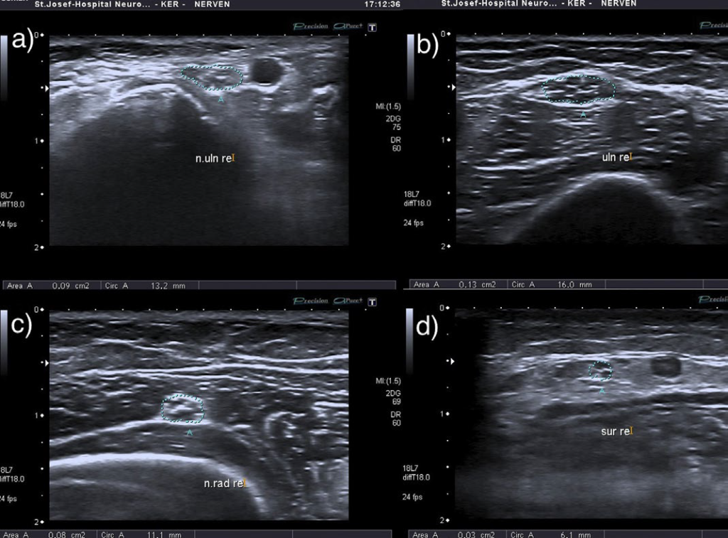
Although the role of neuromuscular ultrasound in the diagnostic workup of immune-mediated polyneuropathies remains less well defined, the recent introduction of a new ultrasound score (Bochum Ultrasound Score-BUS) allowed the differentiation of sub- acute CIDP from AIDP35 (Figure 1).

Figure 1: Overview of the ‘‘Bochum ultrasound score’’ for distinguishing CIDP from AIDP. Cross sectional area of the ulnar nerve in Guyon’s canal (a), upper-arm (b), of the radial nerve in spiral groove (c) and of the sural nerve between the gastrocnemius muscle (d) in a patient with CIDP. The patient showed in all anatomic sites a pathological cross sectional area enlargement, when compared to controls (5), receiving a maximum sum score of 4 points

NOJ-1-101Fig1

In view of our recent report, the anatomical sites summarised under the “Bochum Ultrasound Score’’ included the cross sectional area of: a) the ulnar nerve in Guyon’s canal, b) the ulnar nerve in upper arm, c) the radial nerve in spiral groove and d) the sural nerve between the lateral and medial head of the gastrocnemius muscle (Figure 1). The scoring system included two simple rules 1) the patient received 1 point, for each of the above anatomic sites, where he showed a pathological cross sectional area enlargement, compared the reference values of our lab,5 2) if the patient showed on both sides of the body a pathological cross sectional area nerve enlargement of the concrete nerve, he also received only 1 point.35 Considering the above, each patient could receive a minimum sum score of 0 points and a maximum sum score of 4 points. Following the previous report in the literature we used as a cut-off value for differentiating a sub- acute CIDP from AIDP a sum score of ≥ 2 points.

Among the advantages of the Bochum ultrasound score are a) easy administration, as it summarizes 4 anatomical sites that can be easily sonographically examined, b) economy of time, as it can be performed quickly (about 10 mins), c) high sensitivity and specificity in patients with no difference in disease duration, and d) lack of side effects or pain for the patients, while performing nerve ultrasound. “Bochum Ultrasound Score” should undergo multicentre, prospective evaluation in a larger sample of patients presenting with an (sub-)acute or chronic polyneuropathy to prove its specificity for CIDP.

1. Solbiati L, De Pra L, Ierace T et al. High-resolution sonography of the recurrent laryngeal nerve: anatomic and pathologic considerations. AJR Am J Roentgenol. 1985; 145(5): 989-993.doi: 10.2214/ajr.145.5.989

2. Fornage BD, Rifkin MD. Ultrasound examination of the hand and foot. Radiol Clin North Am. 1988; 26(1): 109-129.

3. Hughes RAC. Peripheral neuropathy. BMJ. 2002; 324: 466- 469.doi: 10.1136/bmj.324.7335.466

4. Cartwright MS, Passmore LV, Yoon JS, Brown ME, Caress JB, Walker FO. Cross-sectional area reference values for nerve ultrasonography. Muscle Nerve. 2008; 37(5): 566-571. doi: 10.1002/mus.21009

5. Kerasnoudis A, Pitarokoili K, Behrendt V, Gold R, Yoon MS. Cross sectional area reference values for sonography of peripheral nerves and brachial plexus. Clin Neurophysiol. 2013; 124(9): 1881-1888. doi: 10.1016/j.clinph.2013.03.007

6. Padua L, Martinoli C, Pazzaglia C, et al. Intra- and internerve cross-sectional area variability: new ultrasound measures. Muscle Nerve. 2012; 45(5): 730-733. doi: 10.1002/ mus.23252

7. Kerasnoudis A, Klasing A, Behrendt V, Gold R, Yoon MS. Intra- and internerve cross-sectional area variability: new ultrasound measures. Muscle Nerve. 2012; 47(1): 146-147.doi: 10.1002/mus.23520

8. Chiò A, Cocito D, Bottacchi E, et al. Idiopathic chronic inflammatory demyelinating polyneuropathy: an epidemiologi cal study in Italy. J Neurol Neurosurg Psychiatry. 2007; 78: 1349-1353.doi: 10.1136/jnnp.2007.114868

9. Iijima M, Koike H, Hattori N, et al. Prevalence and inci–dence rates of chronic inflammatory demyelinating polyneuropathy in the Japanese population. J Neurol Neurosurg Psychiatry. 2008; 79: 1040-1043. doi: 10.1136/jnnp.2007.128132

10. Hughes R. Chronic Inflammatory Demyelinating Polyradiculoneuropathy. J Clin Immunol. 2010.doi: 10.1007/s10875-010-9399-0

11. Taniguchi N, Itoh K, Wang Y, et al. Sonographic detection of diffuse peripheral nerve hypertrophy in chronic inflammatory demyelinating polyneuropathy. J Clin Ultrasound. 2000; 28(9): 488-491. doi: 10.1002/1097-0096(200011/12)28:9<488::aid-jcu7>3.0.co;2-7

12. Duggins AJ, McLeod JG, Pollard JD, et al. Spinal root and plexus hypertrophy in chronic inflammatory demyelinating polyneuropathy. Brain. 1999; 122(Pt7): 1383-1390.doi: 10.1093/brain/122.7.1383

13. Mizuno K, Nagamatsu M, Hattori N, et al. Chronic inflammatory demyelinating polyradiculoneuropathy with diffuse and massive peripheral nerve hypertrophy: distinctive clinical and magnetic resonance imaging features. Muscle Nerve. 1998; 21(6): 805-808. doi: 10.1002/(SICI)1097-4598- (199806)21:6<_x0038_05:_x003a_AID-MUS16>3.0.CO;2-R

14. Naganuma M, Doi S, Shima K, et al. Chronic inflammatory demyelinating polyradiculoneuropathy associated with multifocal nerve hypertrophy: report of a case with MRI study. Rinsho Shinkeigaku. 1991; 31(11): 1186-1191.

15. Matsuda M, Ikeda S, Sakurai S, et al. Hypertrophic neuritis due to chronic inflammatory demyelinating polyradiculoneuropathy (CIDP): a postmortem pathological study. Muscle Nerve. 1996; 19(2): 163-169. doi: 10.1002/(SICI)1097-4598(199602)19:2<163::AID-MUS6>3.0.CO;2-C

16. Matsuoka N, Kohriyama T, Ochi K, et al. Detection of cervical nerve root hypertrophy by ultrasonography in chronic inflammatory demyelinating polyradiculoneuropathy. J Neurol Sci. 2004; 219(1-2): 15-21. doi: http://dx.doi.org/10.1016/j. jns.2003.11.011

17. Imamura K, Tajiri Y, Kowa H, Nakashima K. Peripheral nerve hypertrophy in chronic inflammatory demyelinating polyradiculoneuropathy detected by ultrasonography. Intern Med. 2009; 48(7): 581-582. doi: http://dx.doi.org/10.2169/internalmedicine.48.1924

18. Jang JH, Yoo JK, Kim BJ. Ultrasonographic findings in chronic inflammatory demyelinating polyneuropathy. Am J Phys Med Rehabil. 2012; 91(6): 546-547.doi: 10.1097/PHM.0b013e31824121f1

19. Kerasnoudis A. Ultrasound findings in a case of chronic inflammatory demyelinating polyneuropathy. Muscle Nerve. 2013; 47(3): 443-446.

20. Kerasnoudis A. Ultrasonographic findings in chronic inflammatory demyelinating polyneuropathy. Am J Phys Med Rehabil. 2014; 93(1): 94.

21. Zaidman CM, Al-Lozi M, Pestronk A. Peripheral nerve size in normal and patients with polyneuropathy: an ultrasound study. Muscle Nerve. 2009; 40(6): 960-966.doi: 10.1002/mus.21431

22. Kerasnoudis A, Gold R, Klasing A, Behrendt V, Yoon MS. Correlation of morphological changes in CIDP with the disease duration. 85. Kongress der Deutschen Geselschaft für Neurologie (DGN). 27.09.12, Hamburg Germany. P555.

23. Kerasnoudis A, Pitarokoili K, Behrendt V, Gold R, Yoon MS. Correlation of Nerve Ultrasound, Electrophysiological and Clinical Findings in Chronic Inflammatory Demyelinating Polyneuropathy. J Neuroimaging. 2014. doi: 10.1111/jon.12079

24. Smith EC, Hobson-Webb LD, Massey E. Nerve ultrasound in motor conduction block: pre- and post treatment findings. Muscle Nerve. 2008; 38: 1369.

25. Granata G, Pazzaglia C, Calandro P, et al. Ultrasound visualization of nerve morphological alteration at the site of conduction block. Muscle Nerve. 2009; 40(6): 1068-1070. doi: 10.1002/mus.21449

26. Imamura K, Tajiri Y, Kowa H, et al. Peripheral nerve hypertrophy in chronic inflammatory demyelinating polyradiculoneuropathy detected by ultrasonography. Intern Med. 2009; 48(7): 581-582.doi: 10.2169/internalmedicine.48.1924

27. Jang JH, Cho CS, Yang KS, Seok HY, Kim BJ. Pattern analysis of nerve enlargement using ultrasonography in chronic inflammatory demyelinating polyneuropathy. Clin Neurophysiol. 2014. doi: 10.1016/j.clinph.2013.12.115

28. Padua L, Granata G, Sabatelli M, et al. Heterogeneity of root and nerve ultrasound pattern in CIDP patients. Clin Neurophysiol. 2014; 125(1): 160-165. doi: 10.1016/j.clinph.2013.07.023

29. Koller H, Kieseier BC, Jander S, Hartung P. Chronic inflammatory demyelinating polyneuropathy. N Engl J Med. 2005; 352(13): 1343-1356.doi: 10.1056/NEJMra041347

30. Vallat JM, Sommer C, Magy L. Chronic inflammatory demyelinating polyradiculoneuropathy: diagnostic and therapeutic challenges for a treatable condition. Lancet Neurol. 2010; 9(4): 402-412.doi: 10.1016/S1474-4422(10)70041-7

31. Dalakas MC. Advances in the diagnosis, pathogenesis and treatment of CIDP. Nat Rev Neurol. 2011; 7(9): 507-517. doi: 10.1038/nrneurol.2011.121

32. Hughes RA, Donofrio P, Bril V, et al. ICE Study Group. Intravenous immune globulin (10% caprylate-chromatography purified) for the treatment of chronic inflammatory demyelinating polyradiculoneuropathy (ICE study): a randomised placebo-controlled trial. Lancet Neurol. 2008; 7(2): 136-144. doi: 10.1016/S14744422(07)70329-0

33. Dionne A, Nicolle MW, Hahn AF. Clinical and electrophysiological parameters distinguishing acute-onset chronic inflammatory demyelinating polyneuropathy from acute inflammatory demyelinating polyneuropathy. Muscle Nerve. 2010; 41(2): 202- 207. doi: 10.1002/mus.21480

34. Ruts L, Drenthen J, Jacobs BC, Van Doorn PA. Dutch GBS Study Group. Distinguishing acute-onset CIDP from fluctuating Guillain-Barre syndrome: a prospective study. Neurology. 2010; 74(21): 1680-1686. doi: 10.1212/WNL.0b013e3181e07d14

35. Kerasnoudis A, Pitarokoili K, Behrendt V, Gold R, Yoon MS. Nerve ultrasound score in distinguishing chronic from acute inflammatory demyelinating polyneuropathy. Clin Neurophysiol. 2014; 125(3): 635-641.doi: 10.1016/j.clinph.2013.08.014

LATEST ARTICLES

Unraveling the Mysteries of Type-A Aortic Dissection Using POCUS/Echocardiography

Syeda Rukh*, Sathyanarayana Machani and Milind Awale

doi.

Blood Sample from the Patient

Hypertriglyceridemia-Induced Pancreatitis: A Case Report and Literature Review

Maarten Bulterys, Melvin Willems* and Agnes Meersman

doi.

From Neck Pain to a Life-Threatening Condition: A Case Report

Floris Vandewoude* and Sören Verstraete

doi.

LATEST ARTICLES