INTRODUCTION
Dystocia means difficulty in parturition to the point of needing human intervention. Dystocia also defined as the failure of transmission of stage one to stage two labor or when thirty minutes spend to start of stage two of labor or parturition.1,2,3,4 The first stage of parturition (labor) is usually characterized by decreased appetite, preparing a birthing area, isolation from the herd, restlessness, and leading up to the early uterine contractions but it end-up with forceful abdominal contractions. Stage two of parturition is fetal expulsion or delivery of neonate which usually occurs within 15-30-minutes. The third stage of parturition follows and ends by the expulsion of the fetal membranes which usually takes place within 4-6 hours.2,3,5,6
Dystocia in small ruminants can be resulting from either maternal or fetal pre-disposing factors. Among fetal-related factors the most common causes of dystocia were oversized fetus, lamb mal presentation, malposition, postural defects, and congenital abnormalities.3,7 whereas, over feeding of dam during pregnancy, uterine inertia in polytocous ewes, incomplete cervical dilatation (ring womb) and small diameter of pelvic canal are maternal related factors of causes dystocia.7,8 Other factors that may contribute for the occurrence of dystocia are age of ewe, gender of offspring, large or small birth weights.1 Dystocia constitutes a major reproductive problem among small ruminants and can hinder or affect their productivity because of newborns and dams mortality with severe economic losses.7 Close observation of the ewe and doe throughout parturition is crucial for well-timed interference due to the fact that early recognition and interference in cases of dystocia both manually or surgically will bring about uneventful final results each for the dam and the neonate.3 Bhattacharyya et al5 stated that survival of the animals (and their fetuses) presented for treatment of dystocia irrespective of whether they are managed manually or surgically is directly related to their clinical status. Survival of each the dam and new born lamb were drastically laid low with the duration of time delay among the start of labor and the time of presentation for surgical intervention.9,10 Hence, the outcome and success rate of caesarean section is higher if surgery was performed early when the fetus is alive or freshly dead.9,11 Generally, the effectiveness of the type of the treatment for delivering the fetus in case of dystocia depends upon the type of dystocia and its cause.11,12 Therefore, the aim of this case series is to presents the surgical management of dystocia in ewes.
CASE HISTORY AND EXAMINATION
Case 2
A five-years old local breed sheep weighing 16 kg was brought to the VTH, College of Veterinary Medicine and Agriculture, Addis Ababa University with the history of three-month gestation in sternal recumbency. Sheep was in 4th parity and on the past all three parities the animals was given a twins without history of any difficulty in birth. The history also revealed that five days before admitted to the VTH the abdomen was distended and the distension was gradually increased. The ewe was also treated with Oxytetracycline injection (10 mg/kg) intramuscularly for three days at one-day interval by calling animal health professional from Denkaka animal health clinic at home but after four-days, animal doesn’t get relief. Up on physical examination, the animal become anorectic, lethargy, had ruff hair coat, recumbent with attempt to stand, distended abdomen and granting sound was heard. On clinical examination, body temperature and respiration were within normal range and also the ewe had tachycardia and pale mucous membrane with normal range of capillary refill time. There was no rupture of the fetal membrane and no vaginal discharge was observed. No fetal part was observed in the birth canal. To detect the status of fetus abdominal palpation used but there was no active fetal movements were felt. Depending on history and abdominal palpation the condition was diagnosed as dystocia as a result of fetal death in utero, and animal was suggested to caesarian section to safe the dam.
Pre-operative animal preparation:
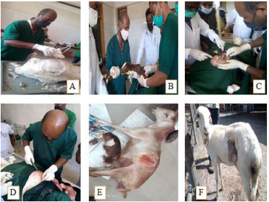
Ewes were immobilized through using physical method by lying down on the surgical table. Then, the left paralumbar fossa area was prepared aseptically by washing with soap and tap water. The hair was shaved by using scalpel blade no. 23 from the skin that covers the left paralumbar fossa area (Figure 1A). Then, the area was again washed by using antiseptic solution (savlon) to remove any fall of hair. Finally, surgical site was scrubbed with 1% povidone iodine in circular manner starting from the center to prefer and made it ready for aseptic surgery. In both case animals were received diclofenac sulfate at 2 mg/kg through intramuscularly (IM) injection. Ringer lactate solution was also administered using 18 G IV cannula as intravenous fluid at a rate of 10 ml/kg/hour to prevent hypo-volumic shock as a result of bleeding and to correct the dehydration status.
Anesthesia and animal control:
Both ewes were controlled in right lateral recumbent position and the head was positioned straight forward by assistant person to facilitate the flow of IV fluid. Field block anesthesias were used by making line infiltration in case 1 and inverted “L” blocks in case 2 with 2% lidocaine hydrochloride along the surgical incision site. Surgical site was draped without any movement and secured to skin with towel forceps. Finally, the surgical site was given a final scrub with 0.1% povidone iodine in circular manner starting from the center to prefer prior to proceeding to surgery.
Surgical procedure:
After surgical site was prepared aseptically and appropriated analgesia was achieved, a celiotomy was performed by incising the skin, starting from 10 cm away from transverse process of lumbar vertebrae and 5 cm distant from the last rib, and ending a 20 cm ventrally from starting point on left paralumbar fossa. The subcutaneous fascia was bluntly dissected by using blunt Mayo scissors. The three abdominal muscles were gently incised by using scalpel blade no. 20 that was loaded on scalpel handle no. 4. Peritoneum was grasped by using serrated tissue forceps and tented up. Then, small incision was made on the peritoneum by using scalpel blade. Finally, two fingers were inserted through the small incision and peritoneum was tented up to extend the incision in between the two fingers down and up by using blunt Mayo scissors and the abdominal contents were exposed. To control bleeding simple swabbing and ligation and twisting of blood vessel with heamostatic forceps was performed depending on the size of blood vessel and amount of blood oozing out to obtain a very clean surgical field. After inserting gloved hand through incision, the gravid uterus was searched and gently pulled out on the surgical field. Then, the uterus was swapped by using gauze and incision was made on greater curvature in parallel to blood vessel. After excision of the uterus was completed live fetus in case 1 (Figure 1B) and died fetus in case 2 (Figure 1C) were removed. Independently trans-fixation of the umbilical cord was done by using absorbable suture material (Chromic surgical cut gut 2-0). The uterus was lavaged externally with warm normal saline and powder of fortified procaine penicillin was poured onto the incision site in both cases immediately after fetus was removed out. Then, uterus was sutured with a continuous inverting pattern (Cushing) followed by lambert suture pattern. After suturing of uterus was completed any bleeding was checked, cleaned and returning it into abdominal cavity. Finally, surgical field was cleaned and celiotomy was closed in three layers in case 1 and four layers in case 2 with chromic surgical cut gut 2-0 and synthetic non-absorbable suture material (silk) for soft tissues and skin suture (Figure 1D), respectively in both cases.
Post-operative care:
Case 1
A two-years old local breed female sheep weighing 18 kg was brought to the Veterinary Teaching Hospital (VTH), College of Veterinary Medicine and Agriculture, Addis Ababa University with the history of full-term gestation in lateral recumbency. Sheep was in 3rd parity and on the past all two parities the animals was given a single lamb without history of any difficulty in birth. Further, history revealed that the parturition process commenced 12-hour before presentation to the hospital and also the animal was anorexic since two-days. On clinical examination, body temperature and respiration were within normal range and also the ewe had tachycardia and pale mucous membrane with capillary refill time of 3 seconds. The ewe was weak and unable to stand. There was no rupture of the fetal membrane and no vaginal discharge was observed. No fetal part was observed in the birth canal. To detect the status of fetus abdominal palpation was used and there was an active fetal movement was felt. Up on vaginal examination there was incomplete dilation of cervix. Therefore, the condition was diagnosed as maternal dystocia due to incomplete dilation of cervix, and animal was admitted to caesarian section to safe the fetus.
Post-operative treatment included administration of Fortified procaine penicillin (22,000 IU/kg) through I.M. for five-days. The owner was also advised to supplement good nutrition to facilitate wound healing (Figure 1E). The wound was properly dressed with 1% Povidone iodine solution and applying wound spray (cyclo spray) for five days daily. Finally, skin sutures were removed on 15th post-operative day (Figure 1F) and uneventful recovery was noticed in case 2, but the dam of the case 1 was died after 18-hours of surgery.
Figure 1. Surgical Manipulations of Caesarean Section in Ewes
DISCUSSION
Dystocia or difficult birth is a major cause of lamb loss in the flock and may result in great economic loss to the farmers.7 The causes of dystocia are either maternal or fetal origin. In this particular case, the causes of dystocia were maternal origin. This in agreement with the previous studies by Sharma et al10 who reported the occurrence of dystocia due to maternal factors (63.33%) were higher than fetal factors (36.67%). In contrary to present case, other researchers Ahmed et al1 ; Bhattacharyya et al5 and Elchikh et al7 reported that the occurrence of dystocia as a result of fetal related factors were higher than maternal related factors. From maternal causes of dystocia, in present case, insufficient dilation of cervix was identified in case 1, which is in agreement with similar case report by Dutt et al9 and Sharma et al10, whereas, premature fetal death in utero was identified in case 2, which is agrees with Jacobson et al.13
Dystocia can be managed through various ways either non-surgically (medically or manual traction) or surgically depending upon time of initiation of labour pains, time of rupture of water bags, and per vaginal examinations.1,10,11 The most common indications for non-surgical management of dystocia in small ruminants are lateral deviation of the head and neck, and bilateral shoulder flexion associated with absolute fetal oversize,3 but their uses were limited due to the small diameter of the pelvis.1 However, cesarean section was an effective method for treatment of most types of dystocia and was safe for dam as well as fetus, especially when it is performed as early as possible after onset of labor when the fetus is alive or freshly dead.1,10,11,14 The most common causes of dystocia that recruit caesarean section in ewe are failure of cervix to dilate and relative or absolute oversize of fetus.9 Similarly, in present case, dystocia due to failure of cervix to dilate was diagnosed and managed through cesarean section.
In small ruminants, cesarean section usually performed while the animal is in right lateral recumbency under local analgesia, with2,14 or without5,9,12,15 sedation. Perhaps, in present case, cesarean section was performed while the animal was in right lateral recumbency under line infiltration5,9 and inverted “L” blocks2,15 of local anesthesia using 2% lidocaine without sedation in case 1 and case 2, respectively. Uterine wall closure, in present case, was done with Cushing followed by lambert suture pattern by using surgical chromic catgut no. 2 without penetration of the mucosal layer, which is in agreement with similar case report by Aliciah et al15; Verma et al.16 In contrast to present case, Kisani et al14 and Bhattacharyya et al5 used two layers of Lambert suture pattern with chromic catgut size 1-0, Iqbal et al2 used single layer Cushing suture pattern with chromic cat gut number 2-0 and Dutt et al9 used continuous interlock suture patterns with chromic catgut-1 to close the uterine wall.
The length of time delay between the start of labor and the time of presentation for surgical intervention are significantly affects the survival rates of both the dam and newborn.1,2,5 This in agreement with the present case, where ewe was straining for at least 12-hours in case 1 and 3-days in case 2. It was reported that fetal and dam survival rates in sheep and goats undergoing the relief of dystocia with caesarean section was 94.12%.2 After operation was conducted, in present case, a single live fetus and a twin dead fetuses were removed from case 1 and 2, respectively. However, the dam of case 1 was died after 18-hours of post-operation. This agrees with the reports of Odedara et al,12 the survival rate of dam was lower following surgical fetal delivery.
The most intra- and post-operative complication following caesarian section was shock, adhesion formation, peritonitis and death.5 This agrees with the present case, where the dam of case 1 was died after 18-hours of post-surgery. Therefore, pre-operative preparation of the patient by administration of intravenous fluids, analgesics and antibiotic has a proven effect in preventing hypo-volumic shock, pain and swelling, and secondary bacterial infection of wound.5,15,16 Similarly, in present case, intravenous fluids, analgesic and antibiotic were administered before operation and as a post-operative care for five-days done.
CONCLUSION
It is concluded from the present case report that caesarean section is a fairly common surgical procedure to resolve dystocia due to incomplete dilation of cervix and fetal death in utero. Doublelayer closure of uterus with Cushing followed by lambert suture pattern should be performed to prevent the leakage of fluid from the uterus and with stands strength from the contraction of uterus, and also result into normal healing for cesarean section.
The effective pre- and post-operative management and anti-septic dressing of surgical wound for animals undergo C-section is very important in preventing bacterial contamination of the wound.
ACKNOWLEDGEMENTS
The authors are thankful to Dr. Shiferaw Moltumo for his help through provision of all necessary materials to perform surgical procedure and Dereje Gudeta for his assistance with the post-operative management of this case.
ETHICS CONSIDERATION
The approval of ethical committee and the Institutional Review Board (IRB) was taken for conducting this study and followed all the animal ethics and welfare guidelines.
CONFLICTS OF INTEREST
The authors declare that they have no conflicts of interest.


