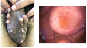
A 35-year-old man consulted for calcified nodules of the scrotal skin. He complained of an impact on his sexual performance. The nodules progressively increased in size. He had no history of trauma or genitourinary or hormonal disorders. His physical examination revealed multiple bilateral calcified cutaneous lesions of the scrotum with no associated inflammation (Figure 1A). The dermatoscopic examination noted erythema with telangiectatic vessels (Figure 1B).
Figure 1. A. Multiple and Bilateral Calcifiedcutaneous Lesions of the Scrotum with No Inflammatory Lesions. B. The Dermatoscopic
Examination Noted Erythema with Telang Ectatic Vessels

The described lesions were exclusively scrotal; there were no extensions to the penis. As this was a young patient with multiple lesions, blood and urine tests were performed, including calcium, phosphorus, and parathyroid hormone, which were normal. He underwent surgery. Complete resection of the lesions was performed.
Scrotal calcinosis is a benign condition first described by Lewinski over a century ago.1 Lesions are nodular and indolent, often bilateral and present without any discharge or inflammation. The question about the etiopathogenesis is not resolved.
Some authors concluded that the common characteristics are calcified dystrophy of epidermal cysts.2 A penile location has been reported for multiple scrotal calcinosis. Nodules may cause sexual discomfort.3 Scrotal calcinosis is not associated with any metabolic, hormonal or endocrine disorder and is thus considered idiopathic in origin.4 Surgery is usually performed in patients presenting with voluminous lesions causing psychological and sexual distress.4
CONSENT
The authors have received written informed consent from the patient.
CONFLICTS OF INTEREST
The authors declare that they have no conflicts of interest.


