Cancer Studies and Molecular Medicine

Open journal

ISSN 2377-1518

Role of Molecular Imaging in Oncology

Pradeep K. Garg*, Surinder K. Batra and Sudha Garg

Pradeep K. Garg, PhD

Executive Director Center for Molecular Imaging and Therapy, Biomedical Research Foundation of NW Louisiana Shreveport, LA 71106, USA; Tel: 318-675-4002; E-mail: pgarg@biomed.org

INTRODUCTION

Molecular Imaging (MI) is an emerging field that combines the non-invasive monitoring of in vivo biological processes at the cellular level and the anatomic information associated with tissues undergoing such transformations. MI therefore has evolved as an indispensable technique for the early detection and diseasestate, and for monitoring response to therapy. These modalities offer non-invasive detection of in vivo biological functions and processes at the molecular level through the careful selection of targeting molecules which is expressed differentially in diseased vs. healthy cells to interrogate the cell microenvironment. These targeting molecules could consist of small molecules, single amino acid units, low molecular weight peptides, antibodies or antibody fragments. There are now a large array of imaging technologies under the MI umbrella which include Single Photon Emission Computed Tomography (SPECT) and Positron Emission Tomography (PET), Magnetic Resonance Imaging (MRI), Magnetic Resonance Spectroscopy (MRS), Computed Tomography (CT), ultrasound (US), bioluminescence (quantum dots), and fluorescence imaging (optical imaging).1 Amongst the tools required for targeting diseased tissues to obtain clinically relevant information, the key is to match the optimal targeting molecule with the imaging modality of choice.

RESULTS AND DISCUSSIONS

Amongst various options within MI, nuclear imaging is the more common non-invasive detection technique to target tumors. Nuclear imaging employs radiolabeled tracers which concentrate in cancerous tissues and generates the radioactive signal for visualization of tumors. This signal is detected, reconstructed, and analyzed with the resulting image corresponding to the spatial distribution of the radiotracer in the cancer mass and surrounding tissues. The two major nuclear imaging modalities are SPECT and PET.

POSITRON EMISSION TOMOGRAPHY/COMPUTERIZED TOMOGRAPHY (PET/CT)

PET, a non-invasive imaging modality, allows quantitative evaluation of the biological processes in vivo through emission of positrons from nuclear decay of an intravenously injected radiopharmaceutical. PET typically has higher sensitivity than conventional SPECT which allows the targeting of sites at much lower concentrations of radiotracers in the target area, allowing localization of small lesions.

One of the most commonly used radiotracers in MI is F-18 fluorodeoxyglucose (FDG), a compound approved by the FDA for clinical use. Since membrane glucose transporter (GLUTs) expression increases significantly in rapidly dividing cancer cells, FDG enters these cells producing preferential uptake and phosphorylation of FDG and permitting clinically meaningful imaging of the tumor. Besides FDG, there have been many advances in developing new class of radiotracers to diagnose cancer and to interrogate certain neurological processes. O-15 water (blood flow),2 F-18 labeled fatty acids (fatty acid/thiokinase metabolism),3 F-18 FLT (thymidine kinase),4 F-18 fluoroestradiol (estrogen receptor targeting),5 C-11 and F-18 choline (choline kinase/oncological imaging),6– 11 F-18 fluoromisonidazole (F-MISO; hypoxia imaging),12,13 radiolabeled annexin (apoptosis),14,15 F-18 FMDHT (androgen receptor imaging),16,17 and F-18 FHBG and analogues (reporter gene targeting/imaging),18,19,20 are some of the examples illustrating a wide scope and contribution of this imaging modality to molecular imaging field. Similarly, SPECT uses radionuclides which decay through a single photon emission branch. As with PET, SPECT also requires the use of radioactive probe for MI, albeit the chemistry involved in preparing those radioactive molecules could be quite different.

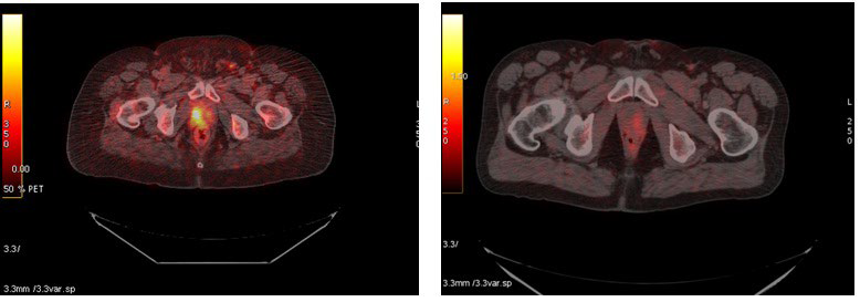
While the choice of radiotracer varies with application, several of these targeting probes are multifunctional and have a widespread utility across many diseases. Most of these probes are small molecules, have rapid clearance from blood pool (circulation), rapid washout from normal tissues and low metabolic disintegration. Our own and other researchers experience with C-11 choline in patients with prostate cancer has been quite impressive.7,21,22,23 Patients with recently diagnosed prostate cancer were scanned using C-11 choline to localize the tumor. At the conclusion of the treatment regimen, the patients returned for a second C-11 choline scan to assess treatment efficacy. As shown in figure 1, PET/CT imaging with C-11 choline has been quite unequivocal in monitoring therapy outcome.6

Figure 1: PET/CT image of a patient with prostate cancer before and after therapy. The left panel shows C-11 choline uptake in the prostate bed (scan acquired prior to therapy). After this scan patient underwent therapy. The right panel shows the PET/CT scan after patient completed therapy. No uptake of C-11 choline is noted in that region supporting the efficacy of C-11 choline to monitor prostate cancer therapy.

CSMMOJ-1-e001Fig1

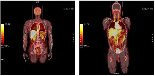
Similarly, C-11 choline plays an impressive role in following treatment response in patients with esophageal cancer.24 Several other PET imaging probes labeled with F-18 have also been developed over the years.6,10,16,17,25,26,27 Whole body PET/CT imaging with 7α-[18F] fluoro 17α-methyl 5αdihydrotestosterone (F-18 FMDHT) in normal healthy volunteers is shown in figure 2. The radiotracer clears from most normal tissues following the hepatobiliary clearance. The initial uptake in the urinary bladder was low but increased after 60 min post injection, indicating its potential to clearly discern primary as well as metastatic prostate cancer from normal tissues.

Figure 2: Distribution of F-18 FMDHT in normal healthy volunteer. Early image (left panel) show uptake of F-18 in the liver, spleen, kidneys. Late image of same patient (2 h post injection) show significant clearance of radioactivity from liver through the hepatobiliary path with radioactivity accumulation in the bladder.

CSMMOJ-1-e001Fig2

While small molecular weight compounds have already been proven effective for molecular imaging applications, the role of large proteins such as antibodies has been steadily gaining momentum. One of the key parameters is to non-invasively visualize target molecules in altered cells by virtue of the target probe interaction at the molecular level. While some evidence suggests the clinical utility of Ab in molecular imaging, because of their large molecular size (150 kDa), the intact immunoglobulins remain in circulation28 and may take a long time to accumulate in tumors (1-2 days) This slow uptake decreases the clinical utility of such a probe and investigators have been less than enthusiastic to consider such molecules as relevant for molecular imaging. Advancement in antibody engineering leading to small molecular weight constructs such as monovalent fragments (variable fragments Fv, single chain variable fragments (scFv), bivalent or bispecific diabodies, tribodies, Fab fragments and minibodies) raised the enthusiasm in the MI community to explore their utility in imaging. In comparison to the intact antibody, these smaller fragments, particularly the single chain antibodies (scFv)2 show faster clearance from the blood and rapid peak tumor localization. Over the years, a number of antibody based tumor biomarkers have been developed for the diagnosis or treatment follow-up of specific cancers.29,30,31 However, challenges to the effective exploitation of differentially expressed markers for the timely visualization of growing tumors remain. With the recent initial successes in preclinical and translational studies, several such antibody based entities are now FDA approved for clinic use. Some of the examples of antibody based radiopharmaceuticals are OncoScint CR/OV (Satumomab Pendetide) which targets the cell surface mucin-like glycoprotein antigen TAG-72 in colorectal and ovarian carcinomas, 111In-labeled Oncoscint for pre-operative evaluation, monitoring recurrence,and detection of extrahepatic metastasis,32 and ProstaScint (CapromabPendetide) for the diagnosis of metastatic prostate cancer.33,34 A radionuclide with a longer half-life such as 124I (4.18 d) has helped to further explore the use of whole IgG because the long half-life radionuclide allows for extended time for uptake in targeted sites while allowing for the clearance from normal tissues and from non-specifically circulating fraction35 and maintaining enough radioactivity to produce a useful imaging signal. More recently, Zirconium-89 (89Zr), a transition metal, has turned into an attractive choice of radionuclide for PET imaging due to its longer half-life (3.3 days) which is compatible with the pharmacokinetics of large molecules such as antibodies and their fragments. 89Zr-labeled mAb has been proven to be stable in vivo and has provided better spatial resolution than that with 18F FDG PET. Use of 89Zr labeled mAbs as a scouting procedure preceding radioimmunotherapy has been proposed.36,37,38 In one of the earliest studies, 89Zr was labeled with mAb U36 recognizing the v6 domain of CD44, a tumor antigen overexpressed in head and neck tumors. This radioimmunoconjugate was used in preclinical models to confirm tumor targeting and estimate the dose deposition to tumor and normal tissues prior to radioimmunotherapy with 90Y-labeled mAbs.39

Another radionuclide with attractive PET properties and compatible with large as well small molecular weight targeting molecules is copper-64 (64Cu). Initial efforts using copper-64 explored the use of macrocyclic chelating agent 1,4,7,10-tetraazacyclododecane-N,N’,N’’, N’’’-tetraacetic acid (DOTA). This radiolabeled small molecular weight antibody (minibody; 80 kDa) cleared rapidly from circulation and exhibited moderate tumor uptake shown by serial imaging of antigen positive tumors using preclinical models and micro PET imaging.

OPTICAL IMAGING

The use of optical imaging is also rapidly being adopted by molecular imaging scientists. Optical imaging includes fluorescence and bioluminescence based imaging probes. Optical imaging modalities are very promising due to their high sensitivity and specificity, low cost, portability of imaging instruments and absence of ionizing radiation. Further with the imaging is showing potential for clinical relevance. In fluorescence imaging, cells are labeled with dyes or proteins which emit light of a limited spectrum when excited. Bioluminescence methods use an enzymatic reaction between a luciferase enzyme and its substrate, luciferin, to produce photons which are converted to electrons and detected by a cooled Charged Couple Detector (CCD). This ultimately results in the detection of visible light through near-infrared light signals. Fluorescent molecules like Green Fluorescent Protein (GFP) and Red Fluorescent Protein (RFP) are used to observe the localization of proteins within a cell, to label specific proteins, or to monitor the production of a gene product.40 For bioluminescence imaging, the bioluminescence reporter Firefly Luciferase (FL) and Renilla Luciferase (RL) are used frequently.

More recently, advances have been made in the field of infrared imaging. Near-Infrared imaging (NIR) has deeper penetration ability and higher sensitivity than the previously discussed optical imaging options. Ogawa et al., analyzed the efficiency of NIR flourophorindocyanine green (ICG) for in vivo imaging and produced encouraging results.41

ULTRASOUND IMAGING (US)

Ultrasound (US) is a cross-sectional imaging modality which uses sound-waves to produce interpretable images. Pulses of sound-waves of appropriate frequencies (usually 1-20 MHz depending on desired imaging depth) are emitted through arrays of transducers (probes).The sound-waves are reflected at tissue boundaries and these returning echoes are captured and reconstructed to produce a two-dimensional image of the plane being scanned. The resulting image is displayed using a grey-scale display corresponding to the intensity of the returning echoes. Ultrasound contrast agents are a relatively new concept and are designed to alter the absorption, reflection or refraction of the sound-waves which enhance the differentiation of the signal from the tissue containing the contrast agent from that of the surrounding sample. Targeted ultrasound contrast agents are site-directed contrast agents which specifically enhance the signal from pathologic tissue which would otherwise be difficult to distinguish from surrounding normal tissue. Microbubbles (MB) are US contrast agents which are gas filled structures encapsulated by lipid shell, polymer or proteins and when injected into the blood stream enhance signals from the target tissues.42,43 Their low cost, easy transportability and utility to provide a discriminated signal from background with high sensitivity has made the MB contrast agent of choice for US imaging. Future research could allow specific ligands to be attached to the MB’s surface which could lead to the accumulation of these agents in the target sites. Because of their large size, MB rarely extravagates from vascular tissue. Also, the MB targeted to the platelet glycoprotein IIb/IIIa integrin44 and to fibrin45 have been used for thrombus imaging. Fortunately, targeted MB based contrast imaging is even making functional characterization of tumor vasculature possible.

Although, there are manuscripts reporting that only a small number of MBs are required to produce adequate contrast for imaging purposes, further research is needed to assess the overall sensitivity of targeted microbbubles, determine the amount of antibody sufficient to get the target microbubble to the desired anatomic location, and to determine the comparative efficacy of MBs in comparison to nuclear and other optical imaging modalities.

MAGNETIC RESONANCE IMAGING (MRI) AND MAGNETIC RESONANCE SPECTROSCOPY (MRS)

MRI primarily uses the magnetic resonance signal produced from the hydrogen atoms in a sample under the influence of strong magnetic field for constructing three dimensional image sets. Positive and negative contrast agents containing metal ions are used to enhance MRI sensitivity. Positive contrast agents (appearing bright on MRI) are small molecular weight compounds usually containing gadolinium (Gd), manganese (Mn) or iron (Fe) as their active element. Negative contrast agents (appearing predominantly dark on MRI) are small particulate aggregates such as Super Paramagnetic Iron Oxide (SPIO) or Ultra-small Super Paramagnetic Iron Oxide (USPIO) particles which have an iron oxide core containing iron atoms covered by a polymer shell usually made of dextran or polyethyleneglycol. Although contrast enhanced MRI is a powerful imaging modality with sub-micrometer resolution (10-100 µm) and exquisite soft tissue contrast, its major drawbacks are low sensitivity, low retention of positive contrast agents, relatively long imaging times, and the necessity to inject large amounts of contrast to obtain quality images.

Since conventionally used contrast agents like Gd lack specificity, targeted contrast agents are being developed by conjugating paramagnetic compounds to various peptides, ligands, antibodies and antibody fragments that target specific tumor cell surface antigens. Superparamagnetic Iron Oxide (SPIO) is another class of negative contrast agents used in MR imaging.

Nanoparticles are nano-sized carriers used for delivery of various pharmaceuticals. In recent years, there has been intense effort to develop multifunctional nano-carriers for the targeted delivery of diagnostic and therapeutic agents. These multifunctional nano-carriers could incorporate antibodies for specific and effective tumor targeting and delivery of therapeutic payloads. For example, nano-shells are optically tunable nanoparticles that contain a dielectric core surrounded by a thin gold shell. These nano-shells may be designed to scatter and/or absorb light over a broad spectral range. Thus depending on wavelength of light chosen, these nanoparticles can be utilized for imaging and for photo thermal therapy.

OTHER IMAGING MODALITIES

Raman spectroscopy is based on the inelastic scattering of light during its interaction with matter. Although the light scattered from biological samples is usually weak, the intensity of scattered light increases tremendously in the vicinity of metallic nanoparticles. These metallic nanoparticles, on excitation at a particular wavelength, exhibit surface plasmon resonance and resonate light. This resonation results in enhanced scattering from these metallic particles which result in increased contrast when compared to the surrounding molecules. The increased contrast is basis for Surface Enhanced Raman Spectroscopy (SERS). Due to its ability to detect picomolar amounts of the targeted molecule, SERS is becoming a predominant imaging modality. SERS provide multiple advantages over existing imaging modalities including the unique spectral properties of SERS nanoparticles with easily resolved narrow peak which allows for multiplex imaging ,lower toxicity due to inert nature of SERS nanoparticles (Gold, silica). Moreover, colloidal gold particles coated with a protective layer of polyethylene glycol (PEG) exhibit excellent in vivo biodistribution and pharmacokinetic properties upon systemic injection, and SERS probes provide enhancement of the Raman scattering of adjoining molecules by as much as 1014-1015 which can lead to the detection of a single molecule.46,47 SERS is becoming the modality of choice when localized and surface tumors. However, its use remains limited in imaging deep tumors due to shallow depth of penetration associated with Raman microscopes and the non-specific uptake of these particles.48

CONCLUSION

A wide array of imaging modalities is available to the preclinical research community and many of these modalities have been adopted by the clinic. Nonetheless, the efforts to further optimize probes to more precisely target cellular level processes with high specificity and selectivity continues to be an area of much research effort. As we gain more knowledge on the molecular interactions of various disease states, new and novel probes are being considered, explored, and designed. Along with probe development, or perhaps because of it, there has been great progress in enhancing the capabilities of imaging instruments and techniques to further help visualize these molecular processes with increased sensitivity and resolution. As the molecular imaging field is evolving and scientists and physicians are recognizing its importance, industries are taking note of the strength of this modality. Several pharmaceutical companies have either entered in collaboration with researchers from various academic institutions or have developed an inhouse molecular imaging program. It is only a matter of time when molecular imaging will become an integral part of drugdevelopment to provide routine standard of care in the clinic.

1. Blasberg RG. Imaging update: new windows, new views. Clin Cancer Res. 2007; 13(12): 3444-3448. doi: 10.1158/1078-0432. CCR-07-0936

2. Herscovitch P, Mintun MA, Raichle ME. Brain oxygen utilization measured with oxygen-15 radiotracers and positron emission tomography: generation of metabolic images. J Nucl Med. 1985; 26: 416-417.

3. Stocklin G. Tracers for metabolic imaging of brain and heart— radiochemistry and radiopharmacology. Eur J Nucl Med. 1992; 19(7): 527-551.doi: 10.1007/BF00185860

4. Agool A, SR, Kluin PM, de Wolf JT, Dierckx RA, Vellenga E. F-18 FLT PET: a non-invasive diagnostic tool for visulaization of the bone marrow compartment in patients with aplastic anemia: a pilot study. Clin Nucl Med. 2011; 36(4): 286-289. doi: 10.1097/RLU.0b013e31820aa1a1

5. Katzenellenbogen JA, Welch MJ, Dehdashti F. The development of estrogen and progestin radiopharmaceuticals for imaging breast cancer. Anticancer Research. 1997; 17(3B): 1573-1576.

6. Fried D, Garg S, Thomas A, et al. Monitoring treatment response inhigh risk prostate cancer patients using C-11 choline PET/CT. J Nucl Medicine Mol Imaging. 2010; 51: 51P.

7. Li X, Liu Q, Wang, et al. C-11 choline PET/CT imaging for differentiating malignant from benign prostate lesions. Clin Nucl Med. 2008; 33(10): 671-676. doi: 10.1097/ RLU.0b013e318184b3a0

8. Rottenburger C, Hentschel M, Kelly T, et al. Comparison of C-11 methionine and C-11 choline for PET imaging of brain metastases: a prospective pilot study. Clin Nucl Med. 2011; 36(8): 639-642. doi: 10.1097/RLU.0b013e3182175840

9. Shinoura N, Nishijima M, Hara T, et al. Brain tumors: detection with C-11 choline PET. Radiology. 1997; 202(2): 497-503. doi: 10.1148/radiology.202.2.9015080

10. Mease RC. Radionuclide based imaging of prostate cancer. Curr Top Med Chem. 2010; 10(16): 1600-1616. doi: http://dx.doi.org/10.2174/156802610793176774

11. Reske SN. Nuclear imaging of prostate cancer: current status. Urologe A. 2007; 46(11): 1485-1499. doi: 10.1007/s00120- 007-1572-6

12. Mendichovszky I, Jackson A. Imaging hypoxia in gliomas. Br J Radiol. 2011; 84 Spec No 2: S145-S158. doi: 10.1259/ bjr/82292521

13. Padhani AR, Krohn KA, Lewis JS, Alber M. Imaging oxygenation of human tumours. Eur Radiol. 2007; 17(4): 861- 872. doi: 10.1007/s00330-006-0431-y

14. Collingridge DR, Glaser M, Osman S, et al. In vitro selectivity, in vivo biodistribution and tumour uptake of annexin V radiolabelled with a positron emitting radioisotope. Br J Cancer. 2003; 89(7): 1327-1333. doi: 10.1038/sj.bjc.6601262

15. Li X, Link JM, Stekhova S, et al. Site-specific labeling of annexin V with F-18 for apoptosis imaging. Bioconjug Chem. 2008; 19(8): 1684-1688. doi: 10.1021/bc800164d

16. Garg PK, Labaree DC, Hoyte RM, Hochberg RB. [7a18F]Fluoro-17a-methyl-5a-dihydrotestosterone: a ligand for androgen receptor-mediated imaging of prostate cancer. Nucl Med Biol. 2001; 28(1): 85-90.doi: 10.1016/s0969-8051(00)00172-4

17. Garg S, Doke A, Black KW, Garg PK. In vivo biodistribution of an androgen receptor avid PET imaging agent 7-alphafluoro-17 alpha-methyl-5-alpha-dihydrotestosterone ([(18)F] FMDHT) in rats pretreated with cetrorelix, a GnRH antagonist. Eur J Nucl Med Mol Imaging. 2008; 35(2): 379-385.doi: 10.1007/s00259-007-0610-3

18. Ponde DE, Dence CS, Schuster DP, Welch MJ. Rapid and reproducible radiosynthesis of [18F] FHBG. Nucl Med Biol. 2004; 31(1): 133-138. doi: 10.1016/S0969-8051(03)00096-9

19. Lee YL, Lee YJ, Ahn SJ, et al. Combined radionuclidechemotherapy and in vivo imaging of hepatocellular carcinoma cells after transfection of a triple-gene construct, NIS, HSV1-sr39tk, and EGFP. Cancer Lett. 2010; 290(1): 129-138. doi: 10.1016/j.canlet.2009.09.004

20. Qiao H, Surti S, Choi SR, et al. Death and proliferation time course of stem cells transplanted in the myocardium. Mol Imaging Biol. 2009; 11(6): 408-414. doi: 10.1007/s11307-009- -0222-3

21. Giovacchini G, Samanes Gajate AM, Messa C, Fazio F. Increased C-11 choline uptake in pagetic bone in a patient with coexisting skeletal metastases from prostate cancer. Clin Nucl Med. 2008; 33(11): 797-798. doi: 10.1097/ RLU.0b013e318187ee35

22. Hara T, Kosaka N, Kishi H. PET imaging of prostate cancer using carbon-11-choline. J Nucl Med. 1998; 39(6): 990-995.

23. Hara T, Kosaka N, Kondo T, et al. Imaging of brain tumor, lung cancer, esophagus cancer, colon cancer, prostate cancer, and bladder cancer with [11C]choline [Abstract]. J Nucl Med. 1997; 38: 250P.

24. Attia A, V GT, Garg PK, et al. The utility of C-11 choline PET/CT for staging advanced esophageal cancer. Int J Radiat Biol Phys. 2011; 81: S314.

25. Larson SM, Morris M, Gunther I, et al. Tumor localization of 16beta-18F-fluoro-5alpha-dihydrotestosterone versus 18F-FDG in patients with progressive, metastatic prostate cancer. J Nucl Med. 2004; 45(3): 366-373.

26. DeGrado TR, Baldwin SW, Wang S, et al. Synthesis and evaluation of (18)F-labeled choline analogs as oncologic PET tracers. J Nucl Med. 2001; 42(12): 1805-1814.

27. Garg S, Lynch AJ, Doke AK, Minton RC, Garg PK. A remote controlled system for the preparation of 7 alpha-[18F]fluoro-17 alpha-methyl 5 alpha-dihydrotestosterone ([18F]FMDHT) using microwave. Appl Radiat Isot. 2008; 66(5): 612-618. doi: 10.1016/j.apradiso.2008.01.017

28. van Dongen GA, Visser GW, Lub-de Hooge MN, de Vries EG, Perk LR. Immuno-PET: a navigator in monoclonal antibody development and applications. Oncologist. 2007; 12(12): 1379- 1389. doi: 10.1634/theoncologist.12-12-1379

29. Fakih MG, Padmanabhan A. CEA monitoring in colorectal cancer. What you should know. Oncology (Williston Park). 2006; 20(6): 579-587; discussion 588, 594, 596 passim.

30. Grote T, Siwak DR, Fritsche HA, et al. Validation of reverse phase protein array for practical screening of potential biomarkers in serum and plasma: accurate detection of CA19-9 levels in pancreatic cancer. Proteomics. 2008; 8(15): 3051-3060. doi: 10.1002/pmic.200700951

31. Wiernik PH. Serum CA125 and PSA concentrations in patients with lymphoma. Clin Adv Hematol Oncol. 2008; 6(7): 527-531.

32. Kalofonos HP, Karamouzis MV, Epenetos AA. Radioimmunoscintigraphy in patients with ovarian cancer. Acta Oncol. 2001; 40(5): 549-557.doi: 10.1080/028418601750444079

33. Keane TE, Rosner IL, Wingo MS, McLeod DG. The emergence of radioimmunoscintigraphy for prostate cancer. Rev Urol. 2006; 8 Suppl 1: S20-S28.

34. Brassell SA, Rosner IL, McLeod DG. Update on magnetic resonance imaging, ProstaScint, and novel imaging in prostate cancer. Curr Opin Urol. 2005; 15(3): 163-166.doi: 10.1097/01.mou.0000165549.94663.2d

35. Sundaresan G, Yazaki PJ, Shively JE, et al. 124I-labeled engineered anti-CEA minibodies and diabodies allow high-contrast, antigen-specific small-animal PET imaging of xenografts in athymic mice. J Nucl Med. 2003; 44(12): 1962-1969.

36. Verel I, Visser GW, van Dongen GA. The promise of immuno-PET in radioimmunotherapy. J Nucl Med. 2005; 46 Suppl 1: 164S-171S.

37. Verel I, Visser GW, Boellaard R, Stigter-van Walsum M, Snow GB, van Dongen GA. 89Zr immuno-PET: comprehensive procedures for the production of 89Zr-labeled monoclonal antibodies. J Nucl Med. 2003; 44(8): 1271-1281.

38. Verel I, Visser GW, Vosjan MJ, Finn R, Boellaard R, van Dongen GA. High-quality 124I-labelled monoclonal antibodies for use as PET scouting agents prior to 131I-radioimmunotherapy. Eur J Nucl Med Mol Imaging. 2004; 31(12): 1645-1652. doi: 10.1007/s00259-004-1632-8

39. Verel I, Visser GW, Boellaard R, et al. Quantitative 89Zr immuno-PET for in vivo scouting of 90Y-labeled monoclonal antibodies in xenograft-bearing nude mice. J Nucl Med. 2003; 44(10): 1663-1670.

40. Penuelas I, Haberkorn U, Yaghoubi S, Gambhir SS. Gene therapy imaging in patients for oncological applications. Eur J Nucl Med Mol Imaging. 2005; 32 Suppl 2: S384-S403. doi: 10.1007/s00259-005-1928-3

41. Ogawa M, Kosaka N, Choyke PL, Kobayashi H. In vivo molecular imaging of cancer with a quenching near-infrared fluorescent probe using conjugates of monoclonal antibodies and indocyanine green. Cancer Res. 2009; 69(4): 1268-1272. doi: 10.1158/0008-5472.CAN-08-3116

42. Dijkmans PA, Juffermans LJ, Musters RJ, et al. Microbubbles and ultrasound: from diagnosis to therapy. Eur J Echocardiogr. 2004; 5(4): 245-256. doi: http://dx.doi.org/10.1016/j. euje.2004.02.001

43. Lindner JR. Microbubbles in medical imaging: current applications and future directions. Nat Rev Drug Discov. 2004; 3(6): 527-532. doi: 10.1038/nrd1417

44. Lanza GM, Wallace KD, Scott MJ, et al. A novel site-targeted ultrasonic contrast agent with broad biomedical application. Circulation. 1996; 94(12): 3334-3340. doi: 10.1161/01. CIR.94.12.3334

45. Unger EC, McCreery TP, Sweitzer RH, Shen D, Wu G. In vitro studies of a new thrombus-specific ultrasound contrast agent. Am J Cardiol. 1998; 81(12A): 58G-61G. doi: 10.1016/ S0002-9149(98)00055-1

46. Niu Z, Fang Y. Surface-enhanced Raman scattering of single-walled carbon nanotubes on silver-coated and gold-coated filter paper. J Colloid Interface Sci. 2006; 303(1): 224-228. doi: 10.1016/j.jcis.2006.07.039

47. Nie S, Emory SR. Probing Single Molecules and Single Nanoparticles by Surface-Enhanced Raman Scattering. Science. 1997; 275(5303): 1102-1106. doi: 10.1126/science.275.5303.1102

48. Zavaleta CL, Smith BR, Walton I, et al. Multiplexed imaging of surface enhanced Raman scattering nanotags in living mice using noninvasive Raman spectroscopy. Proc Natl Acad Sci U S A. 2009; 106(32): 13511-13556.doi: 10.1073/pnas.0813327106

LATEST ARTICLES

Practical Pointers for Drug Development and Medical Affairs

Gerald L. Klein*, Roger E. Morgan, Shabnam Vaezzadeh, Burak Pakkal and Pavle Vukojevic

doi.

10.17140/CTPOJ-7-125

Prevalence and Risk Factors of Subclinical Mastitis of Goats in Banadir Region, Somalia

Omar M. Salah*, Yasin H. Sh-Hassan, Moktar O. S. Mohamed, Mohamed A. Yusuf and Abas S. A. Jimale

doi.10.17140/VMOJ-9-184

Use of Black Soldier Fly (Hermetia illucens) Prepupae Reared on Organic Waste

Maggot Debridement Therapy: A Natural Solution for Wound Healing

Isayas A. Kebede*, Haben F. Gebremeskel and Gelan D. Dahesa,

doi.10.17140/VMOJ-9-183

Figure 11. Risk Map for the Introduction of Ruminant Diseases at Borders

Ovine Network in Morocco: Epizootics Spread Prevention and Identification of the At-Risk Areas for “Peste des Petits Ruminants” and “Foot and Mouth Disease”

Yassir Lezaar*, Mehdi Boumalik, Youssef Lhor, Moha El-Ayachi, Abelilah Araba and Mohammed Bouslikhane

doi.

10.17140/EPOJ-8-131

The Impact of Family Dynamics on Palliative Care at the End-of-Life

Neil A. Nijhawan*, Rasha Mustafa and Aqeela Sheikh

doi.10.17140/PMHCOJ-10-154

Long-Term Follow-Up After Laparoscopic Radical Prostatectomy for Localized and Locally Advanced Prostate Cancer

Shrenik J. Shah*, Abhishek Jha, Chirag Davara, Rushi Mistry and Kapil Kachhadiya

doi.

10.17140/UAOJ-7-147

LATEST ARTICLES

Pie Chart Showing Overall Proportions of Diagnostic Category of FNAC, JUMC

Retrospective Study

2024 Apr

Abel Tefera*, Lemlem Terefe and Kitesa Biresa
Prevalence (%) of Types of Anthropometric Failure among Previous and Present Studied Tribal Children

Original Research, peer reviewed

2024 Apr

Biswajit Mahapatra and Kaushik Bose*

Opinion

2024 Apr

Gerald L. Klein*, Roger E. Morgan, Shabnam Vaezzadeh, Burak Pakkal and Pavle Vukojevic