Clinical Trials and Practice

Open journal

ISSN 2771-7380

Role of Low-Level Laser Therapy as an Adjunct in the Management of Diabetic Ulcer

Shijina Koliyath, Ravi K. Chittoria*, Chirra L. Reddy, Padmalakshmi B. Mohan, Imran Pathan, Neljo Thomas and Nishad Kerakada

Ravi K. Chittoria, MS, MCh, DNB, MNAMS, PhD

Professor, Head of IT Wing and Telemedicine, Department of Plastic Surgery and Telemedicine, Jawaharlal Institute of Postgraduate Medical Education and Research (JIPMER), Pondicherry 605006, India; E-mail: drchittoria@yahoo.com

INTRODUCTION

Diabetic foot ulcers (DFUs) cause morbidity and frequent visit to healthcare professionals and may lead to lower extremity amputation. A multimodality approach is recommended to address potential underlying problems. Proper clinical examination and shoe gear, gait, orthopaedic, neurologic, and vascular examination are also recommended. Appropriate offloading and continuing diabetes education are also an important aspect included in the treatment for all DFUs. Various treatment options are available. This article describes our experience regarding the use of low-level laser therapy (LLLT) as an adjunct in the treatment of diabetic ulcers.

MATERIALS AND METHODS

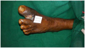
This study was conducted in the plastic surgery department in a tertiary care center in the month of February-March 2020. Informed written consent was taken from the patient. Departmental ethics committee clearance was obtained. The study was a 70-yearold male with known case of hypertension, diabetes mellitus and dyslipidemia. The patient had a non-healing ulcer at the first web space of left foot for 3-months (Figure 1). Wound bed preparation was done by multiple surgical debridement and antibiotics were initiated according to culture sensitivity.

Figure 1. Ulcer at the Time of Presentation

CTPOJ-3-116Fig1

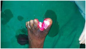
As an adjunct treatment modality LLLT was applied over the ulcer (Figure 2) after debridement weekly twice for a period of 4-weeks. We used Gallium Arsenide (GaAs) diode red laser of wavelength 650 nm, frequency 10 kHz and output power 100 mW. It was a continuous beam laser with an energy density of 4 J/cm2 . The machine delivers laser in scanning mode (non-contact delivery) with a 60 cm distance between the laser source and wound. Wound was given laser therapy for duration of 125 seconds every time

Figure 2. Low-Level Laser Therapy being Applied

CTPOJ-3-116Fig2

RESULTS

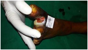
Application of LLLT helped in achieving healthy granulation tissue over the wound bed, thus aiding in the process of wound healing in a chronic diabetic ulcer (Figure 3).

Figure 3. Ulcer at the Time of Discharge

CTPOJ-3-116Fig3

DISCUSSION

Diabetic foot ulcers are the most costly and devastating complication of diabetes mellitus which affects 15% diabetic patients in their lifetime.1Early effective management can reduce the severity of complications such as preventable amputations and reduce the mortality. More than 50% of non-traumatic lower-extremity amputations are related to diabetic foot ulcer infections and 85% of all lower-extremity amputations in patients with diabetes are preceded by an ulcer; up to 70% of diabetic patients with a DFU-related amputation die within 5-years of their amputation.2

The acronym LASER can be abbreviated as “light amplification by stimulated emission of radiation”. Low-level lasers are defined by a power density at less than 500 mW/cm2.3,4 It is defined as low-level laser as the energy used is much less than that is used for cutting, ablation therapy. Low-level laser therapy (LLLT) has been used as an adjuvant to conventional therapy with promising results, especially in patients with acute and bloody ulcers.5 LLLT is a form of phototherapy that employs electromagnetic radiation capable of generating enough energy to interact with living tissues. It produces photochemical and photophysical effects without generating heat, with the intention of re-establishing cell homeostasis. Essentially, light energy is delivered topically in a controlled, safe manner and it is absorbed by photo-absorbers (chromophores) that transform it into chemical energy.6

Positive effects include acceleration of tissue repair, increased formation of granulation tissue, wound contraction, inflammation, modulation, and pain reduction.6

According to the literature, low-energy photoemissions given at a wavelength range of 600 nm to 900 nm accelerates cell proliferation and wound healing processes.7 Its action is thought to:

  • Stimulate respiratory chain components such as flavin and cytochromes which increase adenosine triphosphate (ATP) synthesis,8 thus enhancing the rate of mitoses and increasing fibroblast numbers.8-13
  • Stimulate collagen and elastin production, leading to better re-epithelialisation.14
  • Stimulate microcirculation and dilatation of the capillaries and neovascularisation to increase tissue oxygenation.15
  • Liberate mediator substances such as histamine, serotonin and bradykinin to influence macrophages.
  • Regenerate lymphatic vessels.

Laser therapy is painless, cost effective procedure which induces faster granulation, wound contraction and re-epithelialization, thus accelerates complete wound healing hence avoiding secondary procedures like split skin grafting in many cases. Control of infection is also better in patients whom low-level laser therapy was given.16 In addition to reducing the lesion area and accelerating the healing process, laser therapy has the advantage of being easily administered. These benefits assist in promoting patient quality of life and minimizing possible complications.17

CONCLUSION

In our study we found that LLLT was useful in promoting healthy granulation tissue and in expediting the process of wound healing in diabetic ulcer. The limitation of the study includes that it is a case report and a single centre study with no statistical analysis. Further randomised controlled studies are required to validate the efficacy of the LLLT in the treatment of diabetic ulcers.

CONSENT

The authors have received written informed consent from the
patient.

CONFLICTS OF INTEREST

The authors declare that they have no conflicts of interest.

1. Yazdanpanah L, Nasiri M, Adarvishi S. Literature review on management of diabetic foot ulcer. World J Diabetes. 2015; 6(1): 37-53. doi: 10.4239/wjd.v6.i1.37

2. Mavrogenis AF, Megaloikonomus PD, Antoniadou T, Igoumenou VG, Panagopoulos GN, Dimopoulos L, et al. Currentconcepts for the evaluation and management of diabetic footulcers. EFFORT Open Rev. 2018; 3(9): 513-525. doi: 10.1302/2058- 5241.3.180010

3. Karu TI. Low-power laser therapy. In: Vo-Dinh T, ed. Biomedicalphotonics Handbook. London, UK: CRC Press, 2003: 7-20.

4. David CBG. Therapeutic Lasers. Theory and Practice. London, UK: Churchill Livingstone; 1994.

5. Palagi S, Severo IM, Menegon DB, Lucena AF. Laserterapiaemúlceraporpressão: avaliaçãopelas pressure ulcer scale for healing e nursing outcomes classification. Rev EscolaEnfermagem USP. 2015; 49(5): 826-833. doi: 10.1590/S0080-623420150000500017

6. Andrade FSSD, Clark RMO, Ferreira ML. Effects of low-level laser therapy on wound healing. Rev Col Bras Cir. 2014; 41(2): 129- 133. doi: 10.1590/s0100-69912014000200010

7. Lichtenstein D, Morag B. Low-level laser therapyin ambulatory patients withvenous stasis ulcers. Laser Therapy. 1998; 11: 71-78. doi: 10.5978/islsm.11.71

8. Karu, T. Molecular mechanism of therapeutic effect of low intensity laser irradiation Dokl Akad Nauk SSSR. 1986; 291: 1245- 1249.

9. Karu, T. Photobiology oflow-power laser effects. Health Phys. 1989; 56: 691-704. doi: 10.1097/00004032-198905000-00015

10. Wilden, L., Karthein, R. Import of radiationphenomena of electronsand therapeutic low-levellaser in regard to themitochondrial energytransfer. J Clin Laser Med Surg. 1998; 16: 159-165. doi: 10.1089/clm.1998.16.159

11. Stadler I, Evans R, Kolb B, Naim JO, Narayan V, Buehner N, et al. In vitro effects oflow-level laser irradiation at 660 nm on peripheral bloodly mphocytes. Lasers Surg Med. 2000; 27: 255-261. doi: 10.1002/1096-9101(2000)27:3<_x0032_55:_x003a_aid-lsm7>3.0.co;2-l

12. Yu W, Naim JO, Lanzafame RJ. Effects of photostimulation on wound healing in diabetic mice. Lasers Surg Med. 1997; 20: 56-63. doi: 10.1002/(sici)1096-9101(1997)20:1<_x0035_6:_x003a_aid-lsm9>3.0.co;2-y

13. Yu W, Naim JO, Lanzafame RJ. The effect of laser irradiation on the release of bFGF from 3T3 fibroblasts. Photocem Photobiol. 1994; 59: 167-170. doi: 10.1111/j.1751-1097.1994.tb05017.x

14. Saperia D, Glassberg E, Lyons RF, Abergel RP, Baneux P, Castel JC, et al. Demonstration of elevatedtype I and type III procollagen mRNA levelsin cutaneous wound streated with helium-neonlaser: Proposed mechanismfor enhanced wound healing. Biochem Biophys Res Commun. 1986; 138: 1123-1128. doi: 10.1016/ S0006-291X(86)80399-0

15. Schindl A, Schindl M,Schindl L, Jurecka W, Hönigsmann H, Breier F, et al. Increased dermal angiogenesis after low-intensity laser therapy for a chronic radiation ulcer determined by avideo measuring system. J Am Acad Dermatol. 1999; 40: 481-484. doi: 10.1016/s0190-9622(99)70503-7

16. Priyadarshini LMJ, Kishore Babu EP, Thariq AI. Effect of low level laser therapy on diabetic foot ulcers: A randomized control trial. Int Surg J. 2018; 5(3): 1008-1015. doi: 10.18203/2349-2902. isj20180821

17. Rocha Júnior AM, Vieira BJ, Andrade LCF, Aarestrup FM. Effects of low-level laser therapy on the progress of wound healing in humans: The contribution of in vitro and in vivo experimental studies. J vasc bras. 2007; 6: 257-265. doi: 10.1590/ S1677-54492007000300009

LATEST ARTICLES

Chest X-ray Showed a Hazy Left Upper Lung Infiltrate

A Noteworthy Case of Myasthenic Crisis Induced by Levofloxacin

Ada Young*, Ramya Ramesh and Milind Awale

doi.

The Right Thigh Anterior Compartment was Swollen, and the Skin was Ulcerated due to the Traditional Cautery

Primary Skeletal Muscle Lymphoma: A Case Report and Literature Review

Solomon Bishaw*, Addisu Alemu and Abel Tefera

doi.

An Unusual Presentation of Encephalitis in a Patient with Lyme Neuroborreliosis

Maithily Patel*, Jazmin Jatana, Ramya Ramesh and Milind Awale

doi.

Practical Pointers for Drug Development and Medical Affairs

Gerald L. Klein*, Roger E. Morgan, Shabnam Vaezzadeh, Burak Pakkal and Pavle Vukojevic

doi.

10.17140/CTPOJ-7-125

Prevalence and Risk Factors of Subclinical Mastitis of Goats in Banadir Region, Somalia

Omar M. Salah*, Yasin H. Sh-Hassan, Moktar O. S. Mohamed, Mohamed A. Yusuf and Abas S. A. Jimale

doi.10.17140/VMOJ-9-184

LATEST ARTICLES