INTRODUCTION
Wound measurement is an important step in its management. It helps the clinician in understanding the behavior of the wound and to take necessary action to prevent the increment and enhance the healing. Wound measurement gives an idea for deciding the current treatment efficacy and for changing the current treatment. There are various methods by which wound can be measured like photographic record, comparison, ruler method, graph method.1 The efficacy of computerized planimetry in wound measurement was studied and found digital planimetry as an effective tool. The objective of digital planimetry in this study was to assess the wound size and implement steps to decrease the wound size.
MATERIALS AND METHODS
The study was carried out in a tertiary care hospital in South India after receiving approval from departmental ethical committee. The subject was a 32-years-old male patient with a history of electrical burn 10-years back following which he developed a non-healing ulcer on the back. On examination, the ulcer was on the back- more towards the right of the midline and measures size 8×6 cm, oval in shape, margins are everted (Figure 1). After the initial routine investigation, the patient underwent a computed tomography (CT) dorsolumbar scan to rule out osteomyelitis. The ulcer on the back was excised and sent for histopathology to rule any malignant aetiology (Figure 2). The wound bed was prepared by TIME concept2 and digital planimetry (Figure 3) was performed.
Figure 1. Raw Area on the Back
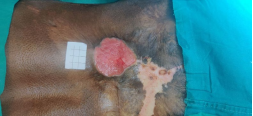
Figure 2. Wide Local Excision of the Ulcer
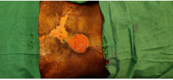
Figure 3. Digital Planimetry with ImageJ
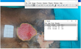
We followed the procedure described by Shetty et al3 for calculation of wound surface area by using imageJ software.
Step 1: Wound was cleaned to define the surroundings.
Step 2: Sterilized grid of 4×4 cm area was kept along the side of the wound.
Step 3: Good quality photograph was taken and saved to the computer. The photograph was analyzed using imageJ free open-source software.
Step 4: The edges of the wound was marked and the area was calculated. As the area of the grid known i.e 16 cm2 the number of pixels falling under the square marked and marked wound were calculated.
Step 5: Wound area was calculated according to following formula.
Area of wound=16 (wound measurement/grid measurement)
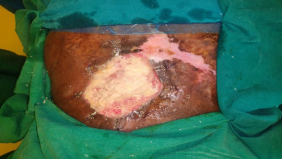
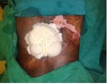
After digital planimetry, the wound measurement was known and an external tissue expansion and wound closure (ETEWC) was performed to decrease the wound size so the smaller flap or graft can be planned (Figure 4).
Figure 4. Clipping in External Tissue Expansion and Wound Closure
RESULT
Digital planimetry helped in accurate measurement of wound size and guided in wound bed preparation. Digital planimetry guided in performing an ETEWC to decrease the wound size (Figure 5).
Figure 5. External Tissue Expansion with Wound Closure
DISCUSSION
Wound measurement is an important part of management. Any change in wound area indirectly provides information about wound healing or response to current treatment. Continuation of ineffective treatment would prolong hospital stay and can be avoided by tracking the wound correctly. Therefore, objective technique of accurate measurement and documentation is needed for wound management.
Digital planimetry is a good tool for wound measurement. It helps the clinician in understanding the behaviour of the wound and to take necessary action to prevent the increment and enhance the healing. Wound measurement using digital planimetry gives an idea for deciding the current treatment efficacy and for changing the current treatment. It’s an effective technique for accurate wound measurement and documentation in wound management.
Traditionally wound measurement was done by photographic record, comparison, ruler method, graph method. Clinical photography is being used by most of the plastic surgeon as a part of record keeping and tracking the progress of the disease/wound. This is relatively simple and easy method, but being a subjective assessment, documentation is not possible for the use of further visits.3,4
A Mayrovitz et al,5 2009 showed efficacy of computerized planimetry in wound measurement. A study done by Wang et al6 compared digital planimetry with other methods and found that digital planimetry as an effective alternative.
ImageJ is an easy, freely available, effective, cheaper tool for measurement of wound surface area in clinical practice.7 It’s a subjective wound measurement tool. With the help of digital planimetry, we could accurately measure the wound size, behaviour of the wound and also enhance the healing by ETEWC.8
Through this article we are highlighting the use of imageJ software as an effective tool for wound management. However, a large sample size, controlled study wound be helpful for more significant results.
CONCLUSION
Digital assessment and imageJ is a simple, easy, cost effective technique of accurate wound measurement for wound management.
DECLARATIONS
Authors’ contributions: All authors made contributions to the
article.
Availability of data and materials: Not applicable.
Financial support and sponsorship: None.
Consent for publication: Not applicable.
CONFLICTS OF INTEREST
This study does not require any institutional approval. The authors declare that they have no conflicts of interest.


