Cancer Studies and Molecular Medicine

Open journal

ISSN 2377-1518

Regulation in Cell Cycle via p53 and PTEN Tumor Suppressors

Yasuko Kitagishi*, Satoru Matsuda*, Akari Minami, Yuna Ono, Atsuko Nakanishi and Yasunori Ogura

Satoru Matsuda, MD

Department of Food Science and Nutrition, Nara Women’s University Kita-Uoya Nishimachi, Nara 630- 8506, Japan; Tel. +81 742 20 3451; E-mail: smatsuda@cc.nara-wu.ac.jp

INTRODUCTION

Mechanisms of cell arrest in cell cycle are predominantly governed by p53 tumor suppressor1 that is a transcription factor. The p53 protein is able to induce G1 arrest of the cell cycle by trans-activating several downstream molecules. Germinal mutations of the p53 gene constitute an etiological genetic base of Li-Fraumeni syndrome, which is a rare hetergoeneous autosomal dominant inherited cancerous disorder.2 The p53 is at a midpoint of cellular signaling networks that are activated by stress signals including DNA damages.3 PTEN (Phosphatase and Tensin homolog deleted in chromosome 10) is also a tumor suppressor gene that is deleted or mutated in a variety of human cancers.4,5 Germ line mutations in PTEN are the cause of PTEN hamartoma syndromes (Cowden syndrome, Bannayan-Riley-Ruvalcaba syndrome, PTEN-related Proteus syndrome, Proteus-like syndrome) with increased risk for a development of cancers.6 Characterization of PTEN protein has showed that it is a phosphatase and can modulate signal-transduction pathways that involve lipid second messengers.7 PTEN prevents an activation of PI3K/AKT pathway by dephosphorylating the membrane phospholipid PIP3, and thus influence cell survival signaling.7 Loss of PTEN results in increased AKT recruitment to the plasma membrane, and activates the signaling pathway.

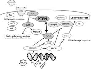
The PTEN has been shown to be involved in a complex network on interactions with p53 (Figure 1). Although they are functionally distinct, reciprocal cooperation has been proposed, as PTEN is thought to regulate p53 stability, and p53 to enhance PTEN transcription. Once PTEN is lost, however, the p53 pathway is strongly activated.8,9 Furthermore, an absence of PTEN cooperates with an absence of p53 to promote cancer,10 supporting a model for cooperative tumor suppression in which p53 is an essential failsafe protein of PTENdeficient tumors.8 Inactivation of tumor suppression may be caused by lack of these key interaction partners. Recent studies have revealed a functional ubiquitin ligase for tumor suppressors play a pivotal role in tumor cell survival.11,12 They may regulate the stability of key tumor suppressors. Mutations found in genes such as p53 and PTEN have emerged as high penetrance susceptibility genes and are clinically relevant for determination of cancer risk. In addition, there are multiple nodes of crosstalk between PI3K/AKT/PTEN and p53 signaling pathways.13 In this review, we summarize the current research and our view of how and when PTEN and p53 with their partners to transduce signals downstream and what are the implications for cell cycle-associated biology in cancer.

Figure 1: Schematic representation of the integrative model of tumor suppressors signaling including PTEN and p53. CSMMOJ-1-101Fig1

Typical examples of molecules known to act on the DNA damage response and cell cycle progression
via the regulatory pathway are shown. Note that some critical pathways have been omitted for clarity.

FUNCTIONAL CHARACTERISTICS OF PTEN AND P53

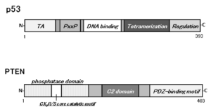
The human genomic PTEN locus consists of 9 exons on chromosome 10q23.3 encoding a 5.5 kb mRNA that specifies a 403 amino-acid open reading frame.14,15 The translation product is a 53 k Da protein with homology to tensin and protein tyrosine phosphatases. PTEN activity can be regulated by posttranslational regulation including phosphorylation, acetylation, and oxidation.16 Methylation of the PTEN promoter region can result in transcriptional silencing of the PTEN gene.17 Schematic structure of the predicted PTEN protein is shown in Figure 2. PTEN negatively regulates the activity of PI3K/AKT signaling through converting Phosphatidylinositol 3,4,5-triphosphate (PIP3) into Phosphatidylinositol 4,5- bisphosphate (PIP2). The PIP3 is the principal second messenger of the PI3K pathway that mediates Receptor Tyrosine Kinase (RTK) signaling to the survival kinase AKT. Activated AKT transfers a phosphate group to target proteins involved in cell survival, cell cycling, proliferation, and cell migration, which are all critical for tumor development.18,19 Generally, AKT is activated by growth factors and the RTK that activates PI3K. Upon activation, PI3K phosphorylates the inositol ring, which in turn serves to anchor AKT to the plasma membrane, where it is phosphorylated and fully activated by the 3-phosphoinositide-dependent kinases PDK1 and PDK2. PTEN acts as regulator of maintaining basal levels of PIP3 below a threshold for those signaling activation. PTEN protein consists of Nterminal phosphatase, and C-terminal C2, and PDZ (PSD-95, DLG1, and ZO-1) binding domains. The PTEN CX5R(S/T) motif resides within an active site that surrounds the catalytic signature with three basic residues, which are critical for PTEN lipid phosphatase activity. The structure provides PTEN with its preference for acidic phospholipid substrates such as PIP3. Overexpression of the PTEN induces growth suppression by promoting cell cycle arrest, which requires lipid phosphatase activity.20,21 Overexpression of PTEN also correlates with decreased levels and nuclear localization of cyclin D122, a cell cycle key molecule regulated by AKT. One mechanism by which PTEN induces cell cycle arrest is by regulating AKT activity so that levels of the cell cycle inhibitor p27kip1 are increased.23 However, despite the main role of PTEN as a negative regulator of the PI3K pathway, studies report many tumor suppressive activities for PTEN that are exerted from within the nucleus, where catalysis of PIP3 does not seem to represent a main function of this enzyme.24 The nuclear PTEN activities may include the regulation of genomic stability and gene expression.

Figure 2: Schematic structures of p53 and PTEN proteins. The predicted consensual domain structures for each protein are depicted. The functionally important sites are shown. TA= transactivation domain; PxxP= proline rich region; C2 domain= a protein structural domain involved in targeting proteins to cell membranes; PDZ= a common structural domain in signaling proteins (PSD95, Dlg, ZO-1, etc)

CSMMOJ-1-101Fig2

The p53 gene, located on chromosome 17p 13.1, encodes a nuclear 393 amino-acids protein which is a transcriptional factor regulating cell cycle and apoptosis when DNA damage occurs (Figure 2). The p53 tumor suppressor plays a pivotal role in regulating cellular processes including cell cycle arrest, apoptosis, cell metabolism and cell senescence. Inactivation of p53 gene is a common event in the development of most types of cancers25, suggesting that p53 plays a critical role in preventing normal cells from becoming malignant cells. The p53 germline mutations may occur in up to 1: 5000 individuals.26 The importance of p53 as an inherited cancer susceptibility gene has also been demonstrated in Li-Fraumeni syndrome.26 Multiple mechanisms have been revealed to collectively achieve the regulation of p53 activity,27,28 which determines the selectivity of p53 for specific transcriptional targets, resulting in precise control of the p53 activity. Release of p53 from repression by factors such as Mdm2 and MdmX may be a key step in the physiological activation of p53.28 Activation of p53 function involves its increased DNA-binding ability, transcriptional activation, increased expression of p53 target genes, associated with cell cycle progression and apoptosis.

FUNCTIONAL INTERPLAY AMONG P53, PTEN, AKT AND MDM2

An important p53 function is to act as a transcription factor by binding to the specific DNA consensus sequence in responsive genes. The PTEN and p53 complex enhances p53 DNAbinding and transcriptional activity,29 which may increase the synthesis of PTEN and p21waf1 that is an important protein involved in cell cycle arrest.30 One way by which p53 inhibits production of PIP3 indirectly is by inducing the expression of this PTEN.31 Under hypoxic conditions, PTEN and p53 form a complex in the nucleus and induce expression of additional tumor suppressor Maspin.32 In other words, the nuclear PTEN and p53 coordinates the induction of maspin. Loss of PTEN attenuates the induction of Maspin even in the presence of wild type p53. Integration of PTEN and p53 into a common pathway for Maspin-induction may constitute a strong tumor suppressors network.33 The ability of p53 to induce cell cycle arrest or apoptosis can be antagonized by survival signals. The PI3K dependent activation of AKT indirectly leads to the inhibition of p53 functions by activating another tumor suppressor MDM2.34 Several studies have implicated AKT in modulating DNA damage responses and genome stability.35 In addition, activation of AKT has the potential of reducing the p53-mediated cell cycle checkpoints through phosphorylation and sequestration of p21waf1, and via the enhanced degradation of p53.36 PTEN also plays a critical role in DNAdamage repair and DNA damage response through its interaction with p53 pathways in an AKT-independent manner.37 Nuclear PTEN is sufficient to reduce tumor progression in a p53 dependent manner. It has also been suggested that nuclear PTEN play a unique role to protect cells upon oxidative damage and to regulate carcinogenesis.38 Studies suggest that nuclear PTEN mediates DNA damage repair through modulating the activity of DNA repair molecules.

MDM2 is an oncoprotein that controls carcinogenesis, whose mRNA level is transcriptionally regulated by the p53 in response to DNA damage such as oxidative stress.39 In addition, the MDM2 protein and subcellular localization are post-translationally modulated by AKT.40 Besides PTEN inhibits the PI3K/AKT signaling, PTEN promotes translocation of MDM2 into the nucleus. In addition, PTEN modulates MDM2 transcription and isoform selection by negatively regul ating its promoter.41 In PTEN-null cells, MDM2 promoter activity is up-regulated, resulting in increased MDM2 expression. Furthermore, PTEN controls MDM2 promoter activity through its lipid phosphatase activity, independent of the p53 activity.29 Although another transcription factors may be able to modulate MDM2 transcription, they have been characterized to work through the p53 responsible promoter.42 MDM2 also regulates the activity of p53 protein by exporting of nuclear p53 protein into the cytoplasm and by promoting the degradation of the p53 protein. PTEN up-regulates the p53 level as well as its activity by down-regulating MDM2 transcription and p53 binding activity.43 However, in the absence of p53, PTEN may have a role inhibiting MDM2-mediated carcinogenesis through regulation of MDM2 transcription. The ability of PTEN to inhibit the nuclear entry of MDM2 increases the cellular content and transactivation of p53 to promote the induction of genes such as p21waf1.44

Consequently, p53 and AKT influence the process of apoptosis in opposite ways. The AKT promotes cell survival by suppressing pro-apoptotic proteins such as Bad through the phosphorylation.45 There are cross talks between p53 and AKT involving gene transcription as well as posttranslational protein modifications. One way by which p53 inhibits indirectly PIP3 production is by repressing the catalytic subunit of PI3K. Subsequent p53-induced expression of PTEN causes the p53- PTEN interaction, which also suppresses the cell survival machinery of AKT pathway. AKT kinase phosphorylates MDM2 to translocate into the nucleus.46 In addition, PTEN associates with p53 and regulates the transcriptional activity of p53 by modulating its DNA binding.29 PTEN is required for the maintenance of p53 acetylation, which is required for target gene transcription.47One side of the PTEN function as a tumor suppressor is achieved through this stabilization of the p53 protein. PTEN has been shown to interact with p53 and prevent its degradation by excluding a portion of p53 protein from the p53 and MDM2 complex. AKT mediates MDM2 nuclear translocation by its phosphorylation. MDM2 negatively regulates p53 by binding for destabilization in the nucleus.48 Attenuation of the AKT pathway by PTEN protects p53 from MDM2 mediated degradation and inactivation. The p53 and MDM2 complex transports from the nucleus into the cytoplasm where MDM2 serves as an E3 ubiquitin ligase.49 Therefore, p53 and MDM2 form a regulatory feedback loop in which p53 positively regulates MDM2 expression, whereas MDM2 negatively regulates the level of p53 protein. Inactivation of either p53 gene or PTEN gene results in lower protein levels of the other gene.

The instability of PTEN correlated with its missense mutations has been shown to involve protein interactions. PTEN may be regulated by ubiquitin-mediated proteasomal degradation, a common mechanism to control protein levels. In cells, ubiquitin ligase NEDD4-1 negatively regulates PTEN stability by catalyzing PTEN ubiquitination.50 Because deletion of the C2 domain of PTEN makes the protein unstable and accelerates the protein degradation, the C2 domain of PTEN seems to regulate itself through maintaining the protein stability.51 In addition, the C-terminus of PTEN contains two PEST (proline, glutamic acid, serine and threonine) sequences involved in ubiquitin protein degradation pathway. Treatment of cells with proteasome inhibitors can cause an increase of PTEN protein level.52,53 So, several NEDD4-like E3 similarly regulate p53. Multiple NEDD4-like E3 show ligase independent function and most of NEDD4-like E3 are commonly regulated by phosphorylation, ubiquitination, translocation, and transcription in cancer cells. Functional interaction of NEDD4-like ubiquitin protein Ligase-1 (NEDL1) with p53 might contribute to the induction of apoptosis in cancerous cells.54,55 Casein kinase II-mediated phosphorylation stabilizes the PTEN protein by preventing the proteasomal degradation, which results in increased PTEN activity and a corresponding reduction in AKT activation.56 Interestingly, inhibitors of Casein kinase II also activate p53 function in wild-type but not in p53 mutant cells, which increases senescence in the p53-dependent manner.57 It seems that Casein kinase II may control the PTEN and the p53 in balance.

INVOLVEMENT OF PTEN-P53-AKT-MDM2 LOOP IN CELL CYCLE REGULATION

It has been proposed that low levels of p53 induce cell cycle arrest, whereas high levels of p53 induce apoptosis.58 Probably, p53 can bind to pro-arrest genes of cell cycle with high affinities but associates with pro-apoptotic genes with low affinities.59 The levels of p53 could vary and is positively related to the amount of DNA damage.60 Activation of AKT, on the other hand, can overcome both the p53-independent G2/M cell cycle checkpoint and apoptosis induced by the DNA damage. In addition, growth factor-activated AKT signaling supports progression of cell cycle by acting on several factors involved in the G1/S or G2/M transitions. Because the ability of p53 to induce cell cycle arrest or apoptosis can be antagonized by survival signals, which indirectly leads to the inhibition of p53 functions by activating its negative regulators.34 Given the ability of PTEN to stabilize p53 protein through antagonizing the AKT-MDM2 pathway29,44 or by directly increasing p53 acetylation,47 decreased p53 activity in PTEN-deficient tumor cells could be expected. Stabilization and constant levels of the p53 may trigger apoptosis in damaged cells. The target genes of p53 are selectively induced to control cell fate. Consequently, the cell fate may be determined by the levels of p53.61,62 Constitutive activation of AKT in PTEN-deficient cells should down-regulate p53 transcriptional activity and block p53-induced p21waf1 induction.44 The PTEN-p53- MDM2-AKT loop in cell cycle regulation now becomes dominant (Figure 3). In addition, PTEN and p53 is known to interact and regulate each other at the transcription as well as protein level, which could be at the important control machinery for switching between survival and death. These cross talks are frequently a combination of reciprocally antagonistic pathways, which may often serve as an added regulatory effect on the expression of key genes involved in cancer. Interestingly, soy isoflavone genistein induces an auto-regulatory loop between PTEN and p53 to promote cell cycle arrest.63 The induction of PTEN expression and nuclear accumulation by genistein elicits a sequence of PTEN-dependent increased nuclear p53 accumulation, enhanced PTEN/p53 physical int eraction, increased recruitment of the PTEN/p53 complex to the p53 binding sites of the PTEN promoter, attenuated expression of proliferative genes cyclin D1and pleiotrophin gene expression, and promotion of cell cycle arrest.63 Genetic variants in the PTEN, p53, AKT, and MDM2 tumor suppressor oncoprotein network may play roles in mediating the susceptibility to cancer.64 It has been shown that zinc deficiency modulates the PTEN-p53-MDM2-AKT signaling axis in normal prostate cells.65

PERSPECTIVE

The tumor suppressor p53 predominantly induces cell cycle arrest or apoptosis in the DNA damage response. In regular unstressed cells, p53 may be kept at low levels by its negative regulator MDM2. This positive feedback loop among PTEN -p53- AKT-MDM2 may function for the precise regulation of the cell cycle (Figure 3). The numerousinteractions may support the biological plausibility that the combination of variants of the PTEN -p53- AKT-MDM2 network could result in more comprehensive and accurate estimates of risk than can be obtained from a single variant. Accordingly, germline genetic testing for mutations in these genes allows for the identification of individuals at increased risk for cancers, which are the state of running off from the cell cycle regulation. However, they may be regulated and interact each other at multiple levels including transcription, protein modulation, and protein stability. For example, increased nuclear localization of PTEN may promote nuclear retention of p53 and the subsequent transactivation by the PTEN and p53 complex of the PTEN promoter. By the way, what are the substrates of PTEN in that situation? Understanding the regulation is crucial for the effective design of novel cancer therapeutics. In addition, it is important to investigate the functional linkage between PTEN, p53 and MDM2 isoforms in human cancer samples, and elucidation of interaction-specific functions may provide insight into regulatory aspects of these tumor suppressors. Further mechanistic studies are needed in order to understand the more precise molecular mechanisms.

ACKNOWLEDGMENTS

This work was supported by grants-in-aid from the Ministry of Education, Culture, Sports, Science and Technology in Japan.

COMPETING INTERESTS STATEMENT

The authors declare that they have no competing financial interests.

1. Perry ME and Levine AJ. Tumor-suppressor p53 and the cell cycle. Curr Opin Genet Dev. 1993; 3(1): 50-54.doi: 10.1016/s0959-437x(05)80340-5

2. Wang B, Xiao Z, Ko HL, and Ren EC. The p53 response element and transcriptional repression. Cell Cycle. 2010; 9(5): 870- 879. doi: 10.4161/cc.9.5.10825

3. Murray-Zmijewski F, Slee EA, and Lu X. A complex barcode underlies the heterogeneous response of p53 to stress. Nat Rev Mol Cell Biol. 2008; 9(9): 702-712. doi: 10.1038/nrm2451

4. Song MS, Salmena L, and Pandolfi PP. The functions and regulation of the PTEN tumour suppressor. Nat Rev Mol Cell Biol. 2012; 13(5): 283-296. doi: 10.1038/nrm3330

5. Merritt MA and Cramer DW. Molecular pathogenesis of endometrial and ovarian cancer. Cancer Biomark. 2010; 9(1-6): 287-305. doi: 10.3233/CBM-2011-0167

6. Hobert JA and Eng C. PTEN hamartoma tumor syndrome: an overview. Genet Med. 2009; 11(10): 687-694. doi: 10.1097/GIM.0b013e3181ac9aea

7. Maehama T and Dixon JE. PTEN: a tumour suppressor that functions as a phospholipid phosphatase. Trends Cell Biol. 1999; 9(4): 125-128. doi: http://dx.doi.org/10.1016/S0962- 8924(99)01519-6

8. Chen Z, Trotman LC, Shaffer D et al. Crucial role of p53- dependent cellular senescence in suppression of Pten-deficient tumorigenesis. Nature. 2005; 436(7051): 725-730. doi: 10.1038/ nature03918

9. Kim J, Eltoum IE, Roh M, Wang J, and Abdulkadir SA. Interactions between cells with distinct mutations in c-MYC and Pten in prostate cancer. PLoS Genet. 2009; 5(7): e1000542. doi: 10.1371/journal.pgen.1000542

10. Blanco-Aparicio C, Renner O, Leal JF, and Carnero A. PTEN, more than the AKT pathway. Carcinogenesis. 2007; 28(7): 1379-1386. doi: 10.1093/carcin/bgm052

11. Faesen AC, Dirac AM, Shanmugham A, Ovaa H, Perrakis A, and Sixma TK. Mechanism of USP7/HAUSP activation by its C-terminal ubiquitin-like domain and allosteric regulation by GMP-synthetase. Mol Cell. 2011; 44(1): 147-159. doi: 10.1016/j.molcel.2011.06.034

12. Sacco JJ, Coulson JM, Clague MJ, and Urbé S. Emerg ing roles of deubiquitinases in cancer-associated pathways. IUBMB Life. 2010; 62(2): 140-157. doi: 10.1002/iub.300

13. Cully M, You H, Levine AJ, and MAKTW. Beyond PTEN mutations: the PI3K pathway as an integrator of multiple inputs during tumorigenesism. Nat Rev Cancer. 2006; 6(3): 184-192. doi: 10.1038/nrc1819

14. Okumura N, Yoshida H, Kitagishi Y, Murakami M, Nishimura Y, and Matsuda S. PI3K/AKT/PTEN Signaling as a Molecular Target in Leukemia Angiogenesis. Adv Hematol. 2012; 2012: 843085. doi: http://dx.doi.org/10.1155/2012/843085

15. Okumura N, Yoshida H, Kitagishi Y, Nishimura Y, and Matsuda S. Alternative splicings on p53, BRCA1 and PTEN genes involved in breast cancer. Biochem Biophys Res Commun. 2011; 413(3): 395-399. doi: 10.1016/j.bbrc.2011.08.098

16. Walden H and Martinez-Torres RJ. Regulation of Parkin E3 ubiquitin ligase activity. Cell Mol Life Sci. 2012; 69(18): 3053- 3067. doi: 10.1007/s00018-012-0978-5

17. Mueller S, Phillips J, Onar-Thomas A et al. PTEN promoter methylation and activation of the PI3K/AKT/mTOR pathway in pediatric gliomas and influence on clinical outcome. Neuro Oncol. 2012; 14(9): 1146-1152. doi: 10.1093/neuonc/nos140

18. Faurschou A, Gniadecki R, Calay D, and Wulf HC. TNFalpha impairs the S-G2/M cell cycle checkpoint and cyclobutane pyrimidine dimer repair in premalignant skin cells: role of the PI3K-AKT pathway. J Invest Dermatol. 2008; 128(8): 2069- 2077. doi: 10.1038/jid.2008.19

19. Chen Y, Wang BC, and Xiao Y. PI3K: a potential therapeutic target for cancer. J Cell Physiol. 2012; 227(7): 2818-2821.doi: 10.1002/jcp.23038

20. Choi Y, Zhang J, Murga C et al. PTEN, but not SHIP and SHIP2, suppresses the PI3K/AKT pathway and induces growth inhibition and apoptosis of myeloma cells. Oncogene. 2002; 21(34): 5289-5300. doi: 10.1038/sj.onc.1205650

21. Petrella BL, and Brinckerhoff CE. PTEN suppression of YY1 induces HIF-2 activity in von-Hippel-Lindau-null renalcell carcinoma. Cancer Biol Ther. 2009; 8(14): 1389-1401. doi: 10.4161/cbt.8.14.8880

22. Yamamoto M, Tamakawa S, Yoshie M, Yaginuma Y, and Ogawa K. Neoplastic hepatocyte growth associated with cy clin D1 redistribution from the cytoplasm to the nucleus in mouse hepatocarcinogenesis. Mol Carcinog. 2006; 45(12): 901-913. doi: 10.1002/mc.20204

23. Andrés-Pons A, Gil A, Oliver MD, Sotelo NS, and Pulido R. Cytoplasmic p27Kip1 counteracts the pro-apoptotic function of the open conformation of PTEN by retention and destabilization of PTEN outside of the nucleus. Cell Signal. 2012; 24(2): 577- 587. doi: 10.1016/j.cellsig.2011.10.012

24. Planchon SM, Waite KA, and Eng C. The nuclear affairs of PTEN. J Cell Sci. 2008; 121(Pt 3): 249-253. doi: 10.1242/ jcs.022459

25. Pei D, Zhang Y, and Zheng J. Regulation of p53: a collaboration between MDM2 and Mdmx. Oncotarget. 2012; 3(3): 228- 235.doi: 10.18632/oncotarget.443

26. Palmero EI, Achatz MI, Ashton-Prolla P, Olivier M, and Hainaut P. Tumor protein 53 mutations and inherited cancer: beyond Li-Fraumeni syndrome. Curr Opin Oncol. 2010; 22(1): 64-69. doi: 10.1097/CCO.0b013e328333bf00

27. Vousden KH and Prives C. Blinded by the Light: The Growing Complexity of p53. Cell. 2009; 137: 413–431. doi: 10.1016/j.cell.2009.04.037

28. Kruse JP and Gu W. Modes of p53 regulation. Cell. 2009; 137: 609–622. doi: 10.1016/j.cell.2009.04.050

29. Freeman DJ, Li AG, Wei G et al. PTEN tumor suppressor regulates p53 protein levels and activity through phosphatasedependent and -independent mechanisms. Cancer Cell. 2003; 3(2): 117-130.

30. Lo PK, Lee JS, and Sukumar S. The p53-p21WAF1 checkpoint pathway plays a protective role in preventing DNA rereplication induced by abrogation of FOXF1 function. Cell Signal. 2012; 24(1): 316-324. doi: 10.1016/j.cellsig.2011.09.017

31. Puszyński K, Hat B, and Lipniacki T. Oscillations and bistability in the stochastic model of p53 regulation. J Theor Biol. 2008; 254(2): 452-465. doi: 10.1016/j.jtbi.2008.05.039

32. Eitel JA, Bijangi-Vishehsaraei K, Saadatzadeh MR et al. EN and p53 are required for hypoxia induced expression of maspin in glioblastoma cells. Cell Cycle. 2009; 8(6): 896-901. doi: 10.4161/cc.8.6.7899

33. Zhang M. PTEN in action: coordinating with p53 to regulate maspin gene expression. Cell Cycle. 2009; 8(8): 1112-1113. doi: 10.4161/cc.8.8.8506

34. Mayo LD and Donner DB. A phosphatidylinositol 3-kinase/ AKT pathway promotes translocation of MDM2 from the cytoplasm to the nucleus. Proc Natl Acad Sci U S A. 2001; 98(20): 11598-11603.doi: 10.1073/pnas.181181198

35. Quevedo C, Kaplan DR, and Derry WB. AKT-1 regulates DNA-damage-induced germline apoptosis in C. elegans. Curr Biol. 2007; 17(3): 286-292. doi: http://dx.doi.org/10.1016/j. cub.2006.12.038

36. Zhou BP, Liao Y, Xia W, Zou Y, Spohn B, and Hung MC. HER-2/neu induces p53 ubiquitination via AKT-mediated MDM2 phosphorylation. Nat Cell Biol. 2001; 3(11): 973-982. doi: 10.1038/ncb1101-973

37. Ming M and He YY. PTEN in DNA damage repair. Cancer Lett. 2012; 319(2): 125-129. doi: 10.1016/j.canlet.2012.01.003

38. Bonavida B and Baritaki S. The novel role of Yin Yang 1 in the regulation of epithelial to mesenchymal transition in cancer via the dysregulated NF-κB/Snail/YY1/RKIP/PTEN Circuitry. Crit Rev Oncog. 2011; 16(3-4): 211-226. doi: 10.1615/CritRevOncog.v16.i3-4.50

39. Wang X and Jiang X. MDM2 and MdmX partner to regulate p53. FEBS Lett. 2012; 586(10): 1390-1396. doi: 10.1016/j.febslet.2012.02.049

40. Levav-Cohen Y, Haupt S, and Haupt Y. MDM2 in growth signaling and cancer. Growth Factors. 2005; 23(3): 183-192. doi: 10.1080/08977190500196218

41. Mayo LD and Donner DB. The PTEN, MDM2, p53 tumor suppressor-oncoprotein network. Trends Biochem Sci. 2002; 27(9): 462-467. doi: http://dx.doi.org/10.1016/S0968-0004- (02)02166-7

42. Kirch HC, Flaswinkel S, Rumpf H, Brockmann D, and Esche H. Expression of human p53 requires synergistic activation of transcription from the p53 promoter by AP-1, NF-kappaB and Myc/Max. Oncogene. 1999; 18(17): 2728-2738. doi: 10.1038/sj.onc.1202626

43. Zheng T, Meng X, Wang J et al. PTEN- and p53-mediated apoptosis and cell cycle arrest by FTY720 in gastric cancer cells and nude mice. J Cell Biochem. 2010; 111(1): 218-228. doi: 10.1002/jcb.22691

44. Mayo LD, Dixon JE, Durden DL, Tonks NK, and Donner DB. PTEN protects p53 from MDM2 and sensitizes cancer cells to chemotherapy. J Biol Chem. 2002; 277(7): 5484-5489. doi: 10.1074/jbc.M108302200

45. Sen P, Mukherjee S, Ray D, and Raha S. Involvement of the AKT/PKB signaling pathway with disease processes. Mol Cell Biochem. 2003; 253(1-2): 241-246. doi: 10.1023/A:1026020101379

46. de Lima Mde D, Marques YM, Alves Sde M Jr et al. MDM2, P53, P21WAF1 and pAKT protein levels in genesis and behaviour of adenoid cystic carcinoma. Cancer Epidemiol. 2009; 33(2): 142-146. doi: 10.1016/j.canep.2009.04.016

47. Li AG, Piluso LG, Cai X, Wei G, Sellers WR, and Liu X. Mechanistic insights into maintenance of high p53 acetylation by PTEN. Mol Cell. 2006; 23(4): 575-587. doi: http://dx.doi.org/10.1016/j.molcel.2006.06.028

48. Vu BT and Vassilev L. Small-molecule inhibitors of the p53- MDM2 interaction. Curr Top Microbiol Immunol. 2011; 348: 151-172. doi: 10.1007/82_2010_110

49. Bixby D, Kujawski L, Wang S, and Malek SN. The pre-cli nical development of MDM2 inhibitors in chronic lymphocytic leukemia uncovers a central role for p53 status in sensitivity to MDM2 inhibitor-mediated apoptosis. Cell Cycle. 2008; 7(8): 971-979. doi: 10.4161/cc.7.8.5754

50. Amodio N, Scrima M, Palaia L et al. Oncogenic role of the E3 ubiquitin ligase NEDD4-1, a PTEN negative regulator, in non-small-cell lung carcinomas. Am J Pathol. 2010; 177(5): 2622-2634. doi: 10.2353/ajpath.2010.091075

51. Valiente M, Andrés-Pons A, Gomar B et al. Binding of PTEN to specific PDZ domains contributes to PTEN protein stability and phosphorylation by microtubule-associated serine/ threonine kinases. J Biol Chem. 2005; 280(32): 28936-28943. doi: 10.1074/jbc.M504761200

52. Torres J and Pulido R. The tumor suppressor PTEN is phosphorylated by the protein kinase CK2 at its C terminus. Implications for PTEN stability to proteasome-mediated degradation. J Biol Chem. 2001; 276(2): 993-998. doi: 10.1074/jbc. M009134200

53. Wu W, Wang X, Zhang W et al. Zinc-induced PTEN protein degradation through the proteasome pathway in human airway epithelial cells. J Biol Chem. 2003; 278(30): 28258-28263. doi: 10.1074/jbc.M303318200

54. Li Y, Ozaki T, Kikuchi H, Yamamoto H, Ohira M, and Nakagawara A. A novel HECT-type E3 ubiquitin protein ligase NEDL1 enhances the p53-mediated apoptotic cell death in its catalytic activity-independent manner. Oncogene. 2008; 27(26): 3700-3709. doi: 10.1038/sj.onc.1211032

55. ShinadAKTsukiyama T, Sho T, Okumura F, Asaka M, and Hatakeyama S. RNF43 interacts with NEDL1 and regulates p53- mediated transcription. Biochem Biophys Res Commun. 2011; 404(1): 143-147. doi: 10.1016/j.bbrc.2010.11.082

56. Barata JT. The impact of PTEN regulation by CK2 on PI3K-dependent signaling and leukemia cell survival. Adv Enzyme Regul. 2011; 51(1): 37-49. doi: 10.1016/j.advenzreg.2010.09.012

57. Kang JY, Kim JJ, Jang SY, and Bae YS. The p53-p21(Cip1/WAF1) pathway is necessary for cellular senescence induced by the inhibition of protein kinase CKII in human colon cancer cells. Mol Cells. 2009; 28(5): 489-494. doi: 10.1007/s10059-009-0141-9

58. Vousden KH and Lane DP. p53 in health and disease. Nat Rev Mol Cell Biol. 2007; 8(4): 275-283. doi: 10.1038/nrm2147

59. Weinberg RL, Veprintsev DB, Bycroft M, and Fersht AR. Comparative binding of p53 to its promoter and DNA recognition elements.J Mol Biol. 2005; 348(3): 589-596. doi: 10.1016/j.jmb.2005.03.014

60. Lahav G. The strength of indecisiveness: oscillatory behavior for better cell fate determination. Sci STKE. 2004; 2004(264): 55. doi: 10.1126/stke.2642004pe55

61. Zhang T, Brazhnik P, and Tyson JJ. Exploring mechanisms of the DNA-damage response: p53 pulses and their possible relevance to apoptosis. Cell Cycle. 2007; 6(1); 85-94. doi: 10.4161/ cc.6.1.3705

62. Zhang XP, Liu F, Cheng Z, and Wang W. Cell fate decision mediated by p53 pulses. Proc Natl Acad Sci U S A. 2009; 106(30): 12245-12250. doi: 10.1073/pnas.0813088106

63. Rahal OM and Simmen RC. PTEN and p53 cross-regulation induced by soy isoflavone genistein promotes mammary epithelial cell cycle arrest and lobuloalveolar differentiation. Carcinogenesis. 2010; 31(8): 1491-1500. doi: 10.1093/carcin/bgq123

64. Zhang X, Chen X, Zhai Y, Cui Y, Cao P, Zhang H, Wu Z, Li P, Yu L, Xia X, He F, and Zhou G. Combined Effects of Genetic Variants of the PTEN, AKT1, MDM2 and p53 Genes on the Risk of Nasopharyngeal Carcinoma. PLoS One. 2014; 9(3): e92135. doi: 10.1371/journal.pone.0092135

65. Han CT, Schoene NW, and Lei KY. Influence of zinc deficiency on Akt-Mdm2-p53 and Akt-p21 signaling axes in normal and malignant human prostate cells. Am J Physiol Cell Physiol. 2009; 297(5): C1188-1199. doi: 10.1152/ajpcell.00042.2009

LATEST ARTICLES

Unraveling the Mysteries of Type-A Aortic Dissection Using POCUS/Echocardiography

Syeda Rukh*, Sathyanarayana Machani and Milind Awale

doi.

Blood Sample from the Patient

Hypertriglyceridemia-Induced Pancreatitis: A Case Report and Literature Review

Maarten Bulterys, Melvin Willems* and Agnes Meersman

doi.

From Neck Pain to a Life-Threatening Condition: A Case Report

Floris Vandewoude* and Sören Verstraete

doi.

LATEST ARTICLES