INTRODUCTION
Obesity is associated with increased rates of surgical complications, wound healing problems, and re-operation.1 Obesity also is associated with increased risk of pressure injuries, obstructive sleep apnea and medical comorbidities like hypertension and diabetes which poses some challenges to these patients’ perioperative anesthesia management. Although others have reported the challenges in providing anesthesia care to obese patients, few have described such challenges in morbidly obese patients.2 To our knowledge, none have described such challenges in a morbidly obese patient with a BMI over 75 undergoing spine surgery that requires the patient to be in a prone position. The patient has given written consent for this report to be published.
CASE DESCRIPTION
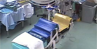
A 41-year-old woman with an enlarging cervical Schwannoma was scheduled to undergo a C2-C3 cervical laminectomy with intradural and extradural exploration with complex muscle flap closure. With a weight of 188 kg and a height of 157 cm, the patient had a body mass index (BMI) of 76.2 kg/m², indicating morbid obesity. Her past medical history included obstructive sleep apnea, for which she used a continuous positive airway pressure device at night. A pulmonology consultation suggested the need for bi-level positive airway pressure (BIPAP) both before surgery for lung recruitment and after surgery to meet the patient’s postoperative respiratory requirements. Because morbidly obese patients have a higher incidence of medical comorbidities such as diabetes, chronic obstructive pulmonary disease, hypertension, and coronary artery disease, an internal medicine consultation was also obtained as per our institutional practice, which helped rule out any cardiac pathology and identified a plan for post-operative glucose management. Since obtaining peripheral intravenous access is challenging in obese patients, to avoid delay on day of surgery a central venous double lumen 8 French line was placed the day before surgery by interventional radiology. (Figure 1)
Figure 1: Operating table set up prior to surgery after the trial of positioning the day before.

We anticipated that placing this patient in a prone position would be very challenging. Therefore, we performed a trial run the day before surgery. We did this primarily to identify the adjustments we would need to make to minimize pressure related injuries, get adequate surgical exposure and to unfold potential pitfalls in delivering this optimal care. We chose to use the Hercules bed 6702 Hercules (Skytron) which has a weight capacity of 1200 pounds (1000 pounds flexed or offset center), and bilateral lateral extension attachments, which made the bed 8 inches wider than a regular surgical bed, to accommodate the patient’s wider torso and the wider knee placement.
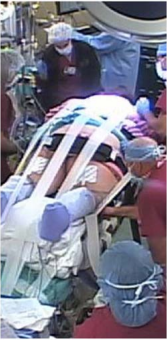
After the goals of the trial run and the plan for the next day’s surgery had been communicated to the nursing, surgery, and anesthesiology teams the patient was brought into the operating room, where she moved herself from the gurney to the operating table and placed herself in the prone position. She provided feedback on roll placement and comfort, and we identified possible pressure areas and aligned the rolls to accommodate her height. For example, when the patient flexed her knees and her pelvis (which was supported on gel rolls), her knees did not reach the bed; therefore, we added padding to support them. Extended straps were attached to the operating table and were determined to be sufficiently long and secure to be effective. (Figure 2)
Figure 2: Patient positioned on operating room table and secured.

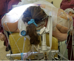
At this time it was discovered the patient’s body habitus and positioning foam brought patients head too far away from the bed for usual Mayfield pinning. We needed an extra 6 inches of Mayfield frame to reach the patients head. By reversing the table attachment and adding a “dog bone” attachment and carefully positioning the locking mechanism to not impinge on the face, we were able to gain the necessary height and anticipated using the Mayfield. (Figure 3)
Figure 3: Patient head in Mayfield pins.

After the patient left the operating room, the anesthesia and nursing teams discussed findings and next-day plans. The operating table was prepared, and additional padding and positioning gel for arm placement on the arm boards were procured. The teams determined that full-length gel padding would be used to prevent sores and that a rolling action would be used to prevent the abrasions that can result from traditional transfers.
The next morning, the patient was brought to the operating room and preoxygenated with continuous positive pressure until her tidal oxygen concentration was >90% to compensate for her very poor pulmonary reserve due to decreased lung volume and functional residual capacity.3,4 Ramped position used to assist with intubation. After intravenous anesthesia induction, with propofol 300 mg, succinylcholine 140 mg and fentanyl 100 mcg, she was intubated using a C-MAC video laryngoscope. To prevent intra-operative atelectasis during mechanical ventilation, we performed recruitment maneuvers using an intermittent positive end-expiratory pressure of up to 20 mmHg very 30-60 minutes throughout the case.5,6,7 Prior to positioning Rocuronium 30 mg was given, after which no further muscle relaxant given due to motor evoked potential monitoring. A slow rolling transfer was performed to place the patient in the prone position. The eyes were taped shut and we ensured that there was no pressure on the eyes.
Total intravenous anesthesia along with multimodal analgesia was utilized as intra-operative neurophysiology monitoring was to be performed. The hemodynamic monitoring included invasive arterial blood pressure and central venous pressure, BIS monitor was utilized to determine depth of anesthesia We used lidocaine (1.5 mg/minute), ketamine (5 mg/hour), dexmedetomidine (0.5 mcg/kg/hour), Sufentanil (0.15 mcg/kg/hour ) and propofol, (75-150 mcg/kg /min) along with intravenous acetaminophen (1 gm every 6 hours) during surgery. We adjusted the doses of the above medication, empirically, for the body weight of 100 kg not utilizing either lean body weight or Allometric scaling but titrating to effect of BIS between 30 to 55.7 Throughout the whole operation her oxygen saturations stayed between 99-100% and end tidal carbon dioxide was between 33 to 38 mmHg. Thus, at the end of the 9.5-hour surgery, we had given Ketamine 30 mg, Sufentanil 130 mcg, hydromorphone 2 mg total, along with a fluid balance of 1700 ml of crystalloid in and Output of 810 ml of urine and 500 ml blood loss. At the end of surgery she was transferred to the bed in a sitting position, there was minimal facial edema and leak around the endotracheal tube was noted and then she was extubated in the operating room. She woke up comfortable and was able to follow commands. We helped prevent atelectasis by using the Boussignac CPAP system (Vitaid), which creates positive end-expiratory pressure that can be adjusted by changing oxygen flows. The patient was taken to the intensive care unit for monitoring and was placed on Bilevel Level Positive Airway Pressure (BIPAP) to prevent atelectasis. This period was crucial and required multimodal analgesia with hydromorphone, intravenous acetaminophen and pregabalin to provide adequate pain control and prevent somnolence. Multi-disciplinary teams assisted in the patient’s recovery.
DISCUSSION
The coordinated efforts of the day before surgery, which included establishing a communication pattern and identifying potential problems, helped facilitate successful anesthesia care.
There are several challenges in the perioperative anesthetic management of the morbidly obese. In the literature are highlighted various strategies of managing the potentially difficult airway, ventilation difficulties and risk of excessive sedation.
In positioning the patient, our primary goals were to expose the surgical site, facilitate ventilation, and stabilize the head and neck; immobilize the body to prevent intra-operative shifting; and perform interventions to prevent pressure sores and/or nerve injury.
Techniques for achieving the above include immobilizing the head in a neutral position, properly placing chest rolls, and providing abdominal support with pelvic gel rolls to eliminate pressure on the abdomen.
Here we showcase the importance of planning ahead of time. With the placement of central line the day before with help of interventional Radiology made that safer for the patient and decreased the time spent in the operating room.
To our knowledge a “trial run” for positioning has not been described for positioning of morbidly obese patients for spine surgery. As stated, we did identify several issues like the knee support and the Mayfield frame fit. As we had done this, the day before surgery, it gave us time to get the additional attachment for the frame.
Another lesson learned was that once the patient had muscle relaxant administered her legs had little tone and tilted to the side in the prone position, this required some support and additional padding.
The prone position is actually advantageous for ventilation, as it increases blood flow to the dependent lung, increases functional residual capacity, better drainage of secretion and the heart is against the sternum thus less of the lung is compressed.8
Thus, planning for positioning in the operating room, judicious fluid management, multimodal pain management and intermittent lung recruitment methods during surgery helped our patient have an uneventful perioperative course.
CONFLICTS OF INTEREST
None.
CONSENT
The patient has provided written permission for publication of the case details.


