Urology and Andrology

Open journal

ISSN 2572-4665

Pleomorphic Giant Cell Carcinoma of Urinary Bladder with Brain Metastases Despite Initial Response to Cisplatin and Gemcitabine: A Case Report

Antonio Vega Vega*, Fraser Simpson, Hemali Samaratunga and Antonio J. Vega-Garcia

Antonio Vega Vega, MBBS, PhD, FRACS

Associate Professor, University of Queensland, Rural Clinical School, Rockhamptom, Australia; E-mail: antonyvegavega@gmail.com

INTRODUCTION

Pleomorphic giant cell carcinoma (PGCC) is a variant of urothelial cell carcinoma which has been recognised by the 2016 World Health Organization (WHO) classification of tumours of the urinary bladder.1,2,3,4 While there are many cases of PGCC of the prostate and other organs described in the literature,4,5 only 25 cases of PGCC of the urinary bladder have been described to date.6,7,8,9,10 PGCC of the urinary bladder is an extremely rare and aggressive urothelial tumour that poses a significant management problem to clinicians as there is very little data on treatment and prognosis of patients with this particular neoplasm.6,11 What little data that is available shows that the outcome for patients with PGCC is worse than comparably staged tumours despite aggressive therapy.12 Additionally, this tumour often presents at an advanced stage which complicates treatment.13,14

This paper reports a rare case of PGCC of the urinary bladder with brain metastasis. Brain metastasis rarely occur in urothelial cell carcinoma,15,16,17,18 and PGCC of the urinary bladder has never been recorded as having spread to the brain. Despite an initial response to cisplatin and gemcitabine as well as surgical removal of the primary tumour, the patient developed brain metastases and passed away 15-months after presentation. Clinicians should be wary of intracranial metastasis when treating patients with this tumour.

CASE REPORT

A 59-year-old male presented to his general practitioner with a history of mild lower back pain for the preceding two months and a single episode of painless macroscopic haematuria. The patient took no regular medications, had no relevant past medical history and had a 20 pack-year history, having had ceased smoking 20-years ago. The patient worked in an office job and denied exposure to any potentially carcinogenic chemicals. Physical examination was normal.

A computerized tomography (CT) intravenous pyelogram was performed which revealed a 17×10 mm polypoidal mass in the right post-erolateral wall of the bladder with radiological features suggestive of encrusted calcification. Additionally, the scan also showed multiple enlarged lymph nodes along the right iliac vessels in the paraaortic and aortocaval regions. Serum biochemistry revealed elevated tumour markers cancer antigen 125 (CA 125) and CA 19-9.

A rigid cystoscopy revealed a macroscopically nodular tumour with anatrophic changes on the surface. This bladder tumour was resected using a transurethral approach (TURBT), after which the bladder was irrigated with Mitomycin C. In light of the patient’s elevated CA125 and CA 19-9, a gastroscopy and colonoscopy were performed to rule out primary bowel malignancy.

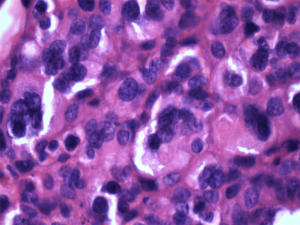
Histology revealed high grade urothelial cell carcinoma with PGCC as well as a focal plasmacytoid urothelial carcinoma (Figure 1). There was extensive invasion into the lamina propria andmuscularis propria. Lymphovascular invasionand urothelial carcinoma in situ were also noted. The carcinoma was positive for CK7, CK20, 34aE12, p63 and racemase consistent with aurothelial origin. Illiac node biopsy showed poorly differentiated carcinoma with morphology to that seen in the bladder. There was normal immunohistochemical staining for MLH-1, MSH2, MSH-6 and PMS-2. FDG PET scan showed extensive fluorodeoxyglucose (FDG) avid malignant nodes above and below both the hemidiaphragms including the left supraclavicular fossa, right internal mammary node as well as retroperitoneal and pelvic nodes. There was no abnormal FDG uptake noted in the brain at this time and no obvious space occupying lesions were present. The patient was started on chemotherapeutic regime consisting of 6 cycles of cisplatin and gemcitabine, which he tolerated well. A follow-up CT scan with IV contrast of the chest, abdomen and pelvis showed a complete response to the chemotherapy, with the previously seen supraclavicular, internal mammary, retroperitoneal and pelvic lymph nodes completely resolved.

Figure 1. Pleomorphic Giant Cell Carcinoma Displaying Bizarre Multinucleated Tumour Giant Cells at X20 Magnification Taken from a Bladder Tumour which was Resected by a TURBT Procedure

Pleomorphic Giant Cell Carcinoma Displaying Bizarre Multinucleated


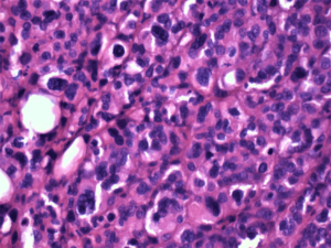
A TURBT procedure was performed again to remove the scar tissue in the bladder from the previous TURBT down to the adventitia of the bladder to look for recurrence of primary tumour cells. Histopathology showed no recurrence of malignant cells. The treating team arranged for radiographic monitoring of the chest, abdomen and pelvis using serial axial CT scans for early detection of relapse or new metastatic disease every 3-months. Additionally, surveillance cystoscopies were arranged at three-monthly intervals. Despite intensive radiographic monitoring of abdomen and pelvis showing no disease, the patient presented with headaches, disorientation and ataxia within six months of initial diagnosis. An magnetic resonance imaging (MRI) of the brain demonstrated multiple metastases, including a right cerebellar lesion measuring 37×28×31 mm which developed a mass effect on the surrounding tissue (Figure 2).

Figure 2. Pleomorphic Giant Cell Carcinoma Displaying Bizarre Multinucleated Tumour Giant Cells at X40 Magnification Taken from a Bladder Tumour which was Resected by a TURBT Procedure

Pleomorphic Giant Cell Carcinoma Displaying Bizarre Multinucleated

To reduce the size of the metastases, the patient received ten sessions of external whole brain radiotherapy which caused significant shrinkage of the metastasis. The patient also received three cycles of immunotherapy with pembrolizumab. Despite this treatment the patient developed obstructive hydrocephalus and began to deteriorate despite neurosurgical insertion of a ventriculoperitoneal shunt. The patient passed away soon after, 15-months after original presentation (Table 1).


Table 1. Clinical Features of Published Cases of Patients and Tumours
Study Sex Age Initial Treatment Stage Outcome
Lopez-Beltran10 Male 61 TURBT T4a, N1 DOD, 6 mo
Male 62 TURBT T3b, N1 DOD, 10 mo
Male 62 Cysto T3b, N1 DOD, 9 mo
Female 75 Cysto T3b, Nx DOD, 14 mo
Female 75 Cysto T4a, N1 DOD, 17 mo
Male 55 Cysto T3a, N0 NED, 74 mo
Male 59 Cysto T3a, N1 AWD, 19 mo
Male 88 TURBT T3b, N1 AWD, 11 mo
Alexiev8 Male 65 Cysto+Omental Resection Not Stated Not Stated
Akkaya7 Male 78 TURBT Not Stated Not Stated
Female 64 TURBT Not Stated Not Stated
Samaratunga25 Male 63 Cysto pT3b, N1 DOD, 10 mo
Female 87 TURBT pT2, N0 DOD, 3 mo
Female 73 TURBT pT1, N0 Nil F/U
Male 69 TURBT pT1, N0 AWD, 17 mo
Female 60 TURBT pT2, N0 AWD, 28 mo
Male 53 TURBT pT1, N0 NED, 46 mo
Male 79 TURBT pT1, N0 DOD, 7 mo
Male 60 TURBT pT2, N0 Nil F/U
Male 60 Cysto pT3b, N1 DOD, 12 mo
Male 75 TURBT pT1, N0 AWD, 15 mo
Female 93 TURBT pT1, N0 DOD, 2 mo
Male 92 TURBT pT1, N0 AWD, 34 mo
Male 72 TURBT pT1, N0 Nil F/U
Barresi9 Male 82 TURBT pT1, N0 NED, 12 mo
TURBT, transurethral resection of the urinary bladder tumour; Cysto, Cystoprostatectomy/Cystohysterectomy; DOD, Died of disease; NED, No evidence of disease; AWD, Alive with disease; Nil F/U, No recorded follow-up; Not stated, no mention of data in published case.


DISCUSSION AND CONCLUSION

Intracranial tumours most commonly arise as a result of spread from primary tumours located in other sites around the body.19 When tumours metastasize to the brain from a peripheral location, the median survival time of the patient is approximately 1-2-months if left untreated.20 Fortunately urothelial cell carcinoma rarely metastasises to the brain21 and so far there have been no reports of PGCC of the urinary bladder spreading to the brain in the literature.

As such, the treating team had not considered the possibility that cerebral metastases may arise in this particular cancer. Consequently, there was no radiographic surveillance of the head or neck. This meant that reimaging of the brain was not performed until 6-months after the initial diagnosis when the patient was presenting with neurological symptoms.By this time the metastases were much less responsive to treatment than they otherwise might have been. It is possible that the survival may have been longer and morbidity reduced if the cerebral metastasis had been detected before the patient experienced neurological symptoms.

The systemic standard treatment for invasive urothelial cell carcinoma arising from the urinary bladderis cisplatin and gemcitabine22,23 and local TURBT or radical cystectomy.3 In this case, cystectomy was rejected in favour of a TURBT as the tumour had already metastasised to lymph nodes and cystectomy is associated with a significant decrease in quality of life.24 Despite a complete response to cisplatin and gemcitabine, new metastases were detected 10-months after initial treatment. As PGCC is very aggressive, it is often fatal despite aggressive treatment.6,13,25

Other cases of PGCC are described in detail in the literature. Samaratunga et al described 13 cases of PGCC of the urinary bladder in adults with the ages of the patients at time of diagnosis ranging from 53 to 92-years, with a mean of 72-years.6,25 Nine of the patients were male and four of the patients were female and all of the cases presented with haematuria.6 Of the 13 cases, 10 were available to follow-up and five had died to the disease within 12-months of diagnosis.6 One patient had developed metastatic disease despite the author noting that the initial staging of the disease was pT1.6 Three patients had recurrent high grade urothelial cell carcinoma in the bladder despite TURBT. The final patient available to follow-up was reported to be alive and well after 46-months of follow-up following a cystoprostatectomy after an initial TURBT.6

Lopez-Beltran et al described eight patients with PGCC of the urinary bladder whose ages ranged from 55 to 88-years, with a mean of 67-years.10 Six of the patients were male and two were female and all presented with haematuria.10 Five of the patients were dead of disease at 17-months, while two were alive with disease at 11-months and 19-months.10 One patient had no evidence of disease despite being staged as T3aN0M0 at diagnosis after 74-months of follow-up.10

Alexiev et al described a single case of PGCC of the urinary bladder associated with polymavirusin a 65-year-old male who had a renal transplant 12-years previously.8 This patient presented with distant metastases despite cystoprostatectomy and omental resection.8 Akkaya et al7 presented two cases (a 78-year-old male and a 64-year-old female) that were treated with TURBT. Out comes were not reported. Finally, Barresi reported a single case of an 82-year-old male with PGCC of the urinary bladder staged at pT1 who was treated with TURBT and had no reoccurrences or metastatic disease 12-months after diagnosis.9

Of interest in this case is the fact that the patient presented with significant metastases (in the form of multiple extra-pelvic lymph node metastasis) despite the primary tumour only being staged as pT2. This is unusual in that this is the first published account of a patient presenting with pT2 PGCC of the urinary bladder and nodal metastasis as other cases only report nodal metastasis with a local stage of greater than T3a.6,10

While previous studies have reported metastasis of PGCC to other organs,6,10 this case is the first case in the literature which reports metastasis of PGCC of the urinary bladder to the brain. This is relevant as clinicians managing this variant of urothelial cell carcinoma need to be aware that metastatic spread to the brain is possible and will require frequent intracranial monitoring to ensure the best possible patient outcomes. Examining all 19 cases of PGCC of the urinary bladder that report on outcomes, only 3 cases report survival of the patient with no recurrence of disease.6,9,10 Additionally as PGCC of the urinary bladder has significantly poorer survival outcomes compared with conventional urothelial carcinoma of similar staging,2 it is important that clinicians are aware of possible complications of the disease. Clinicians treating PGCC must consider surveillance radiological imaging of the head to ensure cranial metastasis have not developed.

CONSENT

The authors have received written informed consent from the patient.

CONFLICTS OF INTEREST

The authors declare that they have no conflicts of interest.

1. Humphrey PA, Moch H, Cubilla AL, Ulbright TM, Reuter VE. The 2016 WHO classification of tumours of the urinary system and male genital organs-part B: Prostate and bladder tumours. Eur Urol. 2016; 70: 106-119, doi: 10.1016/j.eururo.2016.02.028

2. Samaratunga H, Delahunt B. Recently described and unusual variants of urothelial carcinoma of the urinary bladder. Pathology. 2012; 44: 407-418, doi: 10.1097/PAT.0b013e3283560172

3. Lopez-Beltran A, Eble JN, Bostwick D. G. Pleomorphic giant cell carcinoma of the prostate. Arch Pathol Lab Med. 2005; 129(5): 683-685. doi: 10.1043/1543-2165(2005)1292.0.CO;2

4. Kir G, Seneldi, H, Topal CS, Tosun M. Pleomorphic giant cell carcinoma of the prostate: A case report. Virchows Arch. 2015; 467: S277-S277.

5. Parwani AV, Herawi M, Epstein JI. Pleomorphic giant cell adenocarcinoma of the prostate: report of 6 cases. Am J Surg Pathol. 2006; 30: 1254-1259. doi: 10.1097/01.pas.0000209841.77595.4b

6. Samaratunga H, Delahunt B, Egevad L, Adamson M, Hussey D, Malone G, et al. Pleomorphic giant cell carcinoma of the urinary bladder: An extreme form of tumour de-differentiation. Histopathology. 2016; 68: 533-540. doi: 10.1111/his.12785

7. Akkaya H, Ipekci T, Hoscan MB, Ozdemir H. Pleomorphic giant cell carcinoma of the urinary bladder: Report of two cases. Virchows Arch. 2015; 467: S263-S264.

8. Alexiev BA, Papadimitriou JC, Chai TC, Ramos E, Staats PN, Drachenberg CB, et al. Polyomavirus (BK)-associated pleomorphic giant cell carcinoma of the urinary bladder: A case report. Pathol Res Pract. 2013; 209: 255259. doi: 10.1016/j.prp.2013.01.008

9. Barresi V. HOXB13 is not expressed in pleomorphic giant cell carcinoma of the bladder. Virchows Arch. 2018; 473: 259-260. doi: 10.1007/s00428-018-2394-1

10. Lopez-Beltran A, Blanca A, Montironi R, Cheng L, López JCR. Pleomorphic giant cell carcinoma of the urinary bladder. Human Pathology. 2009; 40: 1461-1466. doi: 10.1016/j.humpath.2009.02.016

11. Amin MB. Histological variants of urothelial carcinoma: diagnostic, therapeutic and prognostic implications. Modern Pathology. 2009; 22: S96. doi: 10.1038/modpathol.2009.26

12. Lopez-Beltran A, Cheng L, Comperat E, Rouprêt M, Blanca A, Menendez CL, et al. Large cell undifferentiated carcinoma of the urinary bladder. Pathology. 2010; 42: 364-368. doi: 10.3109/00313021003767363

13. Lopez-Beltran A, Requena MJ, Cheng L, Montironi R. Pathological variants of invasive bladder cancer according to their suggested clinical significance. B J U Int. 2008; 101: 275-281. doi: 10.1111/j.1464-410X.2007.07271.x

14. Mahmoud-Ahmed AS, Suh JH, Kupelian PA, Klein EA, Peereboom DM, Dreicer R, et al. Brain metastases from bladder carcinoma: Presentation, treatment and survival. J Urol. 2002; 167: 2419-2422. doi: 10.1016/S0022-5347(05)64996-8

15. Chung JH, Lee JY, Pyo JY, Oh YH, Lee SW, Moon HS, et al. Brain and skin metastasis from urothelial carcinoma of the bladder. Korean J Urol. 2013; 54: 66-68. doi: 10.4111/kju.2013.54.1.66

16. Sarmiento JM, Wi MS, Piao Z, Stiner ES. Solitary cerebral metastasis from transitional cell carcinoma after a 14-year remission of urinary bladder cancer treated with gemcitabine: Case report and literature review. Surg Neurol Int. 2012; 3: 82. doi: 10.4103/2152-7806.99172

17. Vaa B, Kohli M, Price KA, Swetz KM. Solitary cystic cerebellar metastasis in a patient with invasive transitional cell carcinoma of the bladder. BMJ Case Reports. 2014; 2014: bcr2013200137. doi: 10.1136/bcr-2013-200137

18. Anderson RS, el-Mahdi AM, Kuban DA, Higgins EM. Brain metastases from transitional cell carcinoma of urinary bladder. Urology. 1992; 39: 17-20. doi: 10.1016/0090-4295(92)90034-t

19. Johnson JD, Young, B. Demographics of brain metastasis. Neurosurg Clin N Am. 1996; 7: 337-344. doi: 10.1016/S1042- 3680(18)30365-6

20. Yang G, Wang Y, Wang Y, Lin S, Sun D. CyberKnife therapy of 24 multiple brain metastases from lung cancer: A case report. Oncol Lett. 20132; 6: 534-536. doi: 10.3892/ol.2013.1383

21. Shinagare AB, Ramaiya NH, Jagannathan JP, Fennessy FM, Taplin M-E, Van den Abbeele AD. Metastatic pattern of bladder cancer: correlation with the characteristics of the primary tumor. AJR Am J Roentgenol. 2011; 196: 117-122. doi: 10.2214/AJR.10.5036

22. Bamias A, Tiliakos I, Karali MD, Dimopoulos MA. Systemic chemotherapy in inoperable or metastatic bladder cancer. Ann Oncol. 2006; 17: 553-561. doi: 10.1093/annonc/mdj079

23. Bellmunt J, de Wit R, Vaughn DJ, Fradet Y, Lee J-L, Fong L, et al. Pembrolizumab as second-line therapy for advanced urothelial carcinoma. N Engl J Med. 2017; 376: 1015-1026. doi: 10.1056/NEJMoa1613683

24. Winters BR, Wright JL, Holt SK, Dash A, Gore JL, Schade GR. Health related quality of life following radical cystectomy: Comparative analysis from the medicare health outcomes survey. J Urolo. 2018; 199: 669-675. doi: 10.1016/j.juro.2017.08.111

25. Samaratunga H. Pleomorphic giant cell carcinoma of the urinary bladder: A clinicopathological analysis of 13 cases. Mod Pathol. 2015; 28: 255A-256A.

LATEST ARTICLES

An Unusual Presentation of Encephalitis in a Patient with Lyme Neuroborreliosis

Maithily Patel*, Jazmin Jatana, Ramya Ramesh and Milind Awale

doi.

Practical Pointers for Drug Development and Medical Affairs

Gerald L. Klein*, Roger E. Morgan, Shabnam Vaezzadeh, Burak Pakkal and Pavle Vukojevic

doi.

10.17140/CTPOJ-7-125

Prevalence and Risk Factors of Subclinical Mastitis of Goats in Banadir Region, Somalia

Omar M. Salah*, Yasin H. Sh-Hassan, Moktar O. S. Mohamed, Mohamed A. Yusuf and Abas S. A. Jimale

doi.10.17140/VMOJ-9-184

Use of Black Soldier Fly (Hermetia illucens) Prepupae Reared on Organic Waste

Maggot Debridement Therapy: A Natural Solution for Wound Healing

Isayas A. Kebede*, Haben F. Gebremeskel and Gelan D. Dahesa,

doi.10.17140/VMOJ-9-183

Figure 11. Risk Map for the Introduction of Ruminant Diseases at Borders

Ovine Network in Morocco: Epizootics Spread Prevention and Identification of the At-Risk Areas for “Peste des Petits Ruminants” and “Foot and Mouth Disease”

Yassir Lezaar*, Mehdi Boumalik, Youssef Lhor, Moha El-Ayachi, Abelilah Araba and Mohammed Bouslikhane

doi.

10.17140/EPOJ-8-131

The Impact of Family Dynamics on Palliative Care at the End-of-Life

Neil A. Nijhawan*, Rasha Mustafa and Aqeela Sheikh

doi.10.17140/PMHCOJ-10-154

LATEST ARTICLES

Case Report

2024 Apr

Maithily Patel*, Jazmin Jatana, Ramya Ramesh and Milind Awale
Pie Chart Showing Overall Proportions of Diagnostic Category of FNAC, JUMC

Retrospective Study

2024 Apr

Abel Tefera*, Lemlem Terefe and Kitesa Biresa
Prevalence (%) of Types of Anthropometric Failure among Previous and Present Studied Tribal Children

Original Research, peer reviewed

2024 Apr

Biswajit Mahapatra and Kaushik Bose*