Gastro

Open journal

ISSN 2377-8369

Overriding Elements in Colon Cancer Progression: Some Less Known Facts

Rohit Gundamaraju*, Ravichandra Vemuri and Rajaraman Eri

Rohit Gundamaraju, MPharm

Intestinal Mucosal Immunology, Research Group, School of Health Sciences, University of Tasmania, Launceston, Tasmania, Australia; E-mail: rohit.gundamaraju@utas.edu.au

INTRODUCTION

Colorectal cancer (CRC) is a heterogeneous disease causing more than 4 million deaths.1 Apart from dietary factors, lifestyle and genetics, advancements from other ailments like inflammatory bowel disease are reasons for cause of CRC.2 In several studies, it was affirmed that prevalence of CRC in patients with ulcerative colitis greatly varies from place and nature of race.2 Amongst the molecular pathogenesis, inflammation has a direct link to CRC.2 Not only by the assessment of mucosal biopsies revealed escalated levels of cell division and cell death, there were several conditions like epithelial mesenchymal transition (EMT) which had a higher role in cancer invasion rate or metastasis.3

Cytokines which are categorized under small proteins play an important role in cell signaling. In carcinogenic conditions like colitis associated cancers, cytokines were said to have a significant influence. Factors like nuclear factor-kappa B (NF-kB), tumor necrosis factor (TNF) etc. regulate cell cycle, apoptosis, reactive oxygen species and mutations.4,5 Thus the present view point will list few perplexing conditions, inflammatory modulators and receptors which would aid in CRC progression.

HYPOXIA – THE ENVIRONMENT FOR PROGRESSION

Hypoxia plays a huge role in the augmentation of solid tumors. During hypoxia, there is restricted oxygen supply which is regulated by hypoxia-inducible factor (HIF). HIF is not only involved in transcriptional activity of several genes, but its high levels are associated with poor prognosis of CRC.6 It was well evidenced that hypoxia promotes migration ability of cancer cells conditioning EMT.7 Cytokines such as tumor necrosis factor-α (TNF-α) were said to have direct relation with HIF-α levels enhancement via NF-kB activation. Additionally, Forkhead Box M1 (FOXM1) also advances EMT as FOXM1 promotes urokinase-type plasminogen activator receptor (uPAR) and matrix metalloproteinase 2 & 9.8 In the condition of CRC, endoplasmic reticulum (ER) stress is down-regulated by hypoxic condition thereby increasing the aggression of metastasis. It was clearly observed in several solid tumors that FOXM1 transcription factor was over expressed and ER stress was far reduced.8 The condition of hypoxia can be attenuated either by sensitizing the cancerous cells to ER stress or blocking uPAR pathway. Figure 1 depicts the role of hypoxia in regulating mammalian target of rapamycin (mTOR) in articulating vascular proliferation. It is evident that dysfunction in the P13k/mTOR pathway leads to pathogenesis of CRC. The hypoxia inducing factors HIF-α and HIF-β trigger chemokines to initiate epithelial mesenchymal transition which is a lead step in metastasis.


Figure 1: Correlation between hypoxia, chemokines and mTOR pathway and their role in carcinogenesis.

GOJ-2-124 Fig 1


CHEMOKINES AND THEIR RECEPTORS

Clinical evidences state that chemokine receptor 6 (CCR6) expressions was up-regulated in colorectal cancer.9 Chemokines in general can regulate the movement of tumor cells. CCR7, CCR9, C-X-C chemokine receptor type 1 (CXCR1), and C-X-C chemokine receptor type 2 (CXCR2) are also detected in tumor cells and their ligands can induce the chemotaxis of the corresponding receptor-expressing cells. These chemokines act as inducers of invasion within the tumor and also in regards to movement to other organs. When chemokine (c-c motif) ligand 20 (CCL20) triggered p130 phosphorylation, there was an enhanced migration and proliferation of cancer cells. The expression of chemokine receptor C-X-C chemokine receptor type 4 (CXCR4) in CRC metastasis was well established. Factors like high invasion rate, development of liver metastasis and colon carcinoma micro metastasis of liver were reported by CXCR4.9 Direct association of lymph node metastasis in CRC and CCR7 was observed in a study by Gunther et al.10 Not only does chemokine-chemokine receptor interaction supports metastasis in colon cancer, CCR6-CCL20 interaction was found to be involved in various inflammatory and immune disorders.11 CCR6 role in advanced colon cancer was clearly shown and also CCR6-CCL20 was reported to have involvement in EMT which resulted in poor outcome of colon cancer.11 CCR6 was described as independent risk factor in liver metastasis. On the other hand, CCL20 levels were higher in colon cancer patients with liver metastasis compared with controls without metastasis.12 In a study by Kawada and colleagues,13 amongst the colon cancer samples with CXCR3 expression, there was significantly high lymph node metastasis recorded. CXCR3 expression levels were directly proportional to poor prognosis which clearly indicates that CXCR3 activation was concomitant with colon cancer metastasis preferentially to the draining lymph nodes with poorer prognosis.

mTOR’s Role in Colon Cancer

Hyper activation of mechanistic target of rapamycin (mTOR) is due to signaling malfunctions upstream of mTOR in phosphatidylinositol-3-kinase (PI3K)/Akt/mTOR pathway. Mutagenesis in PI3Kα occurs in late tumorigenesis which is generally evident in more than 30% of colon cancers.14 mTORC1 inhibition had a role in improving regeneration capacity of intestinal stem cells but at the same time, abberant stimulation could trigger carcinogenesis. In humans, immunohistochemical studies on colorectal carcinoma samples revealed that mTORC1 signaling takes place in the early onset of tumorigenesis and plays a role in transforming normal cells to neoplastic. mTORC1 and mTORC2 were implicated in colorectal cancer biology to a greater extent.

In the conditions of in vivo mTORC2 was down-regulated, there was a clear trend of reduction of proliferation and also there was reduction in the formation of tumor xenografts. EMT was also regulated by mTORC1 and mTORC2 and thus sequencing metastasis of CRC.15,16,17

Targeting kinases like polo-kinase 1 which is highly expressed in proliferating cells during G2 and M phase of cell cycle was regarded as a potential target. Besides, induction of apoptosis via mTOR suppression is a strategic therapeutic intervention. BI2536 was one amongst the potent polo-kinase 1 inhibitor used in combination with NVP-BEZ235 which is a dual PI3K/mTOR inhibitor.18 These studies not only show the importance of epigenetic mechanisms in cancer signaling but also clearly denote the role of mTOR signaling in CRC progression.

Nod like receptors (Nlrp3) and their Role in CRC

Chronic inflammation is considered as a risk factor for the progression of CRC. Nod like receptor (Nlrp3) is a protein which assembles the inflammasome which is responsible for secretion of pro-inflammatory cytokines like IL-1β. This aids in cellular functions like repair via immune cell activation and triggering pro-inflammatory cytokines. Inoperative NLR activation results in cell proliferation and tumorigenesis.18 Mice deficient of Nlrp3 were prone to dysplasia and tumor formation thus clearly stating the linkage between Nlrp3 deficiency and susceptibility to colitis associated carcinogenesis. Apoptosis associated speck like protein-1 (ASC) and caspase 1 are two major components of Nlrp3 which play a major role in inducing apoptosis of cancer cells. In experimental conditions of knocking out of these two major components of Nlrp3, the mice developed tumorigenesis. Interleukin 18 (IL-18) which is secreted by Nlrp3 exerts extensive anti-tumor activity and aids in epithelium repair. The fact that IL-18 offers its protection against colitis and carcinogenicity was described in mice models where caspase 1 and Nlrp3 were knocked out and there was development of CRC. It was well defined that IL-18 promotes enterocyte proliferation to repair chemically induced injury of colonic epithelium and also inhibits hyperplasia during chronic stages of colitis. Also in the acute conditions of disease, IL-18 offers restoration of barrier integrity by controlled proliferation of stem cells at the base of the intestinal crypt which indirectly helps in intestinal homeostasis. In experimental conditions of azoxymthane (AOM) or dextran sulfate sodium (DSS) induced colitis, phosphorylated levels of STAT1 were reduced in the colon where the restoration is possible by IL-18.19,20,21 Nlrp3 inflammasome not only reduced the tumorigenesis but also suppressed liver colon cancer metastatic growth.22

SPINK1

Serine peptidase inhibitor kazal type-1 (SPINK1) has been investigated for its role in multiple human carcinomas especially CRC.23 SPINK1 mutation was associated with pancreatitis. Epidermal growth factor receptor (EGFR) involves in intra-cellular message transduction. High expression of EGFR is also associated with poor prognosis. SPINK1 and epidermal growth factor (EGF) bind to EGFR and stimulate proliferation via mitogen-activated protein kinases (MAPK) which was recorded in pancreatic adenocarcinoma.24 In a study by Chen et al,24 the SPINK1 protein played a role in tumor proliferation and malignant transformation in CRC through the EGFR pathway. SPINK1 was also used as a marker for predicting tumors in response to anti-EGFR treatment in CRC patients.25 Metalllothionein expression in colon cancer was analogous with poor survival and SPINK1 caused CRC progression by down regulating metallothioneins expression.23

COX2

The interest in cyclooxygenase (COX2) inhibitors as therapeutic interventions rose after its ability as an anti-carcinogenic agent with 40-50% reduction in CRC prevalence in non-steroidal anti-inflammatory drugs (NSAID) users.26 COX2 over expression has been reported in numerous colorectal adenomas and adeno carcinomas. The direct relation of COX2 with intestinal cancers was well demonstrated in a study where the murine COX2 gene was deleted in APC Δ716 mice which resulted in reduction of intestinal polyps. Ideal tumorigenesis mechanisms of COX2 are inhibition of apoptosis, increased effectiveness of invasiveness etc. It is noteworthy that though COX2 is preponderant to the cytoplasm of cancerous epithelial cells, it is negatively expressed in normal epithelium. In regards to the mucosa, CRC samples were found to have more messenger RNA (mRNA) levels than normal mucosa.27,28 In a clinical study involving 76 patients suffering with CRC, COX2 was conferred to advancement of the disease and decreased patient survival.29 The underlying mechanism of COX2 associated carcinogenicity is by attenuation of the mitochondrial apoptotic pathway distinguished by reduced cytochrome C release and diminished caspase activity. There was an increased expression of anti-apoptotic genes Bcl-2 recorded in cell lines like HCT-15.30 COX2 also was involved in death receptor (DR) pathway via DR expression in HCT-15 cells.31 Targeting CRC with COX2 inhibitors may not be a complete solution though NSAID users had less probability of CRC.

CONCLUSION

Biological mechanism underlying CRC have been markedly improved in recent years with respect to therapeutic interventions selecting target receptors, inflammasomes, cytokines and other chemokines. Finding the right agent for CRC treatment was achieved by inducing apoptosis, reducing EMT and decelerating proliferation and invasiveness. Treatment for CRC is not only designed via symptomatic approach but also targeting signaling mechanisms which drive the disease. Thus, this article highlights some of the facets in CRC which when targeted or inhibited can turn out to be a potential treatment escalating the quality of life (QoL) and improved prognosis.

CONFLICTS OF INTEREST

The authors declare that they have no conflicts of interest.

1. Dekker E, Gulik TV. Colorectal cancer: What the clinician wants to know. Cancer Imaging. 2005; 5: S127-S132. doi: 10.1102/1470-7330.2005.0036

2. Triantafillidis JK, Nasioulas G, Kosmidis PA. Colorectal cancer and inflammatory bowel disease: Epidemiology, risk factors, mechanisms of carcinogenesis and prevention strategies. Anticancer Res. 2009; 29: 2727-2738. Web site. http://ar.iiarjournals.org/content/29/7/2727.long. Accessed July 3, 2016

3. Clevers H. Colon cancer-understanding how NSAIDs work. N Engl J Med. 2006; 354: 761-763. doi: 10.1056/NEJMcibr055457

4. Schottelius AJ, Dinter H. Cytokines, NF-ÎB, microenvironment, intestinal inflammation and cancer. Cancer Treat Res. 2006; 130: 67-87. doi: 10.1007/0-387-26283-0_3

5. Yan B, Wang H, Rabbani ZN, et al. Tumor necrosis factor-alpha is a potent endogenous mutagen that promotes cellular transformation. Cancer Res. 2006; 66(24): 11565-11570. doi: 10.1158/0008-5472.CAN-06-2540

6. Müller-Edenborn K, Léger K, Garzon JFG, et al. Hypoxia attenuates the proinflammatory response in colon cancer cells by regulating IκB. Oncotarget. 2015; 6(24): 20288-20301. doi: 10.18632/oncotarget.3961

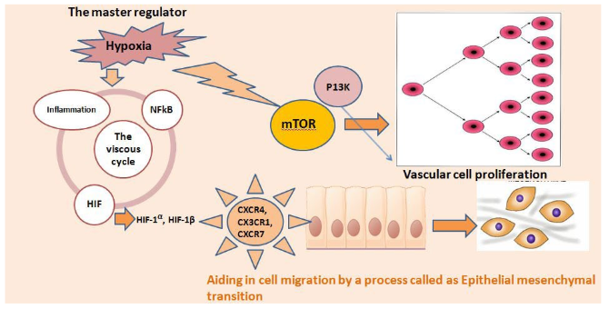
7. Hongo K, Nelson Tsuno H, Kawai K. Hypoxia enhances colon cancer migration and invasion through promotion of epithelial-mesenchymal transition. J Surg Res. 2013; 182: 75-84. doi: 10.1016/j.jss.2012.08.034

8. Luo X, Jinke Yao H, Nie P. FOXM1 promotes invasion and migration of colorectal cancer cells partially dependent on HSPA5 transactivation. Oncotarget. 2016; 7(18): 26480-26495. doi: 10.18632/oncotarget.8419

9. Ghadjar P, Rubie C, Aebersold DM, et al. The chemokine CCL20 and its receptor CCR6 in human malignancy with focus on colorectal cancer. Int J Cancer. 2009; 125(4): 741-745. doi: 10.1002/ijc.24468

10. Gunther K, Leier J, Henning G, et al. Prediction of lymph node metastasis in colorectal carcinoma by expression of chemokine receptor CCR7. Int J Cancer. 2005; 116: 726-733. doi: 10.1002/ijc.21123

11. Kapur N, Mir H, Clark Iii CE, et al. CCR6 expression in colon cancer is associated with advanced disease and supports epithelial-to-mesenchymal transition. Br J Cancer. 2016; 114(12): 1343-1351. doi: 10.1038/bjc.2016.113

12. Rubie C, Oliveira V, Kempf K, et al. Involvement of chemokine receptor CCR6 in colorectal cancer metastasis. Tumor Biol. 2006; 27: 166-174. doi: 10.1159/000092777

13. Kawada K, Hosogi H, Sonoshita M. Chemokine receptor CXCR3 promotes colon cancer metastasis to lymph nodes. Oncogene. 2007; 26(32): 4679-4688. doi: 10.1038/sj.onc.1210267

14. Francipane MG, Lagasse E. mTOR pathway in colorectal cancer: An update. Oncotarget. 2013; 5(1): 49-66. doi: 10.18632/oncotarget.1548

15. Roulin D, Cerantola Y, Dormond-Meuwly A, Demartines N, Dormond O. Targeting mTORC2 inhibits colon cancer cell proliferation in vitro and tumor formation in vivo. Mol Cancer. 2010; 9: 57. doi: 10.1186/1476-4598-9-57

16. Gulhati P, Cai Q, Li J, et al. Targeted inhibition of mammalian target of rapamycin signaling inhibits tumorigenesis of colorectal cancer. Clin Cancer Res. 2009; 15(23): 7207-7216. doi: 10.1158/1078-0432.CCR-09-1249

17. Gulhati P, Bowen KA, Liu J, et al. mTORC1 and mTORC2 regulate EMT, motility, and metastasis of colorectal cancer via RhoA and Rac1 signaling pathways. Cancer Res. 2011; 71(9): 3246-3256. doi: 10.1158/0008-5472.CAN-10-4058

18. Mukherjee B, Tomimatsu N, Amancherla K, et al. The dual PI3K/mTOR inhibitor NVP-BEZ235 is a potent inhibitor of ATM- and DNA-PKCs-mediated DNA damage responses. Neoplasia. 2012; 14(1): 34-43. doi: 10.1593/neo.111512

19. Allen IC, TeKippe EM, Woodford RM, et al. The NLRP3 inflammasome functions as a negative regulator of tumorigenesis during colitis-associated cancer. J Exp Med. 2010; 207(5): 1045-1056. doi: 10.1084/jem.20100050

20. Zaki MH, Boyd KL, Vogel P, Kastan MB, Lamkanfi M, Kanneganti TD. The NLRP3 inflammasome protects against loss of epithelial integrity and mortality during experimental colitis. Immunity. 2010; 32(3): 379-391. doi: 10.1016/j.immuni.2010.03.003

21. Zaki MH, Lamkanfi M, Kanneganti T-D. The Nlrp3 inflammasome in IBD and colorectal tumorigenesis. Trends Immunol. 2011; 32(4): 171-179. doi: 10.1016/j.it.2011.02.002

22. Dupaul-Chicoine J, Arabzadeh A, Dagenais M, et al. The Nlrp3 inflammasome suppresses colorectal cancer metastatic growth in the liver by promoting natural killer cell tumoricidal activity. Immunity. 2015; 43(4): 751-763. doi: 10.1016/j.immuni.2015.08.013

23. Tiwari K, Pandey SK, Goel S. SPINK1 promotes colorectal cancer progression by downregulating Metallothioneins expression. Oncogenesis. 2015; 4: e162. doi: 10.1038/oncsis.2015.23

24. Chen Y-T, Tsao S-C, Shyng-Shiouvyuan F. Serine protease inhibitor kazal type 1 (SPINK1) promotes proliferation of colorectal cancer through the epidermal growth factor as a prognostic marker. Pathol Oncol Res. 2015; 21: 1201-1208. doi: 10.1007/s12253-015-9949-0

25. Chen Y-T, Tsao S-C, Tsai H-P. Serine protease inhibitor kazal type 1 (SPINK1) as a prognostic marker in stage IV colon cancer patients receiving cetuximab based targeted therapy. J Clin Pathol. 2016; 1-5. doi: 10.1136/jclinpath-2016-203638

26. Sinicrope FA, Gill S. Role of cyclooxygenase-2 in colorectal cancer. Cancer Metastasis Rev. 2004; 23: 63-75. doi: 10.1023/A:1025863029529

27. Wang D, Raymond Dubois N. The role of COX-2 in intestinal inflammation and colorectal cancer. Oncogene. 2011; 29(6): 781-788. doi: 10.1038/onc.2009.421

28. Li M, Wu X, Xu X-C. Induction of apoptosis in colon cancer cells by cyclooxygenase-2 inhibitor NS398 through a cytochrome c-dependent pathway. Clin Cancer Res. 2001; 7: 1010-1016. Web site. http://clincancerres.aacrjournals.org/content/7/4/1010.long. Accessed July 3, 2016

29. Sheehan KM, Sheahan K, O’Donoghue DP, et al. The relationship between cyclooxygenase-2 expression and colorectal cancer. JAMA. 1999; 282(13): 1254-1257. doi: 10.1001/jama.282.13.1254

30. Sun Y, Tang XM, Half E, Kuo MT, Sinicrope FA. Cyclooxygenase-2 overexpression reduces apoptotic susceptibility by inhibiting the cytochrome c-dependent apoptotic pathway in human colon cancer cells. Cancer Res. 2002; 62(21): 6323-6328. Web site. http://cancerres.aacrjournals.org/content/62/21/6323.full-text.pdf. Accessed July 3, 2016

31. Tang X, Sun YJ, Half E, Kuo MT, Sinicrope F. Cyclooxygenase-2 overexpression inhibits death receptor 5 expression and confers resistance to tumor necrosis factor-related apoptosis- inducing ligand-induced apoptosis in human colon cancer cells. Cancer Res. 2002; 62(17): 4903-4908. Web site. http://cancerres.aacrjournals.org/content/62/17/4903.full-text.pdf. Accessed July 3, 2016.

LATEST ARTICLES

Unraveling the Mysteries of Type-A Aortic Dissection Using POCUS/Echocardiography

Syeda Rukh*, Sathyanarayana Machani and Milind Awale

doi.

Blood Sample from the Patient

Hypertriglyceridemia-Induced Pancreatitis: A Case Report and Literature Review

Maarten Bulterys, Melvin Willems* and Agnes Meersman

doi.

From Neck Pain to a Life-Threatening Condition: A Case Report

Floris Vandewoude* and Sören Verstraete

doi.

LATEST ARTICLES