INTRODUCTION
Muscle pedicle graft has been advanced as a modality of treatment in Ficat et al1 Grade1 and Grade 2 avascular necrosis (AVN) of the head of the femur.1,2,3 What effect it has on the vasculature of the head has been a matter of speculation.
This surgery has been performed at our institution since 1983, although there is yet to be a validated method to quantify the blood supply to the diseased head. A constant search for some method by which the blood supply in the diseased head could be quantitatively estimated was sought. The one investigation which seemed to hold out promise was the Tc-99 bone scan.4 A paper on perfusion studies by Urologists to study renal perfusion in transplanted kidneys drew attention.5,6 Could the use of radio-isotope Tc-99 and SPECT analysis (Single Photon Emission Computed Tomography) on a triple flow bone scan give an any indication? The SPECT gives a 3D image with cross-sectional slices. This was discussed with the Nuclear Physicist at the scan centre who had been involved in the renal perfusion studies in kidney transplant patients at the Department of Urology, Post Graduate Institute of Medical Education and Research at Chandigarh, India.
Effort was made to develop a standardized procedure of reading the scan for consistency. It was decided that a SPECT analysis with a pixel count would be performed in the anterior and posterior scans defining a constant REGION OF INTEREST with a CONTROL AREA for comparison.
The region of interest was defined on the head of femur and the pixel count used to ensure a constant area. The computer software of the Gamma camera scanner was used to evaluate the region. The control area was like-wise defined on the vertebra. The background ratio was used to eliminate any changes in the pixel count due to any variance in the quantity of isotope injected.
The region of interest was defined on the head of femur and the pixel count used to ensure a constant area. The computer software of the Gamma camera scanner was used to evaluate the region. The control area was like-wise defined on the vertebra. The background ratio was used to eliminate any changes in the pixel count due to any variance in the quantity of isotope injected.
Since the pixel count could not be used as absolute values with reliability, a ratio of the pixel count of target area (H) to the control area (V) was used. Since these were taken from the same phase, they represented similar stages, although even the ratio was not expected to, or could have given a clear cut black and white picture of the outcome.
Consequently, in each patient and in each scan, pixel count in a well-defined region of interest with a pixel count in a control area in L5 vertebra was recorded. Background to target ratio was taken into account while making the pixel counts.
Similar readings were recorded in all subsequent scans. The H/V (hip region of interest to the vertebral control area) ratio in the third phase of the skeletal image was compared. This ratio formed the basis of comparison in the pre-operative and all post-operative bone scans of each patient. In pre-operative scan, H1 was the sum of the pixel count in both the views-anterior and posterior. V1 was the sum of pixel count of both the vertebral control areas in the anterior and posterior readings. X1 was the ratio of H1/V1. Similarly, post-operatively the same ratios were taken, and designated as X2=H2/V2.
OBSERVATIONS
The study was conducted since 2007 in the Department of Orthopaedics, S. M. S. Medical College and Hospital, Jaipur, RJ, India, and subsequently by the author in his practice.
A total of 66 hips in 38 patients have been studied so far (Table 1 and 2). Table 3 shows the side involved. The etiology was steroid induced in 26%, post chronic alcoholism in 26%, postpartum in females in 7% and idiopathic in the remaining 41% (Table 4). Hip function was assessed by Harris’ hip score (Table 5).
| Table 1: Age Distribution. |
|
Age Group
|
Number of cases |
Percentage
|
|
16-20
|
13 |
24.24% |
|
21-25
|
14 |
27.27% |
| 26-30 |
10 |
18.18%
|
| 31-35 |
9 |
16.67%
|
|
36-40
|
7 |
13.64% |
| Total |
53 |
100%
|
| Table 2: Sex Distribution. |
|
Sex
|
Number of cases |
Percentage
|
|
Male
|
42 |
79% |
|
Female
|
11 |
21%
|
| Total |
53 |
100%
|
| Table 3: Side Involved. |
|
Side of Disease
|
Number of cases |
Percentage
|
|
Right
|
22 |
36.37% |
|
Left
|
18 |
30.30% |
| Bilateral |
13 |
33.33%
|
| Total |
53 |
100%
|
| Table 4: Etiological Cause of AVN Hip. |
|
Mode of Disease
|
Number of cases |
Percentage
|
|
Alcohol Induced
|
16 |
30.30% |
|
Steroid Induced
|
18 |
33.33% |
|
Post-partum
|
3 |
6.07%
|
| Idiopathic |
16 |
30.30%
|
| Total |
53 |
100%
|
| Table 5: Preoperative Harris Hip Score. |
|
Harris Hip Score
|
Number of Hips |
Percentage
|
|
Excellent
|
3 |
4.54% |
|
Good
|
8 |
12.12% |
|
Fair
|
12 |
18.18%
|
| Poor |
43 |
65.16%
|
| Total |
66 |
100%
|
The AVN hip was graded according to the grading of and is shown in Table 6.
| Table 6: Stage of AVN.1 |
|
Stage of AVN
|
Number of Hips |
Percentage
|
|
Stage 1
|
5 |
7.58% |
|
Stage 2
|
31 |
46.96% |
|
Stage 3
|
23 |
34.85%
|
| Stage 4 |
7 |
10.61%
|
| Total |
66 |
100%
|
Pre-collapse (Ficat stage 1-2) hips were included in our study. Those patients with a Ficat Stage 3 or 4 hip who were poor and could not afford a joint replacement procedure were also treated with a muscle pedicle graft procedure.
The radiological grading was done as per Marcus et al7 (Table 7).
| Table 7: Radiological Stage.7 |
|
Stage 1
|
9% |
|
Stage 2
|
49% |
|
Stage 3
|
33% |
| Stage 4 |
9%
|
| Stage 5 |
0%
|
| Stage 6 |
0%
|
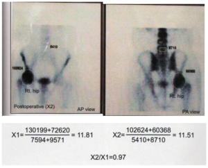
The bone scans were evaluated by performing the pixel counts in the anteroposterior (AP) and posteroanterior (PA) views in the region of interest (Femoral head ‘H’) and the control area (L5 vertebra ‘V’) both pre- and post- operatively as shown in Figures 1 and 2.
Figure 1: The Pre-operative Pixel Counts in AP and PA View in the Region of Interest and Control Area.
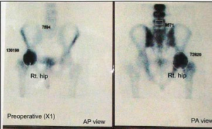
Figure 2: The Post-operative Pixel Counts and the H1/V1 and H2/V2 Values with the X2/X1 Ratio.
The ratio X2 (post-operative)/X1 (pre-operative) were compared (Table 8A and 8B). The pre-operative ratio had a mean of 9.10 while the post-operative mean was 10.62 with a standard deviation of 4.23 and 4.91 respectively. The Paired t-test gave a p-value equal to 0.0104, considered statistically significant after six months’ follow-up.
| Table 8A: X2/X1 Ratio Distribution. |
|
X2/X1
|
Number of Hips |
Percentage
|
|
0.9-1
|
8 |
12% |
|
1.01-1.1
|
28 |
42% |
| 1.11-1.2 |
17 |
26%
|
| >1.2 |
13 |
20%
|
| Table 8B: Statistical Analysis of X2/X1 Ratio. |
|
Statistical Group
|
X1 |
X2
|
|
Mean
|
9.10 |
10.62 |
|
Standard Deviation
|
4.23 |
4.91 |
| SEM |
.90 |
1.04
|
| N |
26 |
26
|
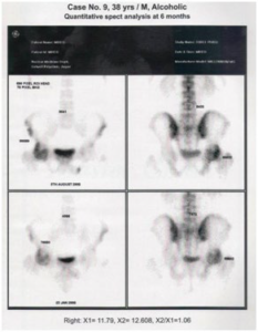
In 87% hips, a tensor fascia femoris graft was done in Grade 2 and Grade 3 AVN with anterior and superior involvement. Sartorius muscle pedicle was used in Grade 1 AVN (8%). In those hips with a posterior quadrant involvement, a quadratus femoris muscle pedicle graft was used through the posterior approach (5%). Figure 3 and 4 shows the X2/X1 ratio after six months of surgery.
Figure 3: Steroid Induced AVN Femoral Head in 18 Year Old Showing the Quantitative Aspect of the SPECT Pre-op and at 6 Months Post-op.
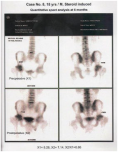
Figure 4: Alcohol Related AVN Hip in a 38-yearOld. Quantitative Aspect Analysis.
The pre- and post-operative scans were compared as the ratio X2/X1. The ratio distribution and statistical analysis of the results is shown in Table 8A and 8B.
The results indicated some surprising values but useful inferences could be drawn. Although it was difficult to comment on revascularization with any degree of certainty, the ratios did reveal a predictive value in the outcome.
The ratio decreased, remained static or marginally increased in those patients where the disease was contained, even reversed to some extent.
The ratio continues to be altered for a period of 2 years.
It significantly increased in those patients where the disease progressed, and eventually led to osteoarthritic changes or the patient was forced to undergo athroplasty. It continued to rise in these patients for a period of up to 2 years after which it became static.
This was inferred on the changes in the Harris hip score clinically (Table 9).
| Table 9: Statistical Analysis of the Pre- and Post-Op Harris Hip Score. |
| Statistical Group |
Pre-Op Harris Hip Score |
Post-Op Harris Hip Score |
|
Mean
|
59.08 |
66.50 |
|
Standard Deviation
|
10.44 |
9.94 |
| SEM |
2.05 |
1.95
|
| N |
26 |
26
|
The Harris hip score showed an improvement in 65% cases, remained unchanged in 15% cases, and showed deterioration in 20% cases, with a p-value of 0.0001 which was statistically significant. Only four out of 66 opted for total hip replacement (THR).
When the radiological status was evaluated post-operatively using the Marcus et al7 grading as used pre-operatively, it showed that there was a change in the radiological status after six to twelve months. Thirty two percent hips improved by one stage. Marcus et al7 stage 3 hips improved to stage 2, stage 2 to stage 1 and stage 1 to no radiological evidence. Fourty three percent hips remained unchanged radiologically. Twenty five percent deteriorated in radiological stage. Six percent required or desired joint replacement.
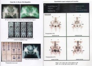
Figure 5 shows the X-ray, MRI and pre-operative bone scan together, with the post-operative SPECT and the pixel counts and the X2/X1 ratio.
Figure 5: Idiopathic AVN in a 20-Year-Old, with X-ray and MRI Showing Bilateral Hip Involvement. The Bone Scan Appearance Pre and Post-op and the Quantitative Aspect Ratio. The Patient’s Symptoms Persisted, and Requested a THR on Both Sides.
In the SPECT analysis in subsequent cases (Figure 6), a target background ratio was also fixed.
Figure 6: Bilateral AVN Operated with Muscle Pedicle graft 6 months’ after Showing the Quantitative Aspects of the 2 Hips 6 months’ post-op.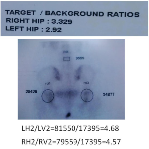 A serial change in the SPECT analysis in a patient with bilateral idiopathic AVN over a period of 6 months, 20 months and 27 months is shown in Figures 6, 7 and 8.
Figure 7: Quantitative Ratios X2/X1 in Same Patient 6 Months after Surgery
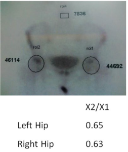
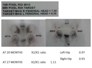
Figure 8: The Quantitative Aspect Ratios X2/X1 of The Right and Left Hip of the Same Patient, with the Target to Background Ratio, at 20 and 27 Months after Surgery, there is a Slight Change in the Ratio, but Patient is Still “Symptom Free” and the Patient Quite Satisfied with the Result.
We could draw the following inferences:
• The ratio decreases as the revascularization occurs or disease is contained.
• It continues to alter over a period of 2 years.
• No further rise occurs after 2 years becomes static.
The prognostic significance as gauged by us from the study data is shown in Table 10.
| Table 10: Prognostic Significance of X1/X2 Ratio. |
|
Significance
|
Ratio |
Percentage of Hips
|
|
Disease arrested and reversed
|
0.9 to 1.0 |
12% |
|
Disease arrested and static
|
>1.0 to 1.1 |
42% |
| Disease progressed but no arthritic evidence |
>1.1 to 1.2 |
26%
|
| Disease progressed to arthritis |
>1.2 |
20%
|
We feel it can be reliably used as a predictor variable with prognostic value.
The post-operative complications were restriction of movement due to presence of the graft, meralgia paraesthetica due to the surgical approach used and low back pain and shortening of >1 cm, which could possibly be a consequence of the disease itself. No patient suffered a fracture of the neck of femur or infection.
CONCLUSION
It would appear that THE IDEAL CANDIDATE for a muscle pedicle graft is a young patient, with no or minimal degeneration of the acetabular cartilage or a crescent sign or collapse of the femoral head. It could also benefit the young patients, without femoral head collapse but with extensive osteonecrotic involvement of the femoral head (a combined necrotic angle of >2000 or femoral head involvement of >50%). Tc-99 bone scan with SPECT analysis could be used as a monitor for the progression of the disease, and could provide a useful indicator of prognostic value as to which patient may eventually require arthroplasty.
ACKNOWLEDGEMENT
The author gratefully acknowledges the help rendered by Dr. Karun Sethi, Nuclear Physicist, Gamma Imaging and Nuclear Medicine section, Get-well Diagnostic Centre, Opposite Jay kay Lon Hospital, Jaipur, in the SPECT analysis of the bone scans, and maintaining their records.


