Vaccination Research

Open journal

ISSN 2771-750X

Model Liposomal Delivery System for Drugs and Vaccines

Farrhana Z.Firdaus, Zeinab G. Khalil, Robert J. Capon, Mariusz Skwarczynski, IstvanToth* and Waleed M. Hussein*

Istvan Toth, PhD

School of Chemistry and Molecular Biosciences, The University of Queensland, Brisbane, QLD 4072, Australia; Tel. +61 7 3346 9892; Fax. +61 7 33654273; E-mail: i.toth@uq.edu.au

Waleed M. Hussein, PhD

School of Chemistry and Molecular Biosciences, The University of Queensland, Brisbane, QLD 4072, Australia; Tel. +61 7 3365 2782; Fax. +61 7 33654273; E-mail: w.hussein@uq.edu.au

INTRODUCTION

Since their discovery by Alec Bangham and colleagues in the 1960s, liposomes have been one of the most investigated forms of nano carriers.1 They were first described as lamellae of swollen or enlarged lipids that could act as a model membrane system.2 Liposomes are spherically shaped amphiphilic lipid bilayers ranging in size from 20 nm to 10 µm.3,4 Their structure is strongly correlated with their unique ability to physically entrap a variety of compounds, including proteins, nucleotides, plasmids, macromolecules and ribonucleic acid/deoxyribonucleic acid (RNA/DNA). Hydrophobic compounds can be embedded in the phospholipid bilayer, whereas hydrophilic compounds are entrapped within the aqueous core of the lipid vesicles.5 Liposomes can increase drug uptake and reduce drug toxicity, while prolonging biological half-life.6 Consequently, liposomes can significantly increase the therapeutic index of the drugs they deliver.6,7

Liposome vesicles can be prepared with synthetic or natural lipids, such as distearoyl phosphatidyl choline (DSPC), dioleoyl phosphatidyl ethanolamine (DOPE) orphosphatidylglycerol (PG). The inclusion of lipids allows liposomes to be biocompatible, non-toxic, flexible, non-immunogenic and biodegradable.8 The physicochemical properties of liposome vesicles depend on the phospholipid (and other components) used in the formulation, and can influence the vesicle’s surface charge, size, permeability and stability.9

Classical liposomes, better known as the first generation of liposome vesicles, were developed to encapsulate compounds for non-specific uptake through endocytosis, adsorption or lipid exchange once administered in a host.4,10 However, these vesicles were found to be rapidly eliminated by the reticuloendothelial system (RES), greatly reducing their therapeutic efficacy.11 Coating vesicles with polyethylene glycol (PEG) is often done to increase the amount of time that the vesicles spent circulating through the blood (i.e. stealth shielding).12,13 PEGylation of liposomes results in the formation of hydrophilic barriers on the surface of the vesicles, which decreases the interaction of the vesicles with RES and increases blood circulation half-life.11,14 The addition of cholesterol to formulations has also been employed to enhance transmembrane permeability and rigidity.5 Adding cholesterol to the liposome membrane can lead to reduced binding of sodium ions to the membrane. This reduces the interaction between vesicles and serum opsonins, and increases the circulation time of the liposomes in the blood stream.15,16

Liposomes are widely used to improve cellular uptake of variety of drugs and antigens. For example, a vaccine against tuberculosis (TB), called bacille calmette-guerin (BCG), was developed and widely distributed more than 100-years-ago. BCG is effective in protecting vaccinated children against acute forms of the disease, but it has shown limited and variable protection in adults, with the protective efficacy ranging from 0% to 77%.17,18 A new TB vaccine and/or more effective treatments are urgently required to fight this disease. TB is caused by mycobacterium tuberculosis (MTB), which infect macrophages. Encapsulation of the anti-TB vaccines or drugs within liposome vesicles can be applied to improve their delivery to the macrophages. Similarly, a variety of strategies has been examined to facilitate uptake of anticancer agents by tumour cells.19,20 As prostate cancer is still the most common cancer diagnosed in men,21 drug delivery systems targeting this cancer have been intensively investigated.22,23 Thus, we developed three liposome formulations to better understand the characteristics needed to improve the uptake by human epithelial prostate cancer (PC-3) cells and mouse macrophages.

MATERIALS AND METHODS

The lipids used in the liposome formulations: DSPC (1,2-distearoyl-sn-glycero-3-phosphocholine), egg PG (L-α-phopshtidyl-DL-glycerol (egg, chicken)), PEG (3000), PE (1,2-distearoyl-sn-glycero-3-phosphoethanolamine-N-[methoxy(polyethylene glycol)-3000] (ammonium salt)), DOPE (1, 2-Dioleoyl-sn-glycero-3-phosphoethanolamine)and cholesterol, were obtained from Avanti Polar Lipids, Inc (Alabaster, Alabama, USA). All chemicals and solvents used were of analytical grade or equivalent. Chloroform, anhydrous chloroform and methanol were supplied by Merck KgaA (Darmstadt, Germany). Buffer HEPES glucose (BHG), phosphate-buffered saline (PBS), L929 cell-conditioned medium (LCM), Roswell Park Memorial Institute (RPMI) medium, foetal bovine serum (FBS), TrypLEexpress, trypanblue stain (0.4%) and Opti-MEM/GlutaMax, Dulbecco’s Modified Eagle’s Medium (DMEM), Newborn Calf Serum (NBCS) and Ethylene diaminetetra acetic acid (EDTA) were acquired from Gibco by Life Technologies. Paraformaldehyde (PFA) was obtained from Scharlau. Staining VybrantTM DIL cell-labelling solution was obtained from InvitrogenTM, and Hoechst 33342 solution was supplied by ThermoScientificTM.

Preparation of Liposomal Formulations

Unilamellar vesicles composed of the selected lipids were prepared through thin-film hydration, following an established procedure.24 Briefly, a mixture of the selected phospholipids (Table 1) were dissolved in chloroform, then dried using nitrogen gas to form a thin lipid film. The film was further dried in a lyophilliser for 14 hours. The homogenous film was hydrated with BHG (pH 7.2) solution filtered through a 0.22 µm filter for sterilisation, then extruded using an extruder device through a 200 nm polycarbonate membrane at room temperature to obtain homogenous-sized vesicles.

Table 1. Composition of the Liposome Formulations Tested

Formulation

Components

Molar Ratio

F1

DSPC:Cholesterol 7:2

F2

Egg PG:Cholesterol

9:1

F3 DOPE:PEG(3000)PE

50:1

Characterisation of Liposomal Formulations

Liposome vesicles (Table 1) were characterised for particle size (nm) and surface charge (zeta potential, mV) using DLS. Particle size dispersion distribution was quantified in terms of polydispersity index (PDI). Disposable cuvettes were used for the measurements at 25 °C and 173° light scattering.

Cell Lines

Human epithelial prostate cancer cells (PC-3 cells) were derived from the metastatic site (bone; grade IV, adenocarcinoma) of a 62-year-old male.25 PC-3 cells are commonly used for research in drug development, as they provide a suitable transfection host.25 Macrophage cells were differentiated from the bone marrow of C57BL/6 mice aged between 2 and 5-weeks.

Flow Cytometry for Cell Uptake Analysis

PC-3 cells:

PC-3 cells were cultured on a 48-well plate for 24-hours and counted with the aid of trypan blue. DIL dye (2 µL/mL) was added to the liposome vesicles. Cells were then plated at a density of 4×10⁵ cells/well in 24 wells of a 48-well plate and incubated for one day. 20 µL of the liposome formulations (F1-F3) was added to the designated wells and 200 µL of RPMI medium was added to each well and the cells were incubated for 3-hours. After incubation, the cells were treated with FACS (x3) and pharmaceutical benefits scheme (PBS) (x1) and then with TrypLE for 10-minutes and FACS buffer was added. Flow cytometry was performed using a red laser (640 nm) to detect the DIL dye (excitation: 549 nm; emission: 565 nm) present in the cells.

Macrophages:

Bone marrow was seeded in 15 cm dishes in DMEM supplemented with 20% LCM. The bone marrow-derivedmacrophages were harvested six-days later and 0.5×10⁶ cells were seeded in sterile 5 mL polystyrene FACS tubes in 1 mL of DMEM supplemented with 10% LCM. The tubes were incubated overnight at 37 °C with 5% CO2 . The next day, a pipette was used to reduce the culture medium to 100 µL and the cells were returned to the incubator for 1-hour. 20 µL of liposome formulation (F1-F3) was added to the designated wells, and the cells were incubated at 37 °C with 5% CO2 for 1-hour. The tubes were placed on ice and 3 mL of ice-cold sterile 3% NBCS in PBS with 0.5 mM EDTA was added to each tube. Cells were centrifuged at 176 xg and 4 °C for 5-minutes, then the supernatant was removed. 3% NBCS in PBS with 0.5 mM EDT (3 mL) (x2) was added to the cells and the supernatant was removed each time. The cells were then resuspended in 3% NBCS in PBS with 0.5 mM EDTA to achieve a total volume of 250 µL. Flow cytometry (BD FACSCantoTM or Beckman Coulter Gallios and analyzed using FlowJo software; TreeStar, Inc., San Carlos, CA, USA) was performed using a red laser (640 nm) to detect the DIL dye (excitation: 549 nm; emission: 565 nm) associated with the cells.

Confocal Microscopy for Cell Uptake Analysis

PC-3 cells:

PC-3 cells were prepared in complete media and seeded on glass coverslips in 12-well plates (1.6×10⁵ cells/well). The cells were incubated for 24-hours at 37 °C to encourage adherence. The liposome vesicles were stained by the addition of DIL dye (2 µL/mL) and the nucleus of the cells was stained by Hoechst (ThermoScientificTM). The cells were washed with PBS buffer (x1) and Opti-MEM/GlutaMaxTM (x1). Then, 20 µL of each liposome formulation (F1-F3) was added to each well and the plates were incubated for three hours. The cells were treated with PBS (x3) and then with 300 µL of 4% paraformaldehyde (PFA) at room temperature for 15-minutes. The PFA was removed, and the cells were washed with PBS buffer (x3). The cover slips were removed and mounted on slides for viewing under confocal microscopy using a green filter (500-570 nm) to detect the 1, 1’-dioctadecyl-3, 3, 3’, 3’-tetramethyl indocarbocyanine perchlorate (DIL) dye (excitation: 549 nm; emission: 565 nm) present in the cells. The assay was carried out in triplicate.

Macrophages:

Bone marrow-derived-macrophages were prepared in complete media and seeded in 24-well plates (2.5×10⁵ cells/well) containing coverslips. The cells were incubated for 24-hours at 37 °C and 5% CO2 . The liposome vesicles were stained by the addition of DIL dye (2 µL/mL) and the nucleus of the cells was stained by Hoechst (ThermoScientificTM). The next day, the medium was removed, and 20 µL of the liposome formulations (F1- F3) were added to cells and incubated for four-hours. The medium was again removed, and the cells were washed with PBS (x2). The cells were then fixed with 300 µL of 4% PFA at room temperature for 15-minutes. The PFA was removed, the cells were washed with PBS buffer (x3), and 300 µL of FACS blocking buffer was added for one hour at room temperature. Cells were viewed under confocal microscopy (ZEISS LSM 510 META) using a green filter (500-570 nm) to detect the DIL dye (excitation: 549 nm; emission: 565 nm) present in the cells. The assay was carried out in triplicate.

RESULTS

Characterisation of the Liposome Vesicles

Liposome formulations F1-F3 were prepared using DSPC, egg PG and DOPE, respectively, as their main lipid components with the addition of cholesterol and PEG (3000) PE. The vesicles were characterised by their mean particle size, PDI and surface charge, as shown in Table 2. All formulations had an average vesicle size between 150 nm and 250 nm. F1 and F2 had PDI values of less than 0.15, suggesting the formation of homogenous vesicles. F3 had a higher PDI value (0.7), indicating high polydispersity within the sample. F1 was neutral, F2 was negatively charged and F3 was positively charged.

Table 2. The Average Particle Size, Polydispersity Index (PDI) Value and Surface Zeta Potential of Three Liposome Formulations Tested for Cell Uptake. The Results are Expressed as the Mean±Standard Deviation (n=5)

Formulation

Components Particle Size (nm) PDI Value

Surface Charge (mV)

F1 DSPC:Cholesterol

243±6

0.04±0.03

-4.0±2.4

F2 Egg PG:Cholesterol

165±2

0.14±0.02

-47.2±0.4

F3 DOPE:PEG(3000)PE

159±7

0.70±0.04

20.8±0.8

Uptake of Liposome Vesicles by PC-3 Cells and Macrophages

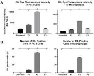
The formulations were tested for their ability to be taken up by PC-3 cells and macrophages (Figure 1) using flow cytometry. PC-3 cells treated with F2 showed the highest fluorescence intensity, followed by the cells treated with F1. The cells treated with F3, which contained DOPE as the main lipid, had similar fluorescence intensity as the untreated cells. F2 was also taken up most effectively by macrophages. In contrast, the macrophages treated with F1 and F3 had low DIL dye fluorescence intensity, comparable to untreated cells. Overall, F2, which was prepared using egg PG, was taken up most effectively by both cell types.

Figure 1. Uptake of Liposome by PC-3 and Macrophage Cells Quantified

VROJ-5-117Fig1

(A) DIL intensity and (B) the number of DIL positive cells as detected by flow cytometry. Each assay was carried out in triplicate and 10,000 events were collected for each measurement. Error bars represent standard deviation. Statistical analysis was performed using one-way ANOVA followed by Tukey’s post hoc multiple comparison test (****, p<0.0001).

Uptake of Liposome Vesicles in PC-3 Cells and Macrophages
Visualised through Confocal Microscopy

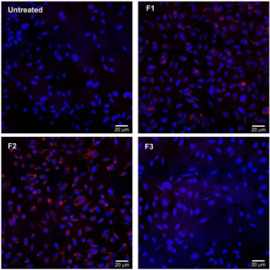
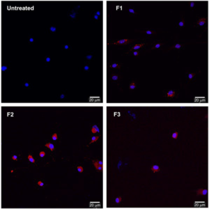
Formulation uptake in PC-3 cells and macrophages was also assessed using confocal microscopy (Figures 2 and 3) as a nonquantitative assay to visualise cell uptake. F1 and F2 were effectively taken up by both PC-3 cells and macrophages.

Figure 2. Microscopy Images of the Cellular Uptake of F1–F3 in PC-3 Cells and with 40x Magnification and the Use of a Green Filter (500 to 570 nm) (excitation: 549 nm; emission: 565 nm). The Nucleus of the Cells was Stained Blue Using Hoechst Solution and the Liposome Vesicles were Stained Red with DIL Dye. The Assay was Carried Out in Triplicate

VROJ-5-117Fig2

Figure 3. Confocal Microscopy Images of the Cellular Uptake of F1- F3 in Macrophages with 40x Magnification and the Use of a Green Filter (500 to 570 nm) (excitation: 549 nm; emission: 565 nm). The Nucleus of the Cells was Stained Blue Using Hoechst Solution and the Liposome Vesicles were Stained Red with DIL Dye. The Assay was Carried out in Triplicate

VROJ-5-117Fig3

DISCUSSION

The three liposome formulations were designed to bear positive, negative and neutral charges. The liposome formulations were characterised and analysed to determine their ability to be taken up by two different cells lines: PC-3 cells, which are a commonly used prostate cancer cell line, and macrophages, as representative cells used for targeting delivery of drugs (e.g. anti-TB). Macrophages are antigen-presenting cells (APCs) and therefore are also crucial for vaccine delivery.26,27 Liposomes can deliver their cargo directly to the cytoplasm of APCs and therefore allow antigen processing and loading on major histocompatibility complex-I (MHC-I). These antigen fragments, known as CD8 epitopes, presented by MHC-I activate cytotoxic T-lymphocytes and cellular immunity in general. Cellular immunity is responsible for immune defence against intracellular pathogens such as viruses and Mtb.28-30 Consequently, liposomes have been widely used for vaccine delivery including subunit vaccines against TB.31-34 Liposomes have also been proposed as a secure drug delivery platform for the treatment of cancers, including prostate cancer.35,36,37

Different lipids were used in the liposome formulations, which could have affected vesicle uptake. The high uptake of F2 could be related to the presence of egg PG. Liposomes are taken up by macrophages through a receptor-mediated process.44,45 Specifically, it was observed that alveolar macrophages have receptors that have higher affinities for liposome vesicles prepared with PG in comparison to phosphatidylcholine (PC).46,47 Therefore, the higher uptake of PG vesicles (F2) compared to PC vesicles (F1) by mouse macrophages in this study is consistent with previous reports.

CONFLICTS OF INTEREST

The authors declare that they have no conflicts of interest.

CONCLUSION

Liposomes have been utilised for the delivery of many biologically active compounds to improve their cellular uptake and reduce toxicity. Liposomal delivery is known to increase the therapeutic index by increasing drug biocompatibility, decreasing drug toxicity and reducing drug exposure to healthy cells. However, many challenges still exist in developing optimised liposome delivery systems for clinical use. Therefore, this study aimed to develop a series of liposome delivery systems for drugs and vaccines. We determined that small, negatively charged liposomes, prepared with egg PG, had high internalisation potential in prostate cancer cell line and macrophages. Further analysis and characterisation of liposomes bearing a drug or an antigen is required to determine the real efficacy of the developed formulations.

1. Bozzuto, G, Molinari, A Liposomes as nanomedical devices. Int J Nanomedicine. 2015; 10: 975-999. doi: 10.2147/Ijn.S68861

2. Bangham A. The 1st description of liposomes – a citation classic commentary on Diffusion of Univalent Ions across the Lamellae of Swollen Phospholipids by Bangham AD, Standish MM, Watkins JC. Current Contents/Life Sciences. 1989(13): 14. Website. http:// garfield.library.upenn.edu/classics1989/A1989T670000001.pdf. Accessed July 5, 2020.

3. Azadi A, Ashrafi H. Cell organelle-shaped liposomes: A novel approach to present the stable intracellular drug delivery systems. Trends in Pharmaceutical Sciences. 2016; 2(2): 85-88.

4. Torchilin VP. Recent advances with liposomes as pharmaceutical carriers. Nature Reviews Drug Discovery. 2005; 4(2): 145-160. doi: 10.1038/nrd1632

5. Akbarzadeh A, Rezaei-Sadabady R, Davaran S, Joo SW, Zarghami N, Hanifehpour Y, et al. Liposome: Classification, preparation, and applications. Nanoscale Res Lett. 2013; 8(1): 102. doi: 10.1186/1556- 276X-8-102

6. Çağdaş M, Sezer AD, Bucak S. Liposomes as potential drug carrier systems for drug delivery. In: Application of Nanotechnology in Drug Delivery. 2014. doi: 10.5772/58459

7. Allen TM, Cullis PR. Liposomal drug delivery systems: From concept to clinical applications. Advanced Drug Delivery Reviews. 2013; 65(1): 36-48. doi: 10.1016/j.addr.2012.09.037

8. Li J, Wang, XL, Zhang, T, Wang C, Huang Z, Luo X, et al. A review on phospholipids and their main applications in drug delivery systems. Asian Journal of Pharmaceutical Sciences. 2015; 10(2): 81-98. doi: 10.1016/j.ajps.2014.09.004

9. Lian, T, Ho, RJ Trends and developments in liposome drug delivery systems. J Pharm Sci. 2001; 90(6): 667-680. doi: 10.1002/ jps.1023

10. Sofou, S, Sgouros, G Antibody-targeted liposomes in cancer therapy and imaging. Expert Opin Drug Deliv. 2008; 5(2): 189-204. doi: 10.1517/17425247.5.2.189

11. Sawant, RM, Hurley, JP, Salmaso, S, Kale A, Tolcheva E, Levchenko TS, et al. “SMART” drug delivery systems: Double-targeted pH-responsive pharmaceutical nanocarriers. Bioconjug Chem. 2006; 17(4): 943-949. doi: 10.1021/bc060080h

12. Pasut, G, Paolino, D, Celia, C, Mero A, Joseph AS, Wolfram J, et al. Polyethylene glycol (PEG)-dendron phospholipids as innovative constructs for the preparation of super stealth liposomes for anticancer therapy. J Control Release. 2015; 199: 106-113. doi: 10.1016/j.jconrel.2014.12.008

13. Pasut G, Veronese FM. State of the art in PEGylation: the great versatility achieved after forty years of research. J Control Release 2012; 161(2): 461-472. doi: 10.1016/j.jconrel.2011.10.037

14. Harris JM, Chess RB. Effect of pegylation on pharmaceuticals. Nat Rev Drug Discov. 2003; 2(3): 214-221. doi: 10.1038/nrd1033

15. Magarkar A, Dhawan V, Kallinteri, P, Viitala T, Elmowafy M, Róg T, et al. Cholesterol level affects surface charge of lipid membranes in saline solution. Sci Rep. 2014; 4: 5005. doi: 10.1038/ srep05005

16. Yan X, Scherphof GL, Kamps JA. Liposome opsonization. J Liposome Res. 2005; 15(1-2): 109-139. doi: 10.1081/lpr-64971

17. Henao-Tamayo, M, Shanley, CA, Verma, D, Zilavy A, Stapleton MC, Furney SK, et al. The efficacy of the BCG vaccine against newly emerging clinical strains of mycobacterium tuberculosis. PLoS One. 2015; 10(9): e0136500. doi: 10.1371/journal. pone.0136500

18. Tameris, MD, Hatherill, M, Landry, BS, Scriba TJ, Snowden MA, Lockhart S, et al. Safety and efficacy of MVA85A, a new tuberculosis vaccine, in infants previously vaccinated with BCG: A randomised, placebo-controlled phase 2b trial. Lancet. 2013; 381(9871): 1021-1028. doi: 10.1016/S0140-6736(13)60177-4

19. Piccolo, M, Misso, G, Ferraro, MG, Riccardi H, Capuozzo A, Zarone MR, et al. Exploring cellular uptake, accumulation and mechanism of action of a cationic Ru-based nanosystem in human preclinical models of breast cancer. Sci Rep. 2019; 9(1): 7006. doi: 10.1038/s41598-019-43411-3

20. Skwarczynski M, Hayashi Y, Kiso Y. Paclitaxel prodrugs: Toward smarter delivery of anticancer agents. J Med Chem. 2006; 49(25): 7253-7269. doi: 10.1021/jm0602155

21. Litwin MS, Tan HJ. The diagnosis and treatment of prostate cancer: A review. JAMA. 2017; 317(24): 2532-2542. doi: 10.1001/jama.2017.7248

22. Hema S, Thambiraj S, Shankaran DR. Nanoformulations for targeted drug delivery to prostate cancer: An overview. J Nanosci Nanotechnol. 2018; 18(8): 5171-5191. doi: 10.1166/jnn.2018.15420

23. Serradell, MC, Rupil, LL, Martino, RA, Prucca CG, Carranza PG, Saura, A et al. Efficient oral vaccination by bioengineering virus-like particles with protozoan surface proteins. Nat Commun. 2019; 10(1): 361. doi: 10.1038/s41467-018-08265-9

24. Samad A, Sultana Y, Aqil M. Liposomal drug delivery systems: an update review. Curr Drug Deliv. 2007; 4(4): 297-305. doi: 10.2174/156720107782151269

25. Kaighn, ME, Narayan, KS, Ohnuki, Y, Lechner JF, Jones LW. Establishment and characterization of a human prostatic carcinoma cell line (PC-3). Invest Urol. 1979; 17(1): 16-23.

26. Skwarczynski M, Toth I. Peptide-based synthetic vaccines. Chem Sci. 2016; 7(2): 842-854. doi: 10.1039/c5sc03892h

27. Skwarczynski, M, Zhao, GZ, Boer, JC, Ozberk V, Azuar A, Cruz JG, et al. Poly(amino acids) as a potent self-adjuvanting delivery system for peptide-based nanovaccines. Sci Adv. 2020; 6(5): eaax2285. doi: 10.1126/sciadv.aax2285

28. Thakur A, Mikkelsen H, Jungersen GIntracellular pathogens: Host immunity and microbial persistence strategies. J Immunol Res. 2019; 2019: 1356540. doi: 10.1155/2019/1356540

29. Al-Attiyah R, El-Shazly A, Mustafa AS. Comparative analysis of spontaneous and mycobacterial antigen-induced secretion of Th1, Th2 and pro-inflammatory cytokines by peripheral blood mononuclear cells of tuberculosis patients. Scand J Immunol. 2012; 75(6): 623-632. doi: 10.1111/j.1365-3083.2012.02692.x

30. Umeshappa, CS, Xie, Y, Xu, S, Nanjundappa RH, Freywald A, Deng Y, et al. Th cells promote CTL survival and memory via acquired pMHC-I and endogenous IL-2 and CD40L signaling and by modulating apoptosis-controlling pathways. PLoS One. 2013; 8(6): e64787. doi: 10.1371/journal.pone.0064787

31. Olusanya, TOB, Haj Ahmad, RR, Ibegbu, DM, Smith JR, Elkordy AA. Liposomal drug delivery systems and anticancer drugs. Molecules. 2018; 23(4): 907. doi: 10.3390/molecules23040907

32. Kroon, J, Metselaar, JM, Storm, G, van der Pluijm G. Liposomal nanomedicines in the treatment of prostate cancer. Cancer Treat Rev. 2014; 40(4): 578-584. doi: 10.1016/j.ctrv.2013.10.005

33. Zhang, W, Song, Y, Eldi, P, Guo X, Hayball JD, Garg S, et al. Targeting prostate cancer cells with hybrid elastin-like polypeptide/liposome nanoparticles. Int J Nanomedicine. 2018; 13: 293-305. doi: 10.2147/IJN.S152485

34. Khademi, F, Taheri, RA, Momtazi-Borojeni, AA, Farnoosh G, Johnston TP, Sahebkar A. Potential of cationic liposomes as adjuvants/delivery systems for tuberculosis subunit vaccines. Rev Physiol Biochem Pharmacol. 2018; 175: 47-69. doi: 10.1007/112_2018_9

35. Marasini N, Ghaffar KA, Skwarczynski M, Toth I. Liposomes as a vaccine delivery system. In: Micro and Nanotechnology in Vaccine Development. Norwich, NY, USA: William Andrew Inc; 2017: 221- 239. doi: 10.1016/B978-0-323-39981-4.00012-9

36. Ghaffar KA, Marasini N, Giddam AK, Batzloff MR, Good MF, Skwarczynski M, et al. Liposome-based intranasal delivery of lipopeptide vaccine candidates against group A streptococcus. Acta Biomater. 2016; 41: 161-168. doi: 10.1016/j.actbio.2016.04.012

37. Ghaffar, KA, Giddam, AK, Zaman, M, Skwarczynski M, Toth I. Liposomes as nanovaccine delivery systems. Current Topics in Medicinal Chemistry. 2014; 14(9): 1194-1208. doi: 10.2174/1568026614 666140329232757

38. Zhang M, Desai T, Ferrari M. Proteins and cells on PEG immobilized silicon surfaces. Biomaterials. 1998; 19(10): 953-60. doi: 10.1016/s0142-9612(98)00026-x

39. Wischerhoff, E, Uhlig, K, Lankenau, A, Börner HG, Laschewsky A, Duschl C, et al. Controlled cell adhesion on PEG-based switchable surfaces. Angew Chem Int Ed Engl. 2008; 47(30): 5666- 568. doi: 10.1002/anie.200801202

40. Immordino, ML, Dosio, F, Cattel, L Stealth liposomes: review of the basic science, rationale, and clinical applications, existing and potential. Int J Nanomedicine. 2006; 1(3): 297-315.

41. Sanchez L, Yi Y, Yu Y. Effect of partial PEGylation on particle uptake by macrophages. Nanoscale. 2017; 9(1): 288-297. doi: 10.1039/c6nr07353k

42. Takano S, Aramaki Y, Tsuchiya S. Physicochemical properties of liposomes affecting apoptosis induced by cationic liposomes in macrophages. Pharm Res. 2003; 20(7): 962-968. doi: 10.1023/a:1024441702398

43. Epstein-Barash H, Gutman D, Markovsky E, Mishan-Eisenberg G, Koroukhov N, Szebeni J, et al. Physicochemical parameters affecting liposomal bisphosphonates bioactivity for restenosis therapy: Internalization, cell inhibition, activation of cytokines and complement, and mechanism of cell death. J Control Release. 2010; 146(2): 182-195. doi: 10.1016/j.jconrel.2010.03.011

44. Straubinger, RM, Hong, K, Friend, DS, Papahadjopoulos D. Endocytosis of liposomes and intracellular fate of encapsulated molecules: Encounter with a low pH compartment after internalization in coated vesicles. Cell. 1983; 32(4): 1069-1079. doi: 10.1016/0092-8674(83)90291-x

45. Muller, WJ, Zen, K, Fisher, AB, Shuman H. Pathways for uptake of fluorescently labeled liposomes by alveolar type II cells in culture. Am J Physiol. 1995; 269(1 Pt 1): L11-L19. doi: 10.1152/ ajplung.1995.269.1.L11

46. Poelma, DL, Ju, MR, Bakker, SC, Zimmermann LJ, Lachmann BF, van Iwaarden JF. A common pathway for the uptake of surfactant lipids by alveolar cells. Am J Respir Cell Mol Biol. 2004; 30(5): 751-758. doi: 10.1165/rcmb.2003-0127OC

47. Quintero OA, Wright JR. Metabolism of phosphatidylglycerol by alveolar macrophages in vitro. Am J Physiol Lung Cell Mol Physiol. 2000; 279(2): L399-L407. doi: 10.1152/ajplung.2000.279.2.L399

LATEST ARTICLES

Chest X-ray Showed a Hazy Left Upper Lung Infiltrate

A Noteworthy Case of Myasthenic Crisis Induced by Levofloxacin

Ada Young*, Ramya Ramesh and Milind Awale

doi.

The Right Thigh Anterior Compartment was Swollen, and the Skin was Ulcerated due to the Traditional Cautery

Primary Skeletal Muscle Lymphoma: A Case Report and Literature Review

Solomon Bishaw*, Addisu Alemu and Abel Tefera

doi.

An Unusual Presentation of Encephalitis in a Patient with Lyme Neuroborreliosis

Maithily Patel*, Jazmin Jatana, Ramya Ramesh and Milind Awale

doi.

Practical Pointers for Drug Development and Medical Affairs

Gerald L. Klein*, Roger E. Morgan, Shabnam Vaezzadeh, Burak Pakkal and Pavle Vukojevic

doi.

10.17140/CTPOJ-7-125

Prevalence and Risk Factors of Subclinical Mastitis of Goats in Banadir Region, Somalia

Omar M. Salah*, Yasin H. Sh-Hassan, Moktar O. S. Mohamed, Mohamed A. Yusuf and Abas S. A. Jimale

doi.10.17140/VMOJ-9-184

LATEST ARTICLES