Advances in Food Technology and Nutritional Sciences

Open journal

ISSN 2377-8350

Leptin Activates NLRP3 Inflammasome-Associated with Type II Diabetes and Obesity

R. Cauble, S. Hamad, E. Herring, C. Lichtenwalter, C. McGuire and Sami Dridi*

Sami Dridi, PhD

Center of Excellence for Poultry Science, University of Arkansas, 1260 W, Maple Street, Fayetteville AR 72701, USA; Tel. (479)-575-2583; Fax: (479)-575-7139; E-mail: dridi@uark.edu

 Obesity is a major health problem worldwide and is often associated with leptin resistance and inflammation.1 In this editorial, we will briefly describe the leptin system and the (NOD)-like receptor protein 3 (NLRP3) inflammasome and discuss recent discoveries related to their interaction and role in the development of metabolic disease mainly obesity and Time-division multiplexing (TDM).

LEPTIN

Leptin is a hormone mainly secreted by adipocytes and is known for its role in long-term energy regulation, food intake, and body weight. As a hormone, its primary responsibility is to report the amount of adipose tissue in the body to the hypothalamus.2 Leptin concentrations in both blood serum and plasma are elevated in direct correlation with a high body mass index (BMI) and percent body fat. A constant elevation of leptin in the body, caused by sustained over eating habits, overloads the hypothalamus and results in leptin resistance; like we observe in obese subjects.3

This resistance is a major factor in causing the chronic inflammatory diseases seen frequently in obese individuals such as asthma, diabetes, and inflammatory bowel disease. In fact, during the active stages of rheumatoid arthritis (an additional condition related to chronic inflammation), leptin levels are elevated. On the other hand, malnourished individuals with decreased leptin levels suffered more infectious diseases, but had significantly lower rates of inflammation. These results are believed to be an indication of leptin’s role in activating the body’s inflammatory immune responses, resulting in these chronic conditions.4

Leptin also operates similarly to pro-inflammatory cytokines known as adipocytokines.4 The hormone has been reported to control energy expenditure and metabolism, and modulate the innate and adaptive immune responses. The stimulation of natural killer cells, chemotaxis of neutrophils, and secretion of tumor necrosis factor (TNF)-α, IL-6 and IL-12 from macrophages4,5 also involves leptin. These increase leptin concentrations in adipose cells and create a cycle of constant stimulation of reactants that encourage inflammation.4

Furthermore, leptin promotes Th17 cell responses6 which are a subset of effector memory T-cells that induce tissue inflammation and destruction that are markers of immune inflammatory diseases7 and decrease the number of T-regulatory cells.8,9 Fat stores release leptin around various lymph nodes signaling to the rest of the immune system if the body has enough energy stored to initiate an immune response which causes continuous leptin circulation in obese or individuals with type II diabetes.3

NLRP3 INFLAMMASOMES

The nucleotide binding domain and leucine-rich repeat containing receptor (NLR) family are proteins that form inflammasomes.10,11 The NLRs are classified and named in accordance with their domain structure.11 The nucleotide-binding oligomerization domain-like receptor family pyrin domain-containing 3 (NLRP3) inflammasome is mainly expressed in macrophages and has a pivotal role in development and maintenance of autoimmunity and inflammation. Research has investigated the role of leptin on NLRP3 inflammasome and found that leptin is an activator and modulator of this inflammasome.12

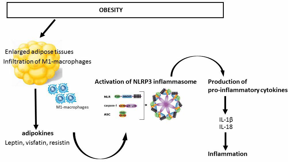
Activation of the NLRP3 inflammasome (Figure 1) is thought to be a two-step process.13 The initial step is a priming step. When exposed to pathogen-associated molecular patterns (PAMPs) or danger associated molecular patterns (DAMPs), phosphorylation of toll-like receptors occurs and NF-κB is activated.14 NF-κB causes an increase in transcription of inactivated forms of NLRP3, proIL-1β, and proIL-18.15 A secondary stimulus causes activation of the inflammasome by oligomerizing inactive NLRP3, apoptosis-associated speck-like protein, and procaspase-1. This structure catalyzes the modification of procaspase-1 to caspase-1. This conversion contributes to the production of functional IL-1β and IL-18.16 The activation of the inflammasome in the second step has been proposed using three different models; potassium efflux as induced by extracellular ATP, the generation of reactive oxygen species (ROS) via PAMP and DAMP, and crystalline structures causing lysosomal rupture and the release of its contents including cathepsin B.17

Figure 1. Potential Interaction between Leptin and NLRP3 Inflammasome in Obesity Pathogenesis As fat cells enlarge when body weight increases (progress from lean to overweight to obese), oxygen availability to cells declines which leads to infiltration of type 1 macrophages. This in turn activates NLRP3 inflammsome and production of mature proinflammatory cytokines (IL-1β and IL-18) leading to inflammation

AFTNSOJ-4-e016Fig1


Leptin promotes IL-18 secretion by activating caspase-1. Caspase-1 in conjunction with NLRP3 inflammasomes regulates the production and secretion of IL-18 via proteolyticall digestion pro-IL-18.8,18 More specifically, the IL-18 promotion via leptin is done so by enhancing reactive oxygen species (ROS) synthesis and K+ efflux. This relationship activates the NLRP3 inflammasome.12

The role of NLRP3 inflammasome in metabolic syndrome and type II diabetes can be split into two subcategories which include mediated roles by sensing endogenous inflammasome activators and indirect roles by inflammasome associated alteration by manipulation of the gut microbiota.19 There are multiple mechanisms that have been investigated that could potentially activate the NLRP3 inflammasome in high fat induced diets; one of which includes a pancreatic hormone that is co-secreted with insulin and triggers IL-1ß secretion by isolate macrophages.20

OBESITY AND TYPE II DIABETES Improvements Inflammation is a player in the pathogenesis of obesity. The chronic overfeeding associated with obesity causes macrophage saturation in the adipose tissue and results in proinflammatory cytokine production.21 This endogenous signaling triggers the intracellular innate immune NLRP3 sensor, results in caspase-1 activation and the production of IL-1ß and IL-18.21 These cytokines are directly related to the development of insulin resistance that we observe in type II diabetes.16,22 Specifically, IL-1ß inhibits adipocyte differentiation23,24 while the absence of IL-18 induces obesity and insulin resistance.25,26 Conversely, the absence of the NLRP3 inflammasome has been shown inhibit the development of obesity-induced insulin resistance27, which would suggest that the inflammasome is a contributor.

Different immune cells, including proinflammatory macrophages, have been shown to penetrate the adipose tissue (AT) and affect its homeostasis by increasing the production of cytokines such as IL-1β, IL-6 and TNF. Macrophages and other innate immune cells can promote inflammatory reactions through detection of pathogen- or danger –associated molecular patterns (PAMPs or DAMPs) using a variety of pattern-recognition receptors (PRRs). One type of PRRs identified are nucleotide-binding oligomerization domain-like receptors (NLRs), specifically looking at NLRP3.28

When PAMPs or DAMPs are activated the NLRP3 interacts with the adapter protein apoptosis-associated speck-like protein (ASC). Then, the caspase recruitment domain (CARD) of ASC binds to the CARD domain on procaspase-1, forming the NLRP3 inflammasome.28 This causes procaspase-1 self-cleavage, creating the active caspase-1, which leads to the conversion of IL-1β and IL-18 immature forms to their active forms that are secreted. NLRP3 inflammasome-activated IL-1β has a vital role in the development of obesity-induced insulin resistance (IR) and type 2 diabetes mellitus (T2DM).

Research involving the relationship between obesity and NLRP3 are working towards finding therapies to decrease obesity along with decreasing the expression of NLRP3. Research has found decreased NLRP3 and IL-1β expressions in subcutaneous adipose tissue (SAT) from T2DM patients after a year of calorie restriction and exercise-mediated weight loss. Scientists have also found that there is a relationship between nutrient excess and inflammation from the initiation of the NLRP3 inflammasome by dietary free fatty acids (FFAs) which are DAMPs. In mice research, scientists have found that high fat diets (HFDs) increase NLRP3 expression in AT, but found that calorie-restricted diets will often decrease the expression of this gene.28 Further research will be needed to find a solution for the human model, but in mice models, the ablation of NLRP3 protected the mice against HFDinduced obesity and IR as well as decreasing the blood glucose and insulin levels.


1. Pan H, Guo J, Su Z. Advances in understanding the interrelations between leptin resistance and obesity. Pysiol Behav. 2014; 130: 157-169. doi: 10.1016/j.physbeh.2014.04.003

2. Allison MB, Myers MG. 20 Years or leptin: Connecting leptin signaling to biological function. J. Endrocrinol. 2014; 223(1): T25- T35. doi: 10.1530/JOE-14-0404

3. Drent ML, Klok MD, Jakobsdottir S. The role of leptin and ghrelin in the regulation of food intake and body weight in humans: A review. Obes Rev. 2007; 8(1): 21-34. doi: 10.1111/j.1467- 789X.2006.00270.x

4. Iikuni N, Lam QLK, Lu L, et al. Leptin and Inflammation. Curr Immunol Rev. 2008; 4(2): 70-79. doi: 10.2174/157339508784325046

5. Groß CJ, Mishra R, Schneider KS, et al. K+ Efflux-Independent NLRP3 Inflammasome activation by small molecules targeting mitochondria. Immunity. 2016; 45: 761-773. doi: 10.1016/j.immuni.2016.08.010

6. Mortimer L,Moreau F, MacDonald JA, Chadee K. NLRP3 inflammasome inhibition is disrupted in a group of auto-inflammatory disease CAPS mutations. Nat Immunol. 2016; 17(10): 1176- 1186. doi: 10.1038/ni.3538

7. Shuttleworth S, Townsend P, Silva F, et al. Progress in the development of small molecule therapeutics targeting Th17 cell function for the treatment of immune-inflammatory diseases. Prog Med Chem. 2011; 50: 109-133. doi: 10.1016/B978-0-12-381290- 2.00003-3

8. Fu S, Xu L, Li S, et al. Baicalin suppresses NLRP3 inflammasome and nuclear factor-kappa B (NF-κB) signaling during Haemophilus parasuis infection. Vet Res. 2016; 47(1): 80. doi: 10.1186/s13567- 016-0359-4

9. De Rosa V, Procaccini VC, Cali G, et al. A key role of leptin in the control of regulatory T cell proliferation. Immunity. 2007; 26(2): 241-255. doi: 10.1016/j.immuni.2007.01.011

10. Reed JC, Doctor K, Rojas A, et al. Comparative analysis of apoptosis and inflammation genes of mice and humans. Genome Res. 2003; 13(6B): 1376-1388. doi: 10.1101/gr.1053803

11. Ting JP, Lovering RC, Alnemri ES, et al. The NLR gene family: A standard nomenclature. Immunity. 2008; 28(3): 285-287. doi: 10.1016/j.immuni.2008.02.005

12. Fu S, Lei L, Lei H, Yiyun Y. Leptin promotes IL-18 secretion by activating the NLRP3 inlammasome in RAW 264.7 cells. Mol Med Rep. 2017; 16(6): 9770-9776. doi: 10.3892/mmr.2017.7797

13. Zhong, Yifei, Anna Kinio, and Maya Saleh. Functions of NOD-like receptors in human disease. Front Immunol. 2013; 4: 333. doi: 10.3389/fimmu.2013.00333

14. Bo-Zong S, Zhe-Qi X, Bin-Ze H, et al. NLRP3 inflammasome and its inhibitors: A review. Front Pharmacol. 2015; 6: 262. doi: 10.3389/fphar.2015.00262

15. Franchi L, Eigenbrod T, Munoz-Panillo R, et al. Cytosolic double-stranded RNA activates the NLRP3 inflammasome via MAVSinduced membrane permeabilization and K+ efflux. J Immunol. 2014; 193(8): 4214-4222. doi: 10.4049/jimmunol.1400582

16. Ozaki E, Campbell M, Doyle SL. Targeting the NLRP3 inflammasome in chronic inflammatory diseases: Current perspectives. J Inflamm Res. 2015; 8: 15-27. doi: 10.2147/JIR.S51250

17. Schroder K. Tschopp J. The inflammasomes. Cell. 2010; 140(6): 821-832. doi: 10.1016/j.cell.2010.01.040

18. Gross O, Thomas CJ, Guarda G, Tschopp J. The inflammasome: An integrated view. Immunol Rev. 2011; 243(1): 136-151. doi: 10.1111/j.1600-065X.2011.01046.x

19. De Zoete MR, Palm NW, Zhu S, Flavell RA. Inflammasomes. Cold Spring Harb Perspect Biol. 2014; 6(12): 1-22. doi: 10.1101/cshperspect.a016287

20. Masters SL, Dunne A, Subramanian SL, et al. Activation of the NLRP3 inflammasome by islet amyloid polypeptide provides a mechanism for enhanced IL-1β in type 2 diabetes. Nat Immunol. 2010; 11(10): 897-904. doi: 10.1038/ni.1935

21. Stienstra R, Tack CJ, Kanneganti TD, et al. The Inflammasome Puts Obesity in the Danger Zone. Cell Metab. 2012; 15:10-18. doi: 10.1016/j.cmet.2011.10.011

22. Olefsky JM, Glass CK. Macrophages, inflammation, and insulin resistance. Annu Rev Physiol. 2010; 72: 219-246. doi: 10.1146/ annurev-physiol-021909-135846

23. Shoelson SE, L. Herrero, and A. Naaz. Obesity, inflammation, and insulin resistance. Gastroenterology. 2007; 132(6): 2169-2180. doi: 10.1053/j.gastro.2007.03.059

24. Jager, J, Gremeaux T, Cormont M, et al. Interleukin-1 betainduced insulin resistance in adipocytes through down-regulation of insulin receptor substrate-1 expression. Endocrinology. 2007; 148: 241-251 doi: 10.1210/en.2006-0692

25. Netea MG, Joosten LA, Lewis E, et al. Deficiency of interleukin-18 in mice leads to hyperphagia, obesity and insulin resistance. Nat Med. 2006; 12(6): 650-656. doi: 10.1038/nm1415

26. Zorrilla EP, Sanchez-Alavez M, Sugama S, et al. Interleukin-18 controls energy homeostasis by suppressing appetite and feed efficiency. Proc Natl Acad Sci USA. 2007; 104(26): 11097-11102. doi: 10.1073/pnas.0611523104

27. Vandanmagsar B, Youm YH, Ravussin A, et al. The NLRP3 inflammasome instigates obesity-induced inflammation and insulin resistance. Nat Med. 2011; 17(2): 179-188. doi: 10.1038/nm.2279

28. Rheinheimer J, De Souza BM, Cardoso NS, et al. Current role of the NLRP3 inflammasome on obesity and insulin resistance: A systemic review. Metabolism. 2017; 74: 1-9. doi: 10.1016/j.metabol.2017.06.002

LATEST ARTICLES

Practical Pointers for Drug Development and Medical Affairs

Gerald L. Klein*, Roger E. Morgan, Shabnam Vaezzadeh, Burak Pakkal and Pavle Vukojevic

doi.

10.17140/CTPOJ-7-125

Prevalence and Risk Factors of Subclinical Mastitis of Goats in Banadir Region, Somalia

Omar M. Salah*, Yasin H. Sh-Hassan, Moktar O. S. Mohamed, Mohamed A. Yusuf and Abas S. A. Jimale

doi.10.17140/VMOJ-9-184

Use of Black Soldier Fly (Hermetia illucens) Prepupae Reared on Organic Waste

Maggot Debridement Therapy: A Natural Solution for Wound Healing

Isayas A. Kebede*, Haben F. Gebremeskel and Gelan D. Dahesa,

doi.10.17140/VMOJ-9-183

Figure 11. Risk Map for the Introduction of Ruminant Diseases at Borders

Ovine Network in Morocco: Epizootics Spread Prevention and Identification of the At-Risk Areas for “Peste des Petits Ruminants” and “Foot and Mouth Disease”

Yassir Lezaar*, Mehdi Boumalik, Youssef Lhor, Moha El-Ayachi, Abelilah Araba and Mohammed Bouslikhane

doi.

10.17140/EPOJ-8-131

The Impact of Family Dynamics on Palliative Care at the End-of-Life

Neil A. Nijhawan*, Rasha Mustafa and Aqeela Sheikh

doi.10.17140/PMHCOJ-10-154

Long-Term Follow-Up After Laparoscopic Radical Prostatectomy for Localized and Locally Advanced Prostate Cancer

Shrenik J. Shah*, Abhishek Jha, Chirag Davara, Rushi Mistry and Kapil Kachhadiya

doi.

10.17140/UAOJ-7-147

LATEST ARTICLES

Pie Chart Showing Overall Proportions of Diagnostic Category of FNAC, JUMC

Retrospective Study

2024 Apr

Abel Tefera*, Lemlem Terefe and Kitesa Biresa
Prevalence (%) of Types of Anthropometric Failure among Previous and Present Studied Tribal Children

Original Research, peer reviewed

2024 Apr

Biswajit Mahapatra and Kaushik Bose*

Opinion

2024 Apr

Gerald L. Klein*, Roger E. Morgan, Shabnam Vaezzadeh, Burak Pakkal and Pavle Vukojevic