Advances in Food Technology and Nutritional Sciences

Open journal

ISSN 2377-8350

Insights into the Immune System and Glaucoma

Jennifer L. Kielczewski*

Received: September 1st, 2017; Accepted: October 4th, 2017; Published: October 9th, 2017

THE IMMUNE SYSTEM HAS BEEN IMPLICATED IN THE PATHOGENESIS OF GLAUCOMA

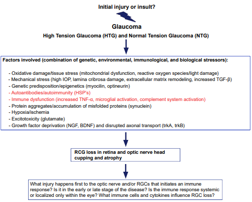
Glaucoma is a neurodegenerative disease of the optic nerve characterized by progressive loss of retinal ganglion cells (RGCs), which can lead to irreversible blindness. Numerous factors have been implicated in the disease, with high intraocular pressure (IOP), coupled with advanced age, being major risk factors. Other factors include ischemia, generation of reactive oxygen species, a genetic pre-disposition and neurotrophin deprivation in the retina or optic nerve (Figure 1).1 The pathogenesis of glaucoma is challenging to understand since it is a multifactorial neurodegenerative disease.2,3 It is even more confounding what causes normal tension glaucoma (NTG). Despite a normal IOP, patients still suffer optic nerve degeneration. The immune system is a probable player in both high tension glaucoma (HTG) and normal tension glaucoma (NTG). Findings from numerous studies support the notion that both the innate and adaptive immune responses are involved in the pathogenesis of glaucoma.1,3-18 Yet, the precise mechanisms of immune responses and the specific cell interactions contributing to the disease process are still not fully understood. Many questions remain about the role of the immune system in glaucoma (Figure 1).

Figure 1: Diagram Shows Factors Contributing to Degeneration of RGCs and the Pathogenesis of Glaucoma. The Pathogenesis of Glaucoma is Multifactorial. It is still unknown which Initial Injury or Damaging Insult Leads to the Onset of Glaucoma and Progression of the Neurodegenerative Disease. The Immune System is Likely to Play a role in Glaucoma, Yet Many Questions Remain about the Timing, Duration, and Where Inflammatory Responses Occur.

OOJ-2-112Fig1

Low-level inflammation is important for biological homeostasis. Immune responses are necessary for proper tissue cleaning, maintenance, and repair.19,20 It has also been proposed that protective or beneficial autoimmunity may help protect against neurodegeneration in the central nervous system (CNS).11,21-25 This concept suggests that the immune system plays a key role in the ability of the CNS, like the optic nerve and retina, to withstand neurodegeneration, by recruiting both innate (resident and blood-borne macrophages) and adaptive (self-antigen specific T-cells) cells that together promote a protective niche and hinder disease progression under a wellcontrolled response.23,24,26-29 However, excessively uncontrolled immune stimulation can lead to a neurotoxic environment in the optic nerve and retina, resulting in the neurodegeneration of RGCs. Immune dysregulation and immune activation in glaucoma pathogenesis are the focus of many studies in both experimental animal models and in human clinical studies.14,20,30

LIMITATIONS OF STUDYING IMMUNE RESPONSES IN EXPERIMENTAL GLAUCOMATOUS ANIMAL MODELS

Experimental glaucoma animal models are quite valuable.31,32 They can be useful in studying the early changes in pathological and molecular changes in the optic nerve and retina. Since glaucoma is considered “the thief of sight” it can go undetected for many years in patients, making it hard to study in the clinic. Likewise, post-mortem eye tissue from human patients is typically limited to late onset and highly diseased glaucoma eyes, while early onset diseased eyes are not abundantly available for research studies. Studying glaucoma in humans is complicated; therefore, animal models are beneficial in dissecting out molecular mechanisms, especially for immune dysregulation and immune responses involved in the early stages of RGC injury.

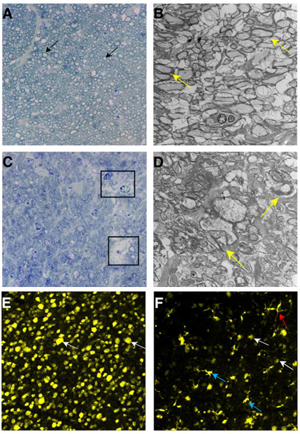
There are several experimental animal models for glaucoma.31,32 Some are based on chemical injury, such as intravitreal injection of neurotoxic concentrations of glutamate, while others utilize elevated pressure with laser injury to the trabecular meshwork or intravitreal injection of microbeads to raise IOP (injection of foreign beads into eye may not be an ideal glaucoma model to study immune responses). There are also animal models for mechanical-induced injury, where the optic nerve is transected or the optic nerve is crushed (Figure 2). Genetic models also exist such as the DBA/2J.5 But all of these models have limitations, especially with studying immune system effects. No animal model can fully recapitulate human glaucoma due to its heterogeneous nature and each model can provide different insight into immune mechanisms and responses. The type of injury, whether biochemical or mechanical, may dictate a different immune response at a different time and location and may not only depend upon the severity, but also the chronicity of the injury. Moreover, some RGC cell types may be more susceptible to a particular injury; hence, certain RGCs may be more prone to immune attack or destruction than others. The immune responses are likely to be different in animal models, where IOP is experimentally raised versus in animals subjected to a non-pressure injury to the RGCs. Strain differences33 can also influence a particular immune response due to differences in the major histocompatibility complex (MHC) and/or human leukocyte antigen (HLA) complexes. Animal models are important in studying immune responses associated with RGC injury and death, but teasing out significant correlations or trends between specific immune responses and RGC degeneration can be daunting, due to the inherent experimental variation present in animal models. Thus, translating the complex immune responses from glaucomatous animal models to human glaucoma is complicated for researchers, yet the immune system has been implicated in the pathogenesis of glaucoma.

Figure 2: Optic Nerve Crush Injury (ONC) is an Experimental Animal Model that Results in Axon Degeneration in the Optic Nerve and Retinal Ganglion Cell Loss.(A) Cross-section Stained with Toluidine Blue Showing a Normal Mouse Optic Nerve. The Black Arrows Depict Healthy Axons. (B) Transmission Electron Microscopy (TEM) Micrograph Illustrating Healthy Axons, which are Myelinated in a Normal Mouse Optic Nerve. (C) Cross-Section Stained with Toluidine Blue Showing a 4 Day Optic Nerve Crush Injured Mouse Nerve. The Black Boxes Represent Areas Where Axons have Undergone Loss and/or Degeneration. (D) TEM Micrograph Representing a 4 Day Optic Nerve Crush Injured Mouse Nerve. The Yellow Arrows Illustrate Sick and/or Dying Axons, which are Demyelinated. (E) Normal Mouse Retinal Flatmount with FluoroGold Labeled RGCs. The White Arrows Show Healthy RGCs. (F) 4 Day Optic Nerve Crush Injured Mouse Retinal Flatmount with Dying RGCs. The Red Arrow Shows a Possible Astrocytic Glia Cell and the Blue Arrows Show Possible Microglia. The White Arrows Show Sick and/or Dying RGCs.

OOJ-2-112Fig2

GLIAL INTERACTIONS IN GLAUCOMA

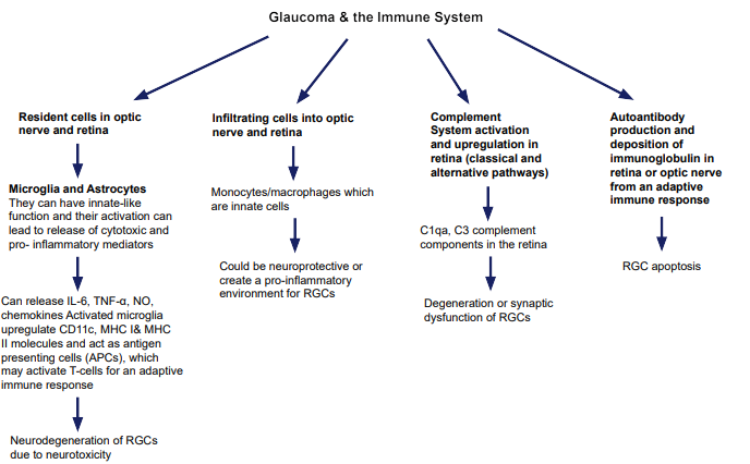
Many studies have shown immune cell responses are involved in glaucoma (Figure 3). This is especially true of glia (astrocytes and microglia), which are resident cells in the retina and optic nerve that can initiate an immune response.19,34,35 Glia are important in immune surveillance, cleaning, as well as removing tissue debris. However, once astrocytes become reactive and microglia take on activated state, both cell types can increase production of cytokines (IL-6), reactive oxygen species (ROS), nitric oxide (NO), and tumor necrosis factor-α (TNF-α), creating a highly neurotoxic environment in the eye.34,36 Microglia can express MHC molecules and function as resident antigen presenting cells.34-36 It is possible microglia become dysregulated due to excessive activation in glaucoma. In the activated state, they recruit other immune cells leading to an uncontrolled adaptive immune response, resulting in increased antigenicity and increased antigen presentation. Microglia could be a therapeutic target and modulating their behavior in glaucoma may decrease RGC loss. Likewise, studying the interactions between RGCs and microglia may shed light on their tropic, cytokine, and immune cell interactions. There are over 20 types of RGCs and certain subtypes may interact more closely with microglia than others, which may influence their immune response to injury or stress.30,37

Figure 3: Diagram Shows Possible Immune Factors Contributing to the Degeneration of RGCs. How the Innate and Adaptive Systems Contribute to Glaucoma, as well as Possible Autoimmune Responses, are Important Questions that Require Further Investigation. There are several Pro-inflammatory Factors that can Contribute to the Neurodegeneration of RGCs, which may be Possible Therapeutic Targets to Prevent or Delay RGC Loss in Glaucoma.

OOJ-2-112Fig3

AUTOIMMUNITY AND GLAUCOMA

There is evidence that autoimmune mechanisms are involved in glaucoma.1,2,17,18 Studies have shown the presence of autoantibodies against ocular antigens such a rhodopsin and glycosaminoglycans (GAGs).1,7,38-40 It has been revealed that patients with normal tension glaucoma have increased levels of heat shock proteins, such as heat shock protein 60 (HSP 60), due to excessive tissue stress and damage.17,18,41-43 But there is no direct evidence to confirm that RGC loss is due to humoral immunity in glaucoma. It is possible that retinal or optic nerve specific autoantigens are present in some glaucoma patients in a manner similar to organ specific autoantigens present in other autoimmune diseases. Autoantibodies in glaucoma patients may be generated as a secondary consequence to disease pathogenesis or they could be generated directly due to RGC death. Likewise, there is evidence aberrant immune activity glaucoma is due to molecular mimicry, through misguided immune responses to self-proteins resulting in injury. A host response is mistakenly directed at a self-protein because it shares high homology to a specific protein found on the surface of a pathogen, like a bacteria or virus.17 One specific study showed increased Heliobacter pylori titers among glaucoma patients, which may represent molecular mimicry to this bacterium.44 Furthermore, epitope mapping has revealed the immunogenicity of rhodopsin antibodies detected in glaucoma patient sera is shared with epitope proteins found on common bacterial and viral pathogens.14,17 Lastly, microbial flora whether oral, gastrointestinal, or ocular, may contribute to the pathogenesis of glaucoma.45 Overall, autoimmunity may play a role in glaucoma, but it is still an area of open investigation.

COMPLEMENT PATHWAY AND GLAUCOMA

The complement system is a key pathway in the innate immune system. Various components in the complement system are upregulated in human glaucoma and in animal models of glaucoma.15,46-49 The complement pathway can be activated in either the optic nerve head or in the inner plexiform layer of the retina.20 It has been shown that RGCs sense damage or stress and respond by activating C1, which is part of the classical complement pathway. Once C1 is activated, C3 convertase is triggered by glial cells, which can amplify RGC damage by recruiting other immune cells like monocytes.20,47 It is still unknown whether the complement pathway is directly responsible for RGC degeneration.

T-CELLS AND GLAUCOMA

Clinical studies have shown that glaucoma patients exhibit differences in immune cell populations, such as T-cells subsets.3,34,50 A study showed significant alterations in Th1 and Th2 cytokine levels in human glaucoma patient serum.50 In animal models, Tcell migration into the optic nerve has been observed. In immune deficient Rag1 knockout mice, which lack mature B- and T-cells, these mice have reduced RGC loss compared to immune competent wild type mice with experimental glaucoma injury.3 But this has not been definitively shown in human glaucoma. T-cell migration into the human retina or optic nerve can be transient and it cannot be ruled out that T-cells can be cytotoxic to RGCs, even with short-term exposure of a small number of T-cells. There may be some type of systemic T-cell response to ocular stress from glaucoma that has yet to be fully unveiled.

NUMEROUS QUESTIONS STILL REMAIN ABOUT THE ROLE OF THE IMMUNE SYSTEM IN GLAUCOMA

Clearly, many unanswered questions remain about the role the immune system plays in glaucoma. First, the timing of the immune response, its duration (acute vs. chronic), and the severity of the immune response. Furthermore, the location of the insult, whether it is in the retina, optic nerve, or even the brain where RGCs project. Although, glaucoma is a disease of the optic nerve with loss of RGCs, it is still not clear where the injury occurs and what specific injury sets off an immune response and whether it is early or late in the disease process. Some have suggested the immune system plays a role in the progressive stages of the disease. But glaucoma pathogenesis involves both primary degeneration and secondary degeneration of RGCs. Primary degeneration occurs after the initial insult to the optic and/or retina, which leads to a chain reaction of cellular and cytokine responses, which creates a neurotoxic environment, resulting in secondary degeneration. The innate and adaptive immune systems are likely to have different roles and responses in the primary degeneration and secondary degeneration stages of glaucoma.

There are key areas in glaucoma research that require further development to study immune responses. For example, high contrast optical coherence tomography (OCT) imaging and adaptive optics imaging techniques of the optic nerve and retina can facilitate studying the role of the immune system in glaucoma patients. Additionally, studies are needed to determine which biomarkers or serum antigens can be indicators of glaucoma.51 It has already been mentioned that HSP are autoantibodies found in glaucoma patients. It is likely there are other autoantibodies generated in response to tissue damage and stress.10,52,53 Macrophages, have been shown to play a role in the pathogenesis of glaucoma, but whether they are protective or beneficial is still open for debate.22,54,55 Identification of specific immune cells, whether they are pathogenic T-cells subsets or monocytes/ macrophages may be prognostic indicators of disease progression.56 Elucidation of epigenetic and genetic alterations, as well as age-related factors and susceptibility factors associated with glaucoma, is another area of open research inquiry.

In summary, glaucoma is likely a disease that is influenced by an uncontrollable immune response, due to an overwhelming burden of constant tissue insults. After a certain length of time and disease progression, the immune system may be unable to provide protection and only offer destruction to neurons. The question is how, when, and what specific immunotherapeutics, cellular therapies (stem cells), or immunomodulators can be used to slow or reduce the loss of RGCs and prevent disease progression, resulting in the precious preservation of sight in glaucoma patients.

1. Wax MB. Is there a role for the immune system in glaucomatous optic neuropathy? Curr Opin Ophthalmol. 2000; 11(2): 145-150. doi: 10.1097/00055735-200004000-00014

2. Russo R, Varano GP, Adornetto A, et al. Retinal ganglion cell death in glaucoma: Exploring the role of neuroinflammation. Eur J Pharmacol. 2016; 787: 134-142. doi: 10.1016/j. ejphar.2016.03.064

3. Tezel G. The immune response in glaucoma: A perspective on the roles of oxidative stress. Exp Eye Res. 2011; 93(2): 178-186. doi: 10.1016/j.exer.2010.07.009

4. Bell K, Gramlich OW, Von Thun Und Hohenstein-Blaul N, et al. Does autoimmunity play a part in the pathogenesis of glaucoma? Prog Retin Eye Res. 2013; 36: 199-216. doi: 10.1016/j. preteyeres.2013.02.003

5. Fan W, Li X, Wang W, Mo JS, Kaplan H, Cooper NG. Early involvement of immune/inflammatory response genes in retinal degeneration in DBA/2J mice. Ophthalmol Eye Dis. 2010; 2: 23-41. doi: 10.4137/oed.s2854

6. Gramlich OW, Bell K, von Thun Und Hohenstein-Blaul N, et al. Autoimmune biomarkers in glaucoma patients. Curr Opin Pharmacol. 2013; 13(1): 90-97. doi: 10.1016/j.coph.2012.09.005

7. Grus F, Sun D. Immunological mechanisms in glaucoma. Semin Immunopathol. 2008; 30(2): 121-126. doi: 10.1007/s00281- 008-0105-8

8. Hendrix S, Nitsch R. The role of T helper cells in neuroprotection and regeneration. J Neuroimmunol. 2007; 184(1-2): 100- 112. doi: 10.1016/j.jneuroim.2006.11.019

9. Huang P, Zhang SS, Zhang C. The two sides of cytokine signaling and glaucomatous optic neuropathy. J Ocul Biol Dis Infor. 2009; 2(2): 78-83. doi: 10.1007/s12177-009-9034-6

10. Joachim SC, Grus FH, Kraft D, et al. Complex antibody profile changes in an experimental autoimmune glaucoma animal model. Invest Ophthalmol Vis Sci. 2009; 50(10): 4734-4742. doi: 10.1167/iovs.08-3144

11. Kipnis J, Schwartz M. Controlled autoimmunity in CNS maintenance and repair: Naturally occurring CD4+CD25+ regulatory T-cells at the crossroads of health and disease. Neuromolecular Med. 2005; 7(3): 197-206. doi: 10.1385/NMM:7:3:197

12. Knier B, Rothhammer V, Heink S, et al. Neutralizing IL-17 protects the optic nerve from autoimmune pathology and prevents retinal nerve fiber layer atrophy during experimental autoimmune encephalomyelitis. J Autoimmun. 2015; 56: 34-44. doi: 10.1016/j.jaut.2014.09.003

13. Moalem G, Gdalyahu A, Shani Y, et al. Production of neurotrophins by activated T cells: Implications for neuroprotective autoimmunity. J Autoimmun. 2000; 15(3): 331-345. doi: 10.1006/jaut.2000.0441

14. Rieck J. The pathogenesis of glaucoma in the interplay with the immune system. Invest Ophthalmol Vis Sci. 2013, 54(3): 2393-2409. doi: 10.1167/iovs.12-9781

15. Tezel G, Yang X, Luo C, et al. Oxidative stress and the regulation of complement activation in human glaucoma. Invest Ophthalmol Vis Sci. 2010, 51(10): 5071-5082. doi: 10.1167/ iovs.10-5289

16. Tezel G, Yang X, Luo C, Peng Y, Sun SL, Sun D. Mechanisms of immune system activation in glaucoma: Oxidative stress-stimulated antigen presentation by the retina and optic nerve head glia. Invest Ophthalmol Vis Sci. 2007; 48(2): 705- 714. doi: 10.1167/iovs.06-0810

17. Wax MB, Tezel G. Immunoregulation of retinal ganglion cell fate in glaucoma. Exp Eye Res. 2009; 88(4): 825-830. doi: 10.1016/j.exer.2009.02.005

18. Wax MB, Tezel G, Yang J, et al. Induced autoimmunity to heat shock proteins elicits glaucomatous loss of retinal ganglion cell neurons via activated T-cell-derived fas-ligand. J Neurosci. 2008; 28(46): 12085-12096. doi: 10.1523/JNEUROSCI.3200-08.2008

19. Ramirez AI, de Hoz R, Salobrar-Garcia E, et al. The role of microglia in retinal neurodegeneration: Alzheimer’s disease, parkinson, and glaucoma. Front Aging Neurosci. 2017; 9: 214. doi: 10.3389/fnagi.2017.00214

20. Williams PA, Marsh-Armstrong N, Howell GR, et al. Neuroinflammation in glaucoma: A new opportunity. Exp Eye Res. 2017; 157: 20-27. doi: 10.1016/j.exer.2017.02.014

21. Kipnis J, Mizrahi T, Hauben E, Shaked I, Shevach E, Schwartz M. Neuroprotective autoimmunity: Naturally occurring CD4+CD25+ regulatory T cells suppress the ability to withstand injury to the central nervous system. Proc Natl Acad Sci U S A. 2002; 99(24): 15620-15625. doi: 10.1073/pnas.232565399

22. London A, Itskovich E, Benhar I, et al. Neuroprotection and progenitor cell renewal in the injured adult murine retina requires healing monocyte-derived macrophages. J Exp Med. 2011; 208(1): 23-39. doi: 10.1084/jem.20101202

23. Schwartz M. Neurodegeneration and neuroprotection in glaucoma: Development of a therapeutic neuroprotective vaccine: The Friedenwald lecture. Invest Ophthalmol Vis Sci. 2003; 44(4): 1407-1411. doi: 10.1167/iovs.02-0594

24. Schwartz M. Modulating the immune system: A vaccine for glaucoma? Can J Ophthalmol. 2007; 42(3): 439-441. doi: 10.3129/can.j.ophthalmol.i07-050

25. Schwartz M, Shaked I, Fisher J, Mizrahi T, Schori H. Protective autoimmunity against the enemy within: Fighting glutamate toxicity. Trends Neurosci. 2003; 26(6): 297-302. doi: 10.1016/ S0166-2236(03)00126-7

26. Schwartz M. Physiological approaches to neuroprotection. boosting of protective autoimmunity. Surv Ophthalmol. 2001; 45(Suppl 3): S256-S260. doi: 10.1016/S0039-6257(01)00208-9

27. Schwartz M. Sell Memorial Lecture. Helping the body to cure itself: Immune modulation by therapeutic vaccination for spinal cord injury. J Spinal Cord Med. 2003; 26(Suppl 1): S6- S10. doi: 10.1080/10790268.2003.11753719

28. Schwartz M, Baruch K. Breaking peripheral immune tolerance to CNS antigens in neurodegenerative diseases: Boosting autoimmunity to fight-off chronic neuroinflammation. J Autoimmun. 2014; 54: 8-14. doi: 10.1016/j.jaut.2014.08.002

29. Schwartz M, Shechter R. Systemic inflammatory cells fight off neurodegenerative disease. Nat Rev Neurol. 2010; 6(7): 405- 410. doi: 10.1038/nrneurol.2010.71

30. Tamm ER, Ethier CR, Lasker IIoA; Glaucomatous Neurodegeneration Participants. Biological aspects of axonal damage in glaucoma: A brief review. Exp Eye Res. 2017; 157: 5-12. doi: 10.1016/j.exer.2017.02.006

31. Johnson TV, Tomarev SI. Rodent models of glaucoma. Brain Res Bull. 2010; 81(2-3): 349-358. doi: 10.1016/j.brainresbull.2009.04.004

32. Struebing FL, Geisert EE. What animal models can tell us about glaucoma. Prog Mol Biol Transl Sci. 2015; 134: 365-380. doi: 10.1016/bs.pmbts.2015.06.003

33. Bakalash S, Kipnis J, Yoles E, Schwartz M. Resistance of retinal ganglion cells to an increase in intraocular pressure is immune-dependent. Invest Ophthalmol Vis Sci. 2002; 43(8): 2648-2653.

34. Tezel G, Fourth APORICWG. The role of glia, mitochondria, and the immune system in glaucoma. Invest Ophthalmol Vis Sci. 2009; 50(3): 1001-1012. doi: 10.1167/iovs.08-2717

35. Vecino E, Rodriguez FD, Ruzafa N, Pereiro X, Sharma SC. Glia-neuron interactions in the mammalian retina. Prog Retin Eye Res. 2015; 51: 1-40. doi: 10.1016/j.preteyeres.2015.06.003

36. Suzumura A, Takeuchi H, Zhang G, Kuno R, Mizuno T. Roles of glia-derived cytokines on neuronal degeneration and regeneration. Ann N Y Acad Sci. 2006; 1088: 219-229. doi: 10.1196/annals.1366.012

37. Vidal-Sanz M, Nadal-Nicolas FM, Valiente-Soriano FJ, Agudo-Barriuso M, Villegas-Perez MP. Identifying specific RGC types may shed light on their idiosyncratic responses to neuroprotection. Neural Regen Res. 2015; 10(8): 1228-1230. doi: 10.4103/1673-5374.162751

38. Romano C, Barrett DA, Li Z, Pestronk A, Wax MB. Antirhodopsin antibodies in sera from patients with normal-pressure glaucoma. Invest Ophthalmol Vis Sci. 1995; 36(10): 1968-1975.

39. Romano C, Li Z, Arendt A, Hargrave PA, Wax MB. Epitope mapping of anti-rhodopsin antibodies from patients with normal pressure glaucoma. Invest Ophthalmol Vis Sci. 1999, 40(6):1275-1280.

40. Tezel G, Edward DP, Wax MB. Serum autoantibodies to optic nerve head glycosaminoglycans in patients with glaucoma. Arch Ophthalmol. 1999; 117(7): 917-924. doi: 10.1001/ archopht.117.7.917

41. Tezel G. Immune regulation toward immunomodulation for neuroprotection in glaucoma. Curr Opin Pharmacol. 2013, 13(1): 23-31. doi: 10.1016/j.coph.2012.09.013

42. Tezel G, Seigel GM, Wax MB. Autoantibodies to small heat shock proteins in glaucoma. Invest Ophthalmol Vis Sci. 1998; 39(12): 2277-2287.

43. Wax MB. The case for autoimmunity in glaucoma. Exp Eye Res. 2011; 93(2): 187-190. doi: 10.1016/j.exer.2010.08.016

44. Kountouras J, Mylopoulos N, Boura P, et al. Relationship between Helicobacter pylori infection and glaucoma. Ophthalmology. 2001;108(3): 599-604. doi: 10.1016/S0161- 6420(00)00598-4

45. Astafurov K, Elhawy E, Ren L, et al. Oral microbiome link to neurodegeneration in glaucoma. PLoS One. 2014; 9(9): e104416. doi: 10.1371/journal.pone.0104416

46. Nikolskaya T, Nikolsky Y, Serebryiskaya T, et al. Network analysis of human glaucomatous optic nerve head astrocytes. BMC Med Genomics. 2009; 2: 24. doi: 10.1186/1755-8794-2-24

47. Stasi K, Nagel D, Yang X, et al. Complement component 1Q (C1Q) upregulation in retina of murine, primate, and human glaucomatous eyes. Invest Ophthalmol Vis Sci. 2006; 47(3): 1024-1029. doi: 10.1167/iovs.05-0830

48. Howell GR, Libby RT, John SW. Mouse genetic models: An ideal system for understanding glaucomatous neurodegeneration and neuroprotection. Prog Brain Res. 2008; 173: 303-321. doi: 10.1016/S0079-6123(08)01122-9

49. Howell GR, Macalinao DG, Sousa GL, et al. Molecular clustering identifies complement and endothelin induction as early events in a mouse model of glaucoma. J Clin Invest. 2011; 121(4): 1429-1444. doi: 10.1172/JCI44646

50. Yang J, Patil RV, Yu H, Gordon M, Wax MB. T cell subsets and sIL-2R/IL-2 levels in patients with glaucoma. Am J Ophthalmol. 2001; 131(4): 421-426. doi: 10.1016/S0002- 9394(00)00862-X

51. Golubnitschaja O, Flammer J. What are the biomarkers for glaucoma? Surv Ophthalmol. 2007; 52(Suppl 2): S155-S161. doi: 10.1016/j.survophthal.2007.08.011

52. Gramlich OW, Beck S, von Thun Und Hohenstein-Blaul N, et al. Enhanced insight into the autoimmune component of glaucoma: IgG autoantibody accumulation and pro-inflammatory conditions in human glaucomatous retina. PLoS One. 2013; 8(2): e57557. doi: 10.1371/journal.pone.0057557

53. Hammam T, Montgomery D, Morris D, Imrie F. Prevalence of serum autoantibodies and paraproteins in patients with glaucoma. Eye (Lond). 2008; 22(3): 349-353. doi: 10.1038/sj.eye.6702613

54. Cui Q, Yin Y, Benowitz LI. The role of macrophages in optic nerve regeneration. Neuroscience. 2009; 158(3): 1039-1048. doi: 10.1016/j.neuroscience.2008.07.036

55. Jung S, Schwartz M. Non-identical twins – microglia and monocyte-derived macrophages in acute injury and autoimmune inflammation. Front Immunol. 2012, 3: 89. doi: 10.3389/ fimmu.2012.00089

56. Yang J, Zhang L, Yu C, Yang XF, Wang H. Monocyte and macrophage differentiation: Circulation inflammatory monocyte as biomarker for inflammatory diseases. Biomark Res. 2014; 2(1): 1. doi: 10.1186/2050-7771-2-1

LATEST ARTICLES

Use of Black Soldier Fly (Hermetia illucens) Prepupae Reared on Organic Waste

review

2024 Mar

Isayas A. Kebede*, Haben F. Gebremeskel and Gelan D. Dahesa,
Figure 11. Risk Map for the Introduction of Ruminant Diseases at Borders

Original Research, peer reviewed

2024 Mar

Yassir Lezaar*, Mehdi Boumalik, Youssef Lhor, Moha El-Ayachi, Abelilah Araba and Mohammed Bouslikhane

Commentary, End-of-Life, Family Dynamics, palliative Care, peer reviewed

2024 Mar

Neil A. Nijhawan*, Rasha Mustafa and Aqeela Sheikh

Retrospective Study

2024 Mar

Shrenik J. Shah*, Abhishek Jha, Chirag Davara, Rushi Mistry and Kapil Kachhadiya

Attention, Disaster, Mindfulness, Original Research, peer reviewed

2024 Mar

Christina Hagen*, Lars Lien, Edvard Hauff and Trond Heir

review

2024 Mar

Isayas A. Kebede* and Gelan D. Dahesa

review

2024 Feb

Bekiyad S. Daro*; Yared A. Deneke, Jiregna G. Negasa and Ishetu N. Dibaba