Advances in Food Technology and Nutritional Sciences

Open journal

ISSN 2377-8350

Innovations in Microbial Biodiscovery, Targeting Silent Metabolism and New Chemical Diversity

Zeinab G. Khalil* and Robert J. Capon

Received: August 29th, 2016 Accepted: September 7th, 2016 Published: September 7th, 2016

INTRODUCTION TO MICROBIAL BIODISCOVERY

Historical Impact of Microbial Natural Products

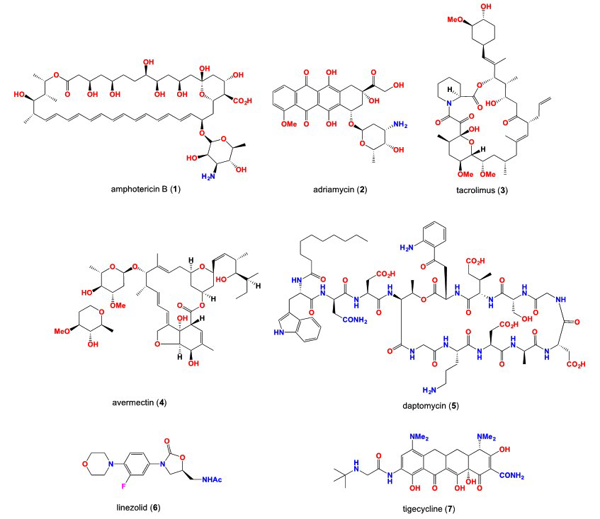
Natural products continue to play a crucial role in the discovery of new drugs and drug leads.1 Biodiscovery from microbial resources can be defined as the exploration of microbial metabolic products that provide important benefits to the fields of medicine, agriculture and biotechnology.2 It has been reported that approximately 50% of approved drugs in the market are derived from microbial origin (Table 1).3 Actinomycetes and fungi are very important groups of microorganisms that are known for their ability to produce secondary metabolites with therapeutic activities.4 Actinobacteria produce over half of the bioactive compounds that are present in the antibiotic literature.5 These compounds include well-known antibacterials such as aminoglycosides and tetracyclines, antifungals such as amphotericin B (1) anticancer agents such as adriamycin (2) immunosuppressants such as tacrolimus (3) and anthelmintics such as avermectin (4) plus numerous other valuable therapeutic drugs. Fungi also play a vital role in the microbial discovery, with the genus Penicillium providing several well-known drugs (e.g. penicillins). Therefore, the drug discovery process is considered as a very critical issue in the industrial field as it is a very expensive process and it requires multiple steps to produce new drug potentials to target specific disease.6

Table 1: Examples of marketed antibiotics originated from microbial origin.3

Original metabolite

Commercial products Producing organism
Penicillins Penicillin G, V, Ampicillin, Methicillin

Penicillium spp., Aspergillus spp.

Cephalosporins

Mefoxin, Ceclor Acremonuim spp.
Thienamycin Primaxin, Invanz

Streptomyces cattleya

Erythromycin

Erythrocin, Zithromax Saccharopolysporaerythraea
Vancomycin Vancocin

Streptomyces orientalis

Fosfomycin

Monuril Streptomyces fradiae
Daptomycin Cubicin

Streptomyces roseosporus


VROJ-1-104Fig1


Despite this great success in the discovery of valuable antibiotics and other pharmaceutical active agents, there has been a dramatic decline in the discovery of new antibiotics in the 21st century. Despite a 2007 review7 revealing that 70% of hospital-acquired infections in the US are resistant to one or more antibiotics, with the exception of the narrow spectrum antibiotic daptomycin (5) and linezolid (6) very few new classes of clinically relevant antibiotics have been approved over the last 40 years. Tigecycline (7) is one of a new class of antibiotics derived from the tetracycline nucleus. This broad-spectrum antibiotic was approved for the treatment of complicated skin infections and is also active against methicillin-resistant staphylococcus aureus (MRSA), vancomycin-resistant Enterococcus faecium and beta-lactamase-producing bacteria as Escherichia coli and Klebsiella pneumonia.8,9

Therapeutic Fields Responsive to Natural Products

Microbial secondary metabolites are highly potent and selective for the treatment of many diseases. Many of these metabolites can be interpreted to be a signal molecule or defense mechanism against competitors or pathogens, an aid to the survival of the microorganism.

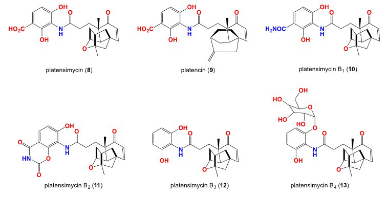
Bacterial pathogens: Natural products are still the main source of promising new antibiotics for the treatment of bacterial diseases. Platensimycin (8) was first reported in 2006 from Streptomyces platensis by Merck researchers, followed by the isolation of platencin (9).11,12,13 In 2008, another platensimycin analogues B1 -B3 (10-12)14 and platensimycin B4 (13).15 This class of antibiotics demonstrated a novel mode of action against gram-positive bacteria through inhibition of cellular lipid biosynthesis. Because of this unique role, it shows no cross resistance to MRSA, vancomycin-resistant enterococcus (VRE) or other antibiotic resistant microbes.

VROJ-1-104Fig2


Fungal pathogens
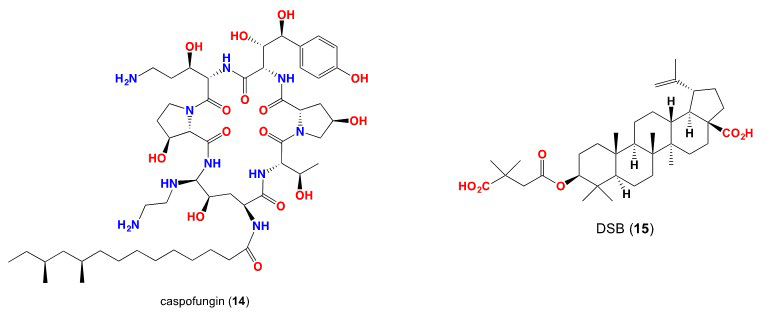
: The need for the discovery of new antifungal agents continues, as we seek to treat the opportunistic infections in immune-compromised patients and the emerging resistance to existing antifungal agents. Jacob et al,16 recently discussed the whole cell screening techniques targeting natural products and their mode of action including the use of genetically modified fungal strains in which the antifungal drug is targeting the cell wall and cell membrane. Caspofungin (14) is a promising new antifungal agents derived from pneumocandin, a natural product produced by Glarealozoyensis.17

Viral pathogens: New anti-HIV (human immunodeficiency virus) drugs are targeting the point of entrance of the virus into the cell such as interferon signaling and cell surface receptors as C-C chemokine receptor type 5 (CCR5)-human cytokine receptor and cluster of differentiation 4 (CD4).18 Many antiviral lectins are from algal origin and they are mainly small proteins that bind to carbohydrates found on the viral envelopes and prevent the transmission of HIV.19 In addition, viral polymerases are the key for viral replication and are considered as promising targets for anti-viral drugs, with inhibitors for viral polymerase and proteases in clinical use for the treatment of acquired immunodeficiency syndrome (AIDS) and chronic hepatitis. All drugs that are in the market for the treatment of HIV are synthetic in origin, although the activities of many natural products have been explored in the recent years.20 Yu et al,21 discovered a plant-derived modified betulinic acid derivative, DSB (15) as a first in class HIV maturation inhibitor. This natural product is currently in Phase II clinical trial.21

Resistance to Microbial Natural Products

Antibiotics and the resistance in nature: The discovery of antibiotics more than 70 years ago initiated an era of drug innovation in human and animal health. These discoveries were interrupted by the emergence of antibiotic resistance, largely due to the widespread of overuse of antibiotics in both medicine and agriculture.22 Not withstanding the outstanding list of antibiotics that were discovered from microbial sources, especially soil microbes, resistance to these antibiotics evolved in the environment long before they were developed as commercial antibiotics.23 The resistance we see in the clinic today is to a great extent similar to the environmental one. Although the resistance in both contexts are similar, clinical resistance tends to increase substantially with time. Therefore there should be a certain mechanism that keeps control of resistance in the environment.24,25,26

VROJ-1-104Fig3


Anticancer resistance
: Cancer cells become resistant to anticancer drugs by several mechanisms. One way is to pump drugs out of cells by increasing the activity of efflux pumps, such as ATP dependent transporters. Alternatively, resistance can occur as a result of reduced drug influx–a mechanism reported for agents that interact with intracellular carriers or enter the cell by means of endocytosis. In cases where drug accumulation is unchanged, activation of detoxifying proteins, such as cytochrome P450 mixed-function oxidases, can promote drug resistance. Cells can also activate mechanisms that repair drug-induced deoxyribonucleic acid (DNA) damage. Finally, disruptions in apoptotic signalling pathways (e.g. p53 or ceramide) allow cells to become resistant to drug-induced cell death.27

In order to overcome these problems, we need to better understand the microbial genomics. In the last 10 years, advances in genomics have revealed new knowledge of a silent microbial secondary metabolism, offering a glimpse of a new source of potentially valuable biomedical agents and tools. To access this genetic resource requires molecular tools capable of activating latent secondary metabolism gene clusters, to build knowledge of microbial systems biology, and facilitate access to new microbial natural products.28,29,30

Silent Resources

After 100 years of discovering microbes, there is great evidence that microbes are capable of producing different classes of bioactive secondary metabolites.31 However, strong evidence revealed that we are only scratching the surface of microbial genome. Microbes share and accumulate multiple secondary metabolite gene clusters, which are capable of producing different bioactive metabolites, with a wide range of biological activities.32 Two challenges limit our ability to access the microbial genome; the first one is that both the bacteria and fungi harbour massive number of genes. These genes usually remain dormant and do not produce any metabolites under normal laboratory conditions until they become activated. It is very probable that these genes are activated by chemical stimuli produced by other competing microbes. Such chemical stimuli represent valuable molecular tools that could be used to unlock the silent secondary metabolism, improving prospective for next generation antibiotics.33

Detecting of silent secondary gene cluster: A 2001 study by Omura et al, on the genome of Streptomyces avermitilis, the microbial source of anthelmintic drug avermectins, revealed an 8.7 Mbp linear chromosome with 25 recognizable secondary metabolite gene clusters accounting for 6.4% of the genome. This study concluded that there are many uncharacterized genes involved in the secondary metabolism. Twenty-five secondary metabolite gene clusters were found in the genome of S. avermitilis. Four of them are responsible for the production of melanin pigment on solid medium, two are derived from tyrosine and one is an aromatic polyketide. Another melanin is an ochronotic pigment, which is derived from homogentiginic acid and produced, in both solid and liquid media.34 S. avermitilis has the highest number of secondary metabolite gene clusters of all bacterial genomes sequenced. The production of different metabolites can be attributed to the presence of many gene clusters, which can encode for enzymes for the activation of different secondary metabolic pathway. The report concluded that there are ”many other uncharacterized genes involved in secondary metabolism”.

In 2002, Bentley et al35 reported that the genome of Streptomyces coelicolor contained 20 secondary metabolite gene clusters and metabolic enzymes of known or predicated secondary metabolites. Furthermore, the genome contained an unprecedented proportion of regulatory genes that are involved in response to external stimuli and stresses. S. coelicolor contains duplicated genes that operate in different phases during colonial development, which if stimulated could lead to new pharmaceutical compounds. The report concluded by noting “The abundance of previously uncharacterized metabolic enzymes, particularly those likely to be involved in the production of natural products, is a resource of enormous potential values”. In 2005, McAlpine et al36 reported the genome analysis of Streptomyces aizunensis to detect the presence of hitherto unexpressed natural product in which different media where used to express microbial secondary metabolites. Udwary et al,37 analysed the genome of Salinospora tropica to show a large percentage of its genome is dedicated to natural product assembly. In addition, this study identified secondary metabolic biosynthetic gene clusters from the complete genome sequence of S. tropica revealing an unrealised secondary metabolism potential and the importance of the understanding and the control for the secondary metabolite pathway.

Therefore, modern genomics has redefined our understanding of microbial secondary metabolism. The computational analysis of microbial metabolism provides the sequencing of thousands of microbial genomes. This knowledge will provide a new pathway to access the microbial molecular diversity and also will help to develop more tools and methodologies to take advantage of this potential. In addition, the previous articles noted a growing appreciation of a global microbial genome that encompasses molecular discovery value than previously appreciated. On the other hand, while learning that microbes possess a secondary metabolism, the challenge is how to activate and benefit from that the activation of silent secondary gene clusters and produce new pharmaceutical compounds.

Activating of Silent Genes

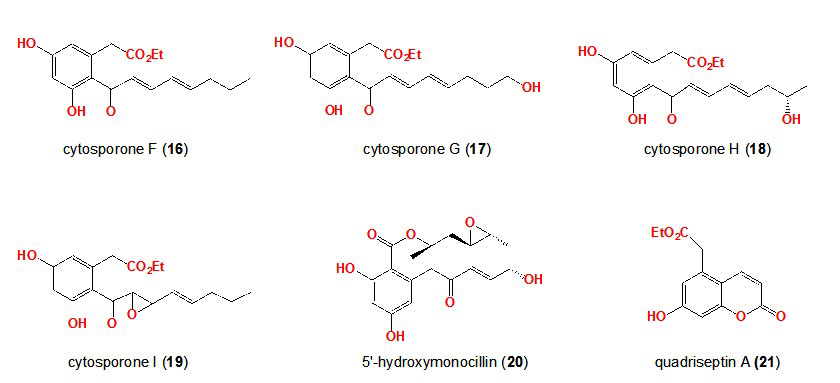
Effect of culturing conditions on secondary metabolite production: The choice of the cultivation parameters is crucial for the production of secondary metabolites by microorganisms. It is well known fact that a change in culture conditions (temperature, pH, oxygen etc.) can affect secondary metabolites production. Any small change in the culture medium may impact not only the level of production of certain compounds, but also the diversity.38 Paranagama et al39 studied the effect of tap water and distilled water on the metabolite profiling of 2 plant associated fungus, Para phaeosphaeria quadrisep tata by the changing the water used in fermentation from tap to distilled resulted in the production of 6 new compounds, cytosporones F-I (16,17,18,19), 5′-hydroxymonocillin (20) and quadriseptin A (21).

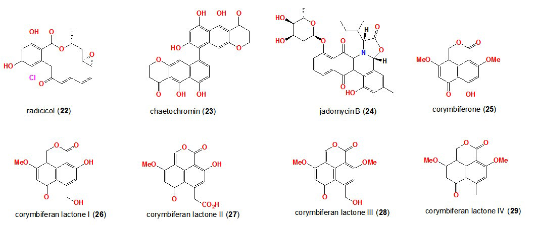
The second fungus, Chaetomium chiversii, producedradicicol (22) on solid phase media, but shifted to chaetochromin (23) in liquid phase.39

In addition to various culture conditions, Ayer et al have studied the impression of the diverse stress conditions on secondary metabolite production. They were able to isolate jadomycin B (24), a glycosylated nezoxazolophenanthridine antibiotic from Streptomyces venezuelae. The aglyconejadomycin was produced in a galactose-isoleucine medium at 37 °C.40 Increasing the temperature to 42 °C increased the level of production of jadomycin B (24). Other parameters were found to increase the production of jadomycin B such as addition of ethanol as well as bacteriophage infection.41 Also, Overy et al42 cultivated different strains of necrotrophic Penicillium strains in different macerated host tissue media. This led to the stimulation of production of corymbiferone (25) and corymbiferan lactones I-IV (26,27,28,29).

Novel metabolites in co-cultures: The functional role of natural products in microbes has long been a topic of discussion. It was found that some natural products result from the interaction of the organism with its own environment. Cueto et al cultured a marine Pestalotia sp. with an unidentified antibiotic resistant marine bacterium, resulting in the biosynthesis of pestalone (30), a new benzophenone. This compound could not be detected when each strain was cultivated independently. Pestalone was found to be active against MRSA and vancomycin-resistant Enterococcus faecium.43 The marine derived fungus Emericella sp. was challenged with the actinomycete Salinispora arenicola. This co-culture activated the production of two new cyclic depsipeptides, emericellamide A (31) and emericellamide B (32).44 Latifiet al. reported another interesting example of the synergistic function of co-culturing in 2006. A co-culture between Pseudomonas aeruginosa and Enterobacter sp. stimulated P. aeruginosa to produce the blue pigment pyocyanin, (33).45 To establish whether any other microorganism can induce the production of pyocyanin, P. aeruginosa was cultivated with different microorganisms. These results showed that P. aeruginosalost its ability to produce pyocyanin when co-cultured with other microbes.46 These related examples highlight the role that co-culture can play in the regulation of secondary metabolite production.

VROJ-1-104Fig4

VROJ-1-104Fig5

Although these strategies have been studied and can lead to promising results, it is nevertheless a time consuming and unpredictable process that while it can express silent metabolites, is not up to the challenge of activating the broad array of silent metabolites hidden within the global microbial genome. In order to access the microbial genome, new approaches informed by the microbial evolution need to be acquired.47

Autoregulators: Autoregulators are compounds that act as triggers to improve cellular development and activate silent secondary metabolite genes, leading to the production of secondary metabolites. Knowing that a microbe is genetically far more capable than previously suspected, reveals a potential, but activating that potential is another challenge entirely. Even without detailed knowledge of the microbial genome, early researchers enjoyed some success in stimulating microbial secondary metabolites. For example, it had been recognized that morphological differentiation during the growth of Streptomyces sp. was closely associated with the secondary metabolites production. More specifically, the transition from primary to secondary metabolism occurred simultaneously with the formation of the aerial hyphae. In exploring these phenomena, a selection of low molecular weight metabolites were found to act as regulators of secondary metabolism.

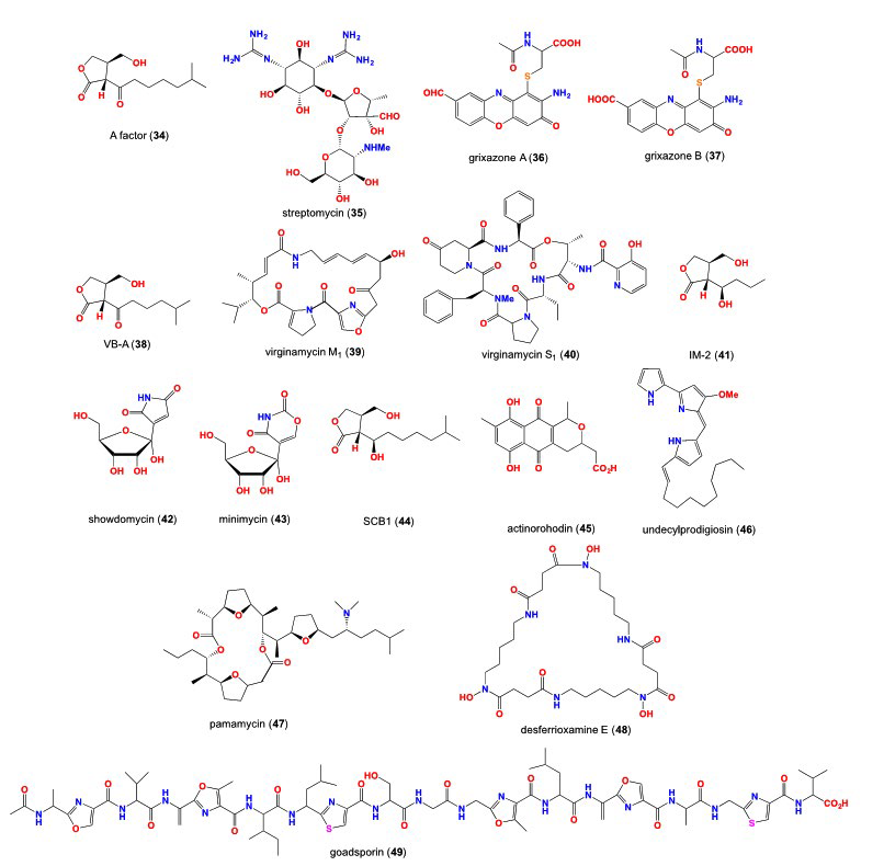
γ–Butyrolactones of actinomycetes: The potential value of autoregulators as tools to regulate antibiotic production was quickly recognized, such that γ–butyrolactones came under intense investigation. An early example, of such a regulator (called an autoregulator) discovered in 1979 was A-factor (34), a γ–butyrolactone produced by Streptomyces griseus. A-factor induced both aerial mycelium formation in S. griseus and production of the antibiotic streptomycin, with A-factor appearing prior to streptomycin production and disappearing before streptomycin reached its maximum level.48

In addition to its effect on streptomycin (35) production,49 other achievements of A-factor included inducing the production of pristinamycins in a mutant strain of S. pristinaespiralis, in which pristinamycins biosynthesis was otherwise blocked, and initiating the production in S. griseus (under phosphate depletion) of the parasiticidal metabolites grixazones A and B (36 and 37). Other closely related naturally occurring microbial γ-butyrolactone autoregulators included the virginiaebutenolides (38) (i.e. VB-A), which activated the production of virginoamycin that consists of 75% virginamycin M1 (39) and 25% virginamycin S1 (40), IM-2 (41), which activated the production of showdomycin (42) and minimycin (43), and SCB1 (44), which activated the production of actinorhodine (45) and undecylprodigiosin (46). IM-2 is noteworthy in that it both activates and suppress secondary metabolism. For instance, biosynthesis of the antituberculosis antibiotic D-cycloserine by S. lavendulae was completely suppressed in the presence of IM-2. Further investigations into the autoregulation of microbial secondary metabolism revealed that this capability was not limited to γ–butyrolactones.

A limited selection of Streptomyces metabolites have been described as promoters of morphogenesis and secondary metabolism, and include the thiazole/oxazole peptide, the polyether pamamycin-607 (47) and the siderophore desferrioxamine E (48). Pamamycin-607 (47) plays an important role in the regulation of aerial mycelium production in 67% of Streptomyces.50 Desferrioxamine E (48) simulates secondary metabolites production in different strains of actinomycetes such as Streptomyces coelicolor.51

However there are some non-butyrolactone Streptomyces metabolites that have been discovered as promoters of secondary metabolites, including thiazole/oxazole peptide goadsporin. Goadsporin (49) is an oligopeptide consisting of 19 amino acids, which acts on the sporulation pathway and regulates secondary metabolites production for Streptomyces.52

Homoserine lactones of gram-negative bacteria: Acyl homoserine lactones (acyl-HSLs) are important intercellular signalling molecules used by many bacteria to monitor their population density in quorum-sensing control of gene expression.53,54 These signals are synthesized by members of the LuxI family of proteins.53 Homoserine lactones (HSLs) function by “quorum sensing” in which they reach a particular extracellular concentration due to high cell density. They act in bioluminescence, antibiotic biosynthesis, animal pathogenicity, plant pathogenicity and extracellular enzyme synthesis.55 HSLs are similar in structure to A-factor. They can also induce exoenzymes, which are plant and animal virulence defense against E. carotovora and P. aeruginosa respectively.56

Genomics-Inspired Screening for Novel Natural Products

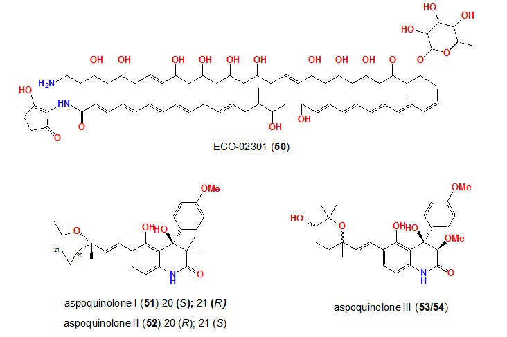
The idea of screening the extract libraries on different types of microorganisms has emerged as an important aspect to determine new metabolites.57 Microbial metabolites (gene activators) may be capable of eliciting the expression of silent secondary metabolism on other microbes. The activation of silent gene clusters will advance the discovery of next generation antibiotics.58,59 Zazopoulos et al60 analyzed the genome of different actinomycetes looking for the genes that were responsible for the production “enydiyne” class of antitumor agents. These loci remain inactive until certain chemical or physical signals activate them. After optimizing the growth conditions, Zazopoulos et al60 were able to induce the expression of the gene cluster and leading to production of “enydiyne”. In addition, McAlpine et al. scanned the genome of Streptomyces aizunensis. The genome scanning identified 11 gene clusters coding for the biosynthesis of wide range of bioactive metabolites. One of these gene clusters was responsible for the production of type I polyketide synthase generating a novel polyketide with characteristic UV absorbance at 300 nm. Varying the culture conditions helped trigger the biosynthesis of compound ECO-02301 (50).36 Scanning the genome sequence of Aspergillus nidulans revealed the presence of three copies of genes that codes for proteins with high similarity to anthranylate synthases (ASs). These enzymes are responsible for the conversion of chorismate to anthranilic acid, which is important for the synthesis of tryptophan. By altering the cultivation conditions, four new prenylated quinoline alkaloids aspoquinolones I, II and III (51,52,53,54) were produced.61

VROJ-1-104Fig6


Epigenetic modifiers
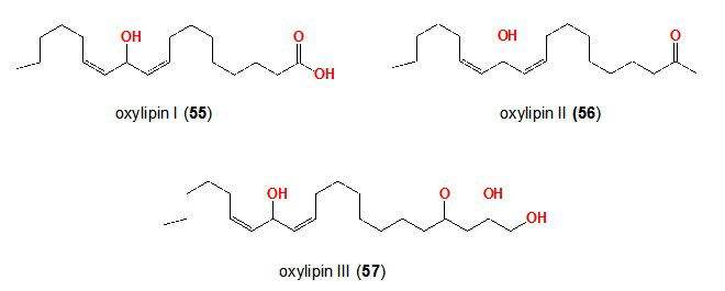
: The strategy of epigenetic modifiers (gene activators) was further studied on different fungi. Some of these studies reported that Aspergillus sp. contains nuclear transcriptional regulator LaeA, which controls secondary metabolite production, suggesting the existence of different regulatory mechanisms that ensure secondary metabolites production at certain developmental stages or under specific environmental conditions.62,63,64 Cichewicz et al65 treated 12 fungi with several DNA methyltransferase and histone deacetylase inhibitors. Eleven strains were found to respond, with the production of new or enhancement of known natural products. One of these strains, Cladosporium cladosporioides, was treated with 5-azacytidine, and led to the production of oxylipins I, II and III (55,56,57).

VROJ-1-104Fig7


VROJ-1-104Fig8


All these observations listed above highlight that the microbial genome offers for greater molecular potential that previously imagined, and that molecular tools are required to access this resource.

Lipopolysaccharide: The lipolysaccharide (LPS) is considered as the main component of the cell wall of all the gram-negative bacteria. The composition of the cell wall of the gram-negative bacteria is made up of an outer membrane, inner plasma membrane and a peptidoglycan layer in the periplasm. The inner cell wall is mainly considered of phospholipids while the outer surface of the outer membrane is composed of 90% LPS, plus the addition of some phospholipids and proteins. LPS consists of phospholipids in which the hydrophilic portion contains different polysaccharide made of core and outer portion.66

LPS is a tripartite molecule comprising a membraneanchored lipid A moiety, a core oligosaccharide and an O-antigen polysaccharide made up of repeating units. 3-deoxy-Dmanno-2-octulosonate (KDO) residues link lipid A to the core oligosaccharide, which can also be decorated with other (often non-stoichiometric) substituents, such as phosphate and phosphoethanolamine. LPS is only found in gram-negative bacteria.67

Bacteria with rough LPS usually have more penetrable cell membranes to hydrophobic antibiotics, since a rough LPS is more hydrophobic.66 The hydrophobic, membrane-anchoring region of LPS is called lipid A. This part of the LPS is well conserved among bacterial species, and is considered as the most active moiety of LPS, responsible for many of the pathophysiological effects associated with infection. Lipid A is composed of phosphorylated glucosamine disaccharide decorated with 6 or even saturated acyl chains linked through amide and ester bonds. When bacterial cells are lysed by the immune system, fragments of membrane containing lipid A are released into the circulation, causing fever, diarrhoea, and even fatal endotoxic shock.67

Khalil et al68 reported on a preliminary investigation of the use the gram-negative bacterial cell wall constituent lipopolysaccharide (LPS) as a natural chemical cue to stimulate and alter the expression of fungal secondary metabolism. IntegratedHTP micro-cultivation and micro-analysis methods determined that 6 of 40 (15%) of fungi tested responded to an optimal exposure to LPS (0.6 ng/mL) by activating, enhancing, or accelerating secondary metabolite production. To explore possible mechanisms behind this effect, we employed light and fluorescent microscopy in conjunction with a nitric oxide (NO) sensitive fluorescent dye and an NO scavenger to provide evidence that LPS stimulation of fungal secondary metabolism coincided with LPS activation of NO. Several case studies demonstrated that LPS stimulation can be scaled from single microplate well (1.5 mL) to preparative (>400 mL) scale cultures. For example, LPS treatment of Penicillium sp. (ACM-4616) enhanced pseurotin A, and activated pseurotin A1 and pseurotin A2 biosynthesis, whereas LPS treatment of Aspergillus sp. (CMB-M81F) substantially accelerated and enhanced the biosynthesis of shornephine A and a series of biosynthetically related ardeemins, and activated production of neoasterriquinone. As an indication of broader potential, we provide evidence that cultivations of Penicillium sp. (CMB-TF0411), Aspergillusniger (ACM-4993F), Rhizopusoryzae (ACM-165F) and Thanatephorus cucumeris (ACM-194F) were responsive to LPS stimulation, the latter 2 examples being particular noteworthy as neither are known to produce secondary metabolites. Our results encourage the view that LPS stimulation can be used as a valuable tool to expand the molecular discovery potential of fungal strains that have either been exhaustively studied by, or that are unresponsive to traditional cultivation methodology.

CONCLUSION

Nature is a good source and producer of small molecules that have a great interaction with biological targets. Despite that natural products continue to provide more than half of all new drugs approved by the US Food and Drug Administration (FDA) during the last century, there is a huge shift in finding a new molecules to overcome the threat coming from multi-drug resistance bacteria and cancer resistant cells.

The decline in the pipeline for the discovery of new microbial metabolites can be attributed to the continuous rediscovery of known metabolites and we believe that the main reason for the decline in the discovery of new biologically active compounds can be that many of the microbial genes can remain dormant under normal laboratory conditions. Therefore, the microbe may require an external stimuli in order to start behave in different way and try to produce different chemistry. In fact, modern genomics have opened the tools and brighten our understanding of the microbial secondary metabolism. Medema et al69 have demonstrated a computational analysis of the microbial metabolism through comprising approximately 300 microbial genome, which lead to the prediction of thousands of microbial genome sequencing. This great outcome provides the opportunity for the acquisition of the full microbial genome and the availability of high throughput sequencing technologies with modern analytical instrumentation allowed to determine or postulate the type of metabolites that can be produced by any microorganisms.

Therefore, we found that it is worthwhile to try to find a new paradigm for drug discovery to help to overcome the limited laboratory conditions and to establish a new technique to target the production of antibiotics. In order to perform such paradigm, we established a combinatorial chemical libraries in order to cover much chemical space to increase the probability of wakening up the silent genes and turning on new secondary metabolites.

CONFLICTS OF INTEREST

The authors declare that they have no conflicts of interest.

1. Newman DJ, Cragg GM, Snader KM. Natural products as sources of new drugs over the period 1981-2002. J Nat Prod. 2003; 66: 1022-1037. doi: 10.1021/np030096l

2. Berdy J. Bioactive microbial metabolites: A personal view. J Antibiot. 2005; 58: 1-26. Website. http://crawl.prod.proquest.com.s3.amazonaws.com/fpcache/e406c496973d03e7326281ef3a3966fe.pdf?AWSAccessKeyId=AKIAJF7V7KNV2KKY2NUQ&Expires=1472901756&Signature=4pJT%2BHcesaq2AouFMa%2FioR47ss0%3D. Accessed August 28, 2016

3. Pelaez F. The historical delivery of antibiotics from microbial natural products-Can history repeat? Biochem Pharmacol. 2006; 71: 981-990. doi: 10.1016/j.bcp.2005.10.010

4. Baker DD, Chu M, Oza U, Rajgarhia V. The value of natural products to future pharmaceutical discovery. Nat Prod Rep. 2007; 24: 1225-1244. doi: 10.1039/b602241n

5. Lazzarini A, Cavaletti L, Toppo G, Marinelli F. Rare genera of actinomycetes as potential producers of new antibiotics. Antonie van Leeuwenhoek. 2000; 78: 399-405. doi: 10.1023/A:1010287600557

6. Capon RJ. Microbial biodiscovery: Back to the future. Curr Top Med Chem. 2012; 12: 1471-1478. doi: 10.2174/156802612802652394

7. Clatworthy AE, Pierson E, Hung DT. Targeting virulence: A new paradigm for antimicrobial therapy. Nat Chem Biol. 2007; 3: 541-548. doi: 10.1038/nchembio.2007.24

8. Hoban DJ, Bouchillon SK, Johnson BM, Johnson JL, Dowzicky MJ. In vitro activity of tigecycline against 6792 Gramnegative and Gram-positive clinical isolates from the global Tigecycline Evaluation and Surveillance Trial (TEST Program, 2004). Diagn. Microbiol Infect Dis. 2005; 52: 215-227. doi: 10.1016/j.diagmicrobio.2005.06.001

9. Draghi DC, Tench S, Dowzicky MJ, Sahm DF. Baseline in vitro activity of tigecycline among key bacterial pathogens exhibiting multidrug resistance. Chemotherapy. 2008; 54: 91-100. doi: 10.1159/000118660

10. Wang J, Soisson SM, Young K, et al. Platensimycin is a selective FabF inhibitor with potent antibiotic properties. Nature. 2006; 441: 358-361. doi: 10.1038/nature04784

11. Jayasuriya H, Herath KB, Zhang C, et al. Isolation and structure of Platencin: A FabH and FabF dual inhibitor with potent broad-spectrum antibiotic activity. Angewandte Chemie. 2007; 46:4684-4688. doi: 10.1002/anie.200701058

12. Singh SB, Jayasuriya H, Ondeyka JG, et al. Isolation, structure, and absolute stereochemistry of platensimycin, a broad spectrum antibiotic discovered using an antisense differential sensitivity strategy. J Am Chem Soc. 2006; 128: 11916-11920. doi: 10.1021/ja062232p

13. Wang J, Kodali S, Lee SH, et al. Discovery of platencin, a dual FabF and FabH inhibitor with in vivo antibiotic properties. Proc. Natl Acad Sci USA. 2007; 104: 7612-7616. doi: 10.1073/pnas.0700746104

14. Zhang C, Ondeyka J, Zink DL, Burgess B, Wang J, Singh SB. Isolation, structure and fatty acid synthesis inhibitory activities of platensimycin B1-B3 from Streptomyces platensis. Chem Commun (Camb). 2008: 5034-5036. doi: 10.1039/b810113b

15. Zhang C, Ondeyka J, Guan Z, et al. Isolation, structure and biological activities of platensimycin B4 from Streptomyces platensis. J Antibiot. 2009; 62: 699-702. doi: 10.1038/ja.2009.106

16. Jacob MR, Walker LA. Natural products and antifungal drug discovery. Methods Mol Med. 2005; 118: 83-109. doi: 10.1385/1-59259-943-5:083

17. Deresinski SC, Stevens DA. Caspofungin. Clinical Infectious Diseases. 2003; 36: 1445-1457. doi: 10.1086/375080

18. Westby M, van Dre. CCR5 antagonists: Host-targeted antivirals for the treatment of HIV infection. Antiviral Chem Chemother. 2005; 16: 339-354. doi: 10.1177/095632020501600601

19. Ziolkowska NE, Wlodawer A. Structural studies of algal lectins with anti-HIV activity. Acta Biochim Pol. 2006; 53: 617-626. Website. http://www.actabp.pl/pdf/4_2006/617.pdf. Accessed August 29, 2016

20. Halfon P. Treatment of hepatitis C virus related diseases using hydroxychloroquine or a combination of hydroxychloroquine and an anti-viral agent. Patent Docs. 2011. Website. http://www.patentsencyclopedia.com/imgfull/20130121965_01. Accessed August 29, 2016

21. Yu D, Morris-Natschke SL, Lee K-H. New developments in natural products-based anti-AIDS research. Med Res Rev. 2007; 27: 108-132. doi: 10.1002/med.20075

22. Taubes G. The bacteria fight back. Science. 2008; 321: 356-361. doi: 10.1126/science.321.5887.356

23. Chait R, Vetsigian K, Kishony R. What counters antibiotic resistance in nature? Nat Chem Biol. 2012; 8: 2-5. doi: 10.1038/nchembio.745

24. D’Costa VM, King CE, Kalan L, et al. Antibiotic resistance is ancient. Nature. 2011; 477: 457-461. doi: 10.1038/nature10388

25. Allen HK, Donato J, Wang HH, Cloud-Hansen KA, Davies J, Handelsman J. Call of the wild: Antibiotic resistance genes in natural environments. Nat Rev Microbiol. 2010; 8: 251-259. doi: 10.1038/nrmicro2312

26. Wright GD. Antibiotic resistance in the environment: A link to the clinic? Curr Opin Microbiol. 2010; 13: 589-594. doi: 10.1016/j.mib.2010.08.005

27. Gottesman MM, Fojo T, Bates SE. Multidrug resistance in cancer: Role of ATP-dependent transporters. Nat Rev Cancer. 2002; 2: 48-58. doi: 10.1038/nrc706

28. Qin T, Skraba-Joiner SL, Khalil ZG, Johnson RP, Capon RJ, Porco JA, Jr. Atropselective syntheses of (-) and (+) rugulotrosin A utilizing point-to-axial chirality transfer. Nat Chem. 2015; 7: 234-240. doi: 10.1038/nchem.2173

29. Shang Z, Khalil Z, Li L, et al. Roseopurpurins: Chemical diversity enhanced by convergent biosynthesis and forward and reverse michael additions. Org Lett. 2016; 18: 4340-4343. doi: 10.1021/acs.orglett.6b02099

30. Shang Z, Salim AA, Khalil Z, Bernhardt PV, Capon RJ. Fungal biotransformation of tetracycline antibiotics. J Org Chem. 2016; 81: 6186-6194. doi: 10.1021/acs.joc.6b01272

31. Khalil ZG, Raju R, Piggott AM, et al. Antimycobacterial Anthracyclines from an Australian Marine-Derived Streptomyces sp. J Nat Prod. 2015; 78: 949-952. doi: 10.1021/acs.jnatprod.5b00095

32. Bok JW, Chiang Y-M, Szewczyk E, et al. Chromatin-level regulation of biosynthetic gene clusters. Nat Chem Biol. 2009; 5: 462-464. doi: 10.1038/nchembio.177

33. Scherlach K, Hertweck C. Triggering cryptic natural product biosynthesis in microorganisms. Org Biomol Chem. 2009; 7: 1753-1760. doi: 10.1039/b821578b

34. Omura S, Ikeda H, Ishikawa J, et al. Genome sequence of an industrial microorganism Streptomyces avermitilis: Deducing the ability of producing secondary metabolites. Proc Natl Acad Sci USA. 2001; 98: 12215-12220. doi: 10.1073/pnas.211433198

35. Bentley SD, Chater KF, Cerdeno-Tarraga AM, et al. Complete genome sequence of the model actinomycete Streptomyces coelicolor A3(2). Nature. 2002; 417: 141-147. doi: 10.1038/417141a

36. McAlpine JB, Bachmann BO, Piraee M, et al. Microbial genomics as a guide to drug discovery and structural elucidation: ECO-02301, a novel antifungal agent, as an example. J Nat Prod. 2005; 68: 493-496. doi: 10.1021/np0401664

37. Udwary DW, Zeigler L, Asolkar RN, et al. Genome sequencing reveals complex secondary metabolome in the marine actinomycete Salinispora tropica. Proc Natl Acad Sci USA. 2007; 104: 10376-10381. doi: 10.1073/pnas.0700962104

38. Elander RP. Industrial production of β-lactam antibiotics. Appl Microbiol Biotechnol. 2003; 61: 385-392. doi: 10.1007/s00253-003-1274-y

39. Paranagama PA,Wijeratne EMK, Gunatilaka AAL. Uncovering biosynthetic potential of plant-associated fungi: Effect of culture conditions on metabolite production by paraphaeosphaeria quadriseptata and chaetomium chiversii. J Nat Prod. 2007; 70: 1939-1945. doi: 10.1021/np070504b

40. Ayer SW, McInnes AG, Thibault P, et al. Jadomycin, a novel 8H-benz[b]oxazolo[3,2-f]phenanthridine antibiotic from Streptomyces venezuelae ISP5230. Tetrahedron Lett. 1991; 32: 6301-6304. doi: 10.1016/0040-4039(91)80152-V

41. Doull JL, Singh AK, Hoare M, Ayer SW. Conditions for the production of jadomycin B by Streptomyces venezuelae ISP5230: Effects of heat shock, ethanol treatment and phage infection. J Ind Microbiol. 1994; 13: 120-125. doi: 10.1007/BF01584109

42. Overy DP, Smedsgaard J, Frisvad JC, Phipps RK, Thrane U. Host-derived media used as a predictor for low abundant, in planta metabolite production from necrotrophic fungi. J Appl Microbiol. 2006; 101: 1292-1300. doi: 10.1111/j.1365-2672.2006.03037.x

43. Cueto M, Jensen PR, Kauffman C, Fenical W, Lobkovsky E, Clardy J. Pestalone, a new antibiotic produced by a marine fungus in response to bacterial challenge. J Nat Prod. 2001; 64:1444-1446. doi: 10.1021/np0102713

44. Chiang Y-M, Szewczyk E, Nayak T, et al. Molecular genetic mining of the aspergillus secondary metabolome: Discovery of the emericellamide biosynthetic pathway. Chem Biol. 2008; 15: 527-532. doi: 10.1016/j.chembiol.2008.05.010

45. Latifi A, Winson MK, Foglino M, et al. Multiple homologs of LuxR and LuxI control expression of virulence determinants and secondary metabolites through quorum sensing in Pseudomonas aeruginosa PAO1. Mol Microbiol. 1995; 17: 333-343. doi: 10.1111/j.1365-2958.1995.mmi_17020333.x

46. Bode HB. No need to be pure: Mix the cultures! Chem Biol. 2006; 13: 1245-1246. doi: 10.1016/j.chembiol.2006.12.001

47. Capon RJ. Microbial biodiscovery: Back to the future. Curr Top Med Chem. 2012; 12: 1471-1478. doi: 10.2174/156802612802652394

48. Horinouchi S, Beppu T. Autoregulatory factors and communication in actinomycetes. Annu Rev Microbiol. 1992; 46: 377-398. doi: 10.1146/annurev.mi.46.100192.002113

49. Kleiner EM, Pliner SA, Soifer VS, et al. Structure of the A factor, a bioregulator from Streptomyces griseus. Bioorg Khim. 1976; 2: 1142-1147.

50. Hashimoto M, Kondo T, Kozone I, Kawaide H, Abe H, Natsume M. Relationship between response to and production of the aerial mycelium-inducing substances pamamycin-607 and A-factor. Biosci Biotechnol Biochem. 2003; 67: 803-808. doi: 10.1271/bbb.67.803

51. Recio E, Colinas A, Rumbero A, Aparicio JF, Martin JF. PI factor, a novel type quorum-sensing inducer elicits pimaricin production in Streptomyces natalensis. J Biol Chem. 2004; 279: 41586-41593. doi: 10.1074/jbc.M402340200

52. Onaka H, Tabata H, Igarashi Y, Sato Y, Furumai T. Goadsporin, a chemical substance which promotes secondary metabolism and morphogenesis in streptomycetes. I. Purification and characterization. J Antibiot. 2001; 54: 1045-1053. doi: 10.7164/antibiotics.54.1045

53. Asad S, Opal SM. Bench-to-bedside review: Quorum sensing and the role of cell-to-cell communication during invasive bacterial infection. Crit Care. 2008; 12: 236. doi: 10.1186/cc7101

54. Antony M, Jayachandran K. Regulation of acyl homoserine lactone synthesis in pseudomonas putida jMQS1 under phenol stress. Water Air and Soil Pollution. 2016; 227: 338. doi: 10.1007/s11270-016-3018-5

55. Hwang I, Cook DM, Farrand SK. A new regulatory element modulates homoserine lactone-mediated autoinduction of Ti plasmid conjugal transfer. J Bacteriol. 1995; 177: 449-458. Website. https://www.ncbi.nlm.nih.gov/pmc/articles/PMC176609/. Accessed August 29, 2016

56. Jones B, Yu B, Bainton NJ, et al. The lux autoinducer regulates the production of exoenzyme virulence determinants in Erwinia carotovora and Pseudomonas aeruginosa. EMBO J. 1993; 12: 2477-2482. Website. https://www.ncbi.nlm.nih.gov/pmc/articles/PMC413484/. Accessed August 29, 2016

57. Xie Y, Liu Z, Zhang G, et al. A rifampicin-resistant (rpoB) mutation in Pseudomonas protegens Pf-5 strain leads to improved antifungal activity and elevated production of secondary metabolites. Res Microbiol. 2016. doi: 10.1016/j.resmic.2016.05.001

58. Cheng J, Zhang X-l, Zhao J-Y, Wen M-l, Ding Z-G, Li M-G. Recent progress of study on secondary metabolites of Streptomyces. Zhongguo Kangshengsu Zazhi. 2015; 40: 791-800.

59. Sengupta S, Pramanik A, Ghosh A, Bhattacharyya M. Antimicrobial activities of actinomycetes isolated from unexplored regions of Sundarbans mangrove ecosystem. BMC Microbiol. 2015; 15: 170. doi: 10.1186/s12866-015-0495-4

60. Zazopoulos E, Huang K, Staffa A, et al. A genomics-guided approach for discovering and expressing cryptic metabolic pathways. Nat Biotechnol. 2003; 21: 187-190. doi: 10.1038/nbt784

61. Scherlach K, Hertweck C. Discovery of aspoquinolones A-D, prenylated quinoline-2-one alkaloids from Aspergillus nidulans, motivated by genome mining. Org Biomol Chem. 2006; 4: 3517-3520. doi: 10.1039/B607011F

62. Perrin RM, Fedorova ND, Bok JW, et al. Transcriptional regulation of chemical diversity in Aspergillus fumigatus by LaeA. PLoS Pathog. 2007; 3: e50. doi: 10.1371/journal.ppat.0030050

63. Bok JW, Hoffmeister D, Maggio-Hall LA, Murillo R, Glasner JD, Keller NP. Genomic mining for aspergillus natural products. Chem Biol. 2006; 13: 31-37. doi: 10.1016/j.chembiol.2005.10.008

64. Shwab EK, Keller NP. Regulation of secondary metabolite production in filamentous ascomycetes. Mycol Res. 2008; 112: 225-230. doi: 10.1016/j.mycres.2007.08.021

65. Williams RB, Henrikson JC, Hoover AR, Lee AE, Cichewicz RH. Epigenetic remodeling of the fungal secondary metabolome. Org Biomol Chem. 2008; 6: 1895-1897. doi: 10.1039/b804701d

66. Raetz CRH, Whitfield C. Lipopolysaccharide endotoxins. Annu Rev Biochem. 2002; 71: 635-700. doi: 10.1146/annurev.biochem.71.110601.135414

67. Rosenfeld Y, Shai Y. Lipopolysaccharide (Endotoxin)-host defense antibacterial peptides interactions: Role in bacterial resistance and prevention of sepsis. Biochim Biophys Acta. 2006; 1758: 1513-1522. doi: 10.1016/j.bbamem.2006.05.017

68. Khalil ZG, Kalansuriya P, Capon RJ. Lipopolysaccharide (LPS) stimulation of fungal secondary metabolism. Mycology. 2014; 5: 168-178. doi: 10.1080/21501203.2014.930530

69. Medema MH, Blin K, Cimermancic P, et al. antiSMASH: Rapid identification, annotation and analysis of secondary metabolite biosynthesis gene clusters in bacterial and fungal genome sequences. Nucleic Acids Res. 2011; 39: W339-W346. doi: 10.1093/nar/gkr466

LATEST ARTICLES

Use of Black Soldier Fly (Hermetia illucens) Prepupae Reared on Organic Waste

review

2024 Mar

Isayas A. Kebede*, Haben F. Gebremeskel and Gelan D. Dahesa,
Figure 11. Risk Map for the Introduction of Ruminant Diseases at Borders

Original Research, peer reviewed

2024 Mar

Yassir Lezaar*, Mehdi Boumalik, Youssef Lhor, Moha El-Ayachi, Abelilah Araba and Mohammed Bouslikhane

Commentary, End-of-Life, Family Dynamics, palliative Care, peer reviewed

2024 Mar

Neil A. Nijhawan*, Rasha Mustafa and Aqeela Sheikh

Retrospective Study

2024 Mar

Shrenik J. Shah*, Abhishek Jha, Chirag Davara, Rushi Mistry and Kapil Kachhadiya

Attention, Disaster, Mindfulness, Original Research, peer reviewed

2024 Mar

Christina Hagen*, Lars Lien, Edvard Hauff and Trond Heir

review

2024 Mar

Isayas A. Kebede* and Gelan D. Dahesa

review

2024 Feb

Bekiyad S. Daro*; Yared A. Deneke, Jiregna G. Negasa and Ishetu N. Dibaba