Advances in Food Technology and Nutritional Sciences

Open journal

ISSN 2377-8350

Innovation of Wound and Burn Care Dressings from Traditional to Nonwoven Polymeric Scaffolds

Sukhwinder K. Bhullar* and Harpal S. Buttar

Received: November 30th, 2016; Accepted: November 30th, 2016; Published: November 30th, 2016

Health care professionals often come across patients inflicted with exuding wounds and burn injuries which get infected with broad range of micro-organisms because wounds often provide a favourable environment for the colonization of microbes. Staphylococcus aureus and Trichophyton rubrum are the most common pathogens which are responsible for skin and nail infection.1 Some deep wounds and burns damage the underlying structures like muscle, tendon, blood vessels, nerves, and bone which need persistent medical care to prevent systemic infection and loss of organ function.2 Sometimes, aggressive wound infection can cause septicemia and death. The remit of this editorial is to highlight the innovation of biocompatible nanofiber dressings for the localized delivery of antiseptics, antibiotics and growth factors which promote wound and burn healing.

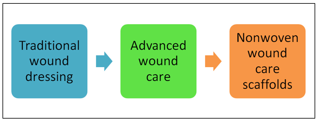
Figure 1 depicts 3 generations of dressings used for wound and burn healing. Traditional dressings consisted of cotton swabs and gauze dressings for managing the chronic and highly exuding wounds over centuries. This practice subsequently led to the development of advanced wound and burn care dressings for the localized delivery of therapeutic products and the modern nonwoven polymeric wound care scaffolds. Advanced antimicrobial wound care dressings or bioactive dressings are comprised of a wide variety of materials such as sodium alginate, ionic silver, chitosan, hydrocolloid, foam, gel or paste, molecular iodine, and have been marketed for several years. The fabrication of modern and novel biocompatible dressings which incorporate bioactive wound healing materials like growth factors are summarized in Table 1.3 Growth factors help to repair the damaged tissues and promote healthy cellular growth.

Figure 1: Three Generations of Dressings for Wound and Burn Care

Three Generations of Dressings

Table 1. Examples of Growth Factros in Wound Healing

Growth Factor or Cytokin

Growth Factor or Cytokin

Current Status

Transforming growth factor β

Re-epitheliasation

Initial studies in venous ulcers encouraging

Neovascularisation

Increased granulation tissue and collagen

Reduced scar formation

Platelet derived growth factor

Re-epithelialisation

Licensed for the treatment of neuropathic diabetic foot ulcers

Neovascularisation

Increased granulation tissue and collagen

Fibroblast growth factor

Re-epitheliasation

Biological effects in pressure ulcers demonstrated to date

Neovascularisation of a provisional matrix

Interleukin 1 β

Healing of infected wounds

Currently under trail for pressure ulcers

Granulocyte macrophage-colony stimulating factor

Improved healing in acute wounds

Pilot studies in infected diabetic foot ulcers encouraging

Examples of a number of modern dressings containing antimicrobial agents and their manufacturers are shown in Table 2.4 Incorporated antiseptic and anti-inflammatory agents are slowly released at the wound surface. Because of its effectiveness against a broad range of micro-organisms, silver is included in many wound and healthcare products. Silver nanoparticles release silver ions in sustainable form to maintain desired concentration for antimicrobial, anti-inflammatory, and wound healing activity, while minimizing the toxic effect of silver. Silver accelerates healing of injured tissue through antimicrobial, antiinflammatory, and antioxidant effect. The emergence of bacterial resistance to silver and its potential to induce cross-resistance to antibiotics has also been reported.5 In cell culture experiments done with human mesenchymal stem cells, silver ions were found to be much more toxic than silver nanoparticles.6 However, despite of these risks, the use of silver-containing dressings (e.g. hydrofiber dressing, polyurethane foams and gauzes) is increasing in wound and burn care products.

Table 2. Examples of Antimicrobial Dressings

Dressing Name Antimicrobial Ingredient Dressing Format Manufacturer
Acticoat absorbent

Ionic silver

Calcium alginate

Smith & Nephew, Inc., Largo, FL, USA
Actisorb Silver 220

Ionic silver and activated charcoal

Silver impregnated activated charcoal cloth

Johnson and Johnson Wound Management, Somerville, NJ, USA
Arglaes

Ionic silver

Transparent film or powder

Medline Industries, Inc., Mundelein, IL, USA
Aquacel AG

Ionic silver

Hydrofiber

Convatec, Skillman, NJ, USA
Contreet H

Ionic silver

Hydrocolloid

Coloplast Corp., Marietta, GA, USA
Contreet F

Ionic silver

Foam

Coloplast Corp., Marietta, GA, USA
Iodosorb

Molecular iodine

Gel or paste

HealthPoint Ltd., Ft. Worth, TX, USA
Silvasorb Antimicrobial Silver Dressing

Ionic silver

Hydrogel sheet or amorphous gel

Medline Industries, Inc., Mundelein, IL, USA
Kerlix AMD Gauze

PHMB

Gauze

Tyco Healthcare/Kendall, Mansfield, MA, USA

Currently, the development of a variety of biocompatible dressings are the focus of attention of biomedical researchers.4,7 These dressings serve as vehicle for the promising delivery of wound or burn care ingredients or even allogenic cells which may provide a specific wound healing benefit. Further, the dressing acts to maintain a locally moist environment needed for wound healing.

Biocompatible and biodegradable polymer micro- and nano-fiber devices fabricated from nanofiber materials with sizes less than 1 µm or 1 nm are especially useful in the field of medicine because these nanomaterials tend to replicate the molecular components of in vivo cellular and bimolecular environment. The synthetic and natural nanofibers of biodegradable and biocomatible polymers are good career vehicles for targeted drug delivery in wound or burn care as well as anti-cancer drugs. Also, the nanofiberous devices are beneficial for burn and wound healing due to their large surface-area-to-volume ratio, high porosity, improved cell adherence, cellular proliferation and migration, as well as controlled in vivo biodegradation rates. The large surface area of polymer nanofiber dressings not only allows increased close interaction of therapeutic agents and exchange of O2 and CO2 with tissues, but also provides a mechanism for sustained release and localized delivery of antiseptic remedies, analgesics, and growth factors needed for burn and wound healing. In addition, the high porosity of nanofiber dressings permits diffusion of nutrients and removal of waste products from the application site. With all these attributes and functions, nanofiber devices promote wound and burn healing. Owing to their multifaceted properties, the nanofiber dressings created from both natural and synthetic polymers have attracted the attention of surgeons, physicians, biomedical researchers, and industry. Their envisioned potential applications are due to the optically transparent functional materials and nano-composites required for making scaffolds to grow stem cells, wound healing dressings and mats, transdermal patches, targeted drug-delivery systems, tissue compatibility and biodegradability, improved cell adherence, and relatively lower manufacturing cost.8,9,10,11

Several studies and experimental evidence suggest that nanofibers with diameter range of 50-100 nm have a great potential for making nanofiber dressings for wound care due to their large surface area, high porosity, and small pore size. Nonwoven nanofibrous scaffolds or patches for wound care have shown to produce skin substitutes with optimal cellular organization, proliferation and to reduce wound contraction. As the skin provides perfect protection from external environment, therefore for wound care, an ideal wound dressing should be compatible with skin and native tissue, should have the capacity to provide thermal insulation, gaseous exchange, and to help drainage and debris removal thus promoting tissue reconstruction processes. Further, an ideal dressing should be biocompatible and not provoke any allergic or immune response reaction, protect the wound from secondary infections, and should be easily removable without causing trauma.12,13,14,15,16,17,18 A variety of wound and burn care dressings in the form of foam, membranous and nonwoven materials with natural or synthetic polymers, as well as combination of both, and impregnated hydrogels are being investigated by researchers worldwide.19,20 In addition, nanofibrous biodegradable polymers impregnated with wound healing materials are also under investigation.21,22,23,24

In summary, it’s estimated that the global wound care products market is expected to reach $18.3 billion by 2019 from $15.6 billion in 2014.25 Such, dollar figures suggest that sophisticated and innovative wound care dressings would form a significant part of the total medical devices market. Therefore, continued attention of the biomedical researchers and industry is needed to address the opportunities and challenges posed by wound and burn healing products and to improve the quality of patient care.

CONFLICTS OF INTEREST

The authors declare that they have no conflicts of interest.

1. Nguyen DT, Orgill DP, Murphy GF. The Pathophysiologic Basis for Wound Healing and Cutaneous Regeneration Biomaterials for Treating Skin Loss. Chap 4. In: Biomaterials for Treating Skin Loss. Cambridge/Boca Raton, FL, USA: Woodhead Publishing (UK/Europe) & CRC Press (US); 2009: 25-57. doi: 10.1533/9781845695545.1.25

2. Wikipedia, the free encyclopedia. Wound healing Web site. https://en.wikipedia.org/wiki/Wound_healing. Accessed November 29, 2016.

3. Harding KG, Morris HL, Patel GK. Science, medicine, and the future: Healing chronic wounds. BMJ. 2002; 324: 160-1633. doi: 10.1136/bmj.324.7330.160

4. Harcup JW, Saul PA. A study of the effect of cadexomer iodine in the treatment of venous leg ulcers. Br J Clin Pract. 1986; 40: 360-364. Web site. http://europepmc.org/abstract/med/3542001. Accessed November 29, 2016.

5. Percival SL, Bowler PG, Russell D. Bacterial resistance to silver in wound care. J Hospital Infection. 2005; 60(1): 1-7. doi: 10.1016/j.jhin.2004.11.014

6. Kittler S, Greulich C, Diendorf J, Koller M, Epple M. Toxicity of silver nanoparticles increases during storage because of slow dissolution under release of silver ions. Chem Mater. 2010; 22: 4548-4554. doi: 10.1021/cm100023p

7. Ormiston MC. Controlled trial of iodosorb in chronic venous ulcers. Br Med J (Clin Res Ed). 1985; 291(6491): 308-310. doi: 10.1136/bmj.291.6491.308

8. Habibi Y, Lucia L, Rojas O. Cellulose nanocrystals: Chemistry, self-assembly, and applications. Chemical Reviews. 2010; 110: 3479-3500. doi: 10.1021/cr900339w

9. Siro I, Plackett D. Microfibrillated cellulose and new nanocomposite materials: A review. Cellulose. 2010; 17: 459-494. doi: 10.1007/s10570-010-9405-y

10. Visakh P, Thomas S. Preparation of bionanomaterials and their polymer nanocomposites. Waste and Biomass Valourization. 2010; 1: 121-134. doi: 10.1007/s12649-010-9009-7

11. Klemm D, Heublein B, Fink HP, Bohn A. Cellulose: Fascinating biopolymer and sustainable raw material. Angewandte Chemie Int. 2005; 44: 3358-3393. doi: 10.1002/anie.200460587

12. Morin RJ, Tomaselli NL. Interactive dressings and topical agents. Clin Plast Surg. 2007; 34(4): 643-658. doi: 10.1016/j.cps.2007.07.004

13. Lloyd LL, Kennedy JF, Methacanon P, Paterson M, Knill CJ. Carbohydrate polymers as wound management aids. Carbohydr Polym. 1998; 37: 315-322. Web site. http://agris.fao.org/agris-search/search.do?recordID=US201302913136. Accessed November 29, 2016.

14. Mulder M. The selection of wound care products for wound bed preparation. Prof Nurs Today. 2011; 15(6): 30-36. Web site. http://www.pntonline.co.za/index.php/PNT/article/viewFile/563/850. Accessed November 29, 2016.

15. Harding KG, Jones V, Price P. Topical treatment: Which dressing to choose. Diabetes Metab Res Rev. 2000; 16(Suppl 1): S47- S50. doi: 10.1002/1520-7560(200009/10)16:1+<::AID-DMRR133>3.0.CO;2-Q

16. Morton LM, Phillips TJ. Wound healing update. Semin Cutan Med Surg. 2012; 31(1): 33-37. doi: 10.1016/j.sder.2011.11.007

17. Wittaya-areekul S, Prahsarn C. Development and in vitro evaluation of chitosan-polysaccharides composite wound dressings. Int J Pharm. 2006; 313(1-2): 123-128. doi: 10.1016/j.ijpharm.2006.01.027

18. Boateng JS, Matthews KH, Stevens HN, Eccleston GM. Wound healing dressings and drug delivery systems: A review. J Pharm Sci. 2008; 97: 2892-2923. doi: 10.1002/jps.21210

19. Zahedi P, Rezaeian I, Ranaei-Siadat S, Jafari S, Supaphol P. A review on wound dressings with an emphasis on electrospun nanofibrous polymeric bandages. Polym Adv Technol. 2010; 21: 77-95. doi: 10.1002/pat.1625

20. Gultekin G, Atalay-Oral C, Erkal S, et al. Fatty acid-based polyurethane films for wound dressing applications. J Mater Sci Mater Med. 2009; 20: 421-431. doi: 10.1007/s10856-008-3572-5

21. Vaseashta A, Erdem A, Stamatin I. Nanobiomaterials for controlled release of drugs and vaccine delivery. Mater Res Soc Symp Proc. 920; 2006: 143-148. Web site. https://www.cambridge.org/core/journals/mrs-online-proceedings-library-archive/article/nanobiomaterials-for-controlled-release-of-drugs-and-vaccine-delivery/A7B20086ED5E0DC405AF9AC53C103962. Accessed November 29, 2016.

22. Dai XY, Nie W, Wang YC, Shen Y, Li Y, Gan SJ. Electrospun emodin polyvinylpyrrolidone blended nanofibrous membrane: A novel medicated biomaterial for drug delivery and accelerated wound healing. J Mater Sci Mater Med. 2012; 23: 2709-2716. doi: 10.1007/s10856-012-4728-x

23. Losi P, Briganti E, Costa M, Sanguinetti E, Soldani G. Silicone-coated non-woven polyester dressing enhances reepithelialisation in a sheep model of dermal wounds. J Mater Sci Mater Med. 2012; 23: 2235-2243. doi: 10.1007/s10856-012-4701-8

24. Nguyen TTT, Ghosh C, Hwang S-G, Tran LD, Park JS. Characteristics of curcumin-loaded poly (lactic acid) nanofibers for wound healing. J Mater Sci. 2013; 48: 7125. doi: 10.1007/s10853-013-7527-y

25. Mateescu M, Baixe S, Garnier T, et al. Antibacterial peptide-based gel for prevention of medical implanted-device infection. PLoS One. 2015; 10(12): e0145143. doi: 10.1371/journal.pone.0145143

LATEST ARTICLES

Use of Black Soldier Fly (Hermetia illucens) Prepupae Reared on Organic Waste

review

2024 Mar

Isayas A. Kebede*, Haben F. Gebremeskel and Gelan D. Dahesa,
Figure 11. Risk Map for the Introduction of Ruminant Diseases at Borders

Original Research, peer reviewed

2024 Mar

Yassir Lezaar*, Mehdi Boumalik, Youssef Lhor, Moha El-Ayachi, Abelilah Araba and Mohammed Bouslikhane

Commentary, End-of-Life, Family Dynamics, palliative Care, peer reviewed

2024 Mar

Neil A. Nijhawan*, Rasha Mustafa and Aqeela Sheikh

Retrospective Study

2024 Mar

Shrenik J. Shah*, Abhishek Jha, Chirag Davara, Rushi Mistry and Kapil Kachhadiya

Attention, Disaster, Mindfulness, Original Research, peer reviewed

2024 Mar

Christina Hagen*, Lars Lien, Edvard Hauff and Trond Heir

review

2024 Mar

Isayas A. Kebede* and Gelan D. Dahesa

review

2024 Feb

Bekiyad S. Daro*; Yared A. Deneke, Jiregna G. Negasa and Ishetu N. Dibaba