Veterinary Medicine

Open journal

ISSN 2475-1286

Impact of Autonomic Tonus Typological Features on the Duodenum Structure of Chickens

Andrij Tybinka, Olexander Zaitsev and Galyna Blishch*

Galyna Blishch, MD

Department of Normal and Pathological Morphology and Forensic Veterinary Medicine, Lviv National University of Veterinary Medicine and Biotechnology, 79010 Lviv, Ukraine
Tel. +380679105825. E-mail: galyna.blishch@gmail.com

INTRODUCTION

Being characterized by a determined cellular and tissue structure the intestine wall is a very dynamic structure of the poultry organism. Adaptation processes that adjust its morphological peculiarities and functional characteristics constantly take place there.1,2,3,4,5,6 These processes are influenced by a variety of internal and external environment factors that are mutually intertwined and exert a complex impact on the organism. Genetic factor occupies a dominant position in determining the morphological and functional intestinal wall profile.7,8,9 Nervous, endocrine and immune systems generate regulatory influence on the intestine in the process of its growth and development.10,11,12,13,14,15 They help form the phenotypic expression of genetic potential according to individual living conditions of the poultry. Forage base and climate are of primary importance here too. Living conditions as well as artificial formation of productive qualities through the development of new breeds and crosses become increasingly important for domestic poultry. The use of different feed supplements, growth promoters, antimicrobial and antibacterial drugs, probiotic preparations and many other compounds exerts a significant impact on the intestinal wall.16,17,18,19,20,21 Constantly adapting intestinal mucosa that supports necessary digestion parameters and performs the barrier function responds to these factors in the most sensitive way.22,23,24 All this eventually impacts the poultry health and productivity.

Autonomic nervous system centers are characterized by a different tonic activity in different organisms. That’s why their cumulative effect on the organs will have some differences too. This study was aimed at showing the relationship between the chickens duodenum morphology and typological peculiarities of the autonomic tonus.

MATERIALS AND METHODS

The study was conducted on laying hens (Gallus domesticus) of ISA Brown cross, aged one year. All poultry was a subject of electrocardiographic investigation. ECG recording was performed with the aid of R4-02 rheograph and N338-6P recording device at a belt speed of 250 millimeters per second. Received cardiograms allowed to analyze a heart rate variability (variation pulsometry) of poultry organism.25,26,27 The research enabled to describe the aggregate tonus of the autonomic centers and divide all the poultry into two groups: sympatheticotonic chickens (ST) and sympathetico-normotonic chickens (ST-NT). The first group is characterized by a distinct predominance of sympathetic centers tonus. The second group has a slightly increased tonus of parasympathetic centers. Sympathetico-normotonic chickens are an intermediate type between two known types of autonomic tonus that are typical for mammals: sympatheticotonics and normotonics.

For the different types of research a certain number of specimens was selected from each group. Euthanasia was performed by an inhalation overdose of chloroform. Intestinal wall tissue samples were taken from the middle third of the duodenum.

To determine the duodenum size 12 ST chickens and 13 ST-NT chickens were selected. With the aid of a caliper an inner intestine perimeter was defined in each investigated group. Absolute duodenum length was determined using a measuring tape. Comparing the latter value with the entire intestine length a relative length of the duodenum was defined (in %). Combining the indicators of absolute duodenum length and its perimeter the area of duodenum mucosa was calculated.

To conduct the study of the duodenum blood supply 4 ST chickens and 5 ST-NT chickens were selected. For the specimens of each group an arterial injection of mesentery with a 10% solution of gelatin colored with red or green pigment (gouache) was performed. After the gelatin hardening a number of intestinal arteries within the duodenum wall was determined.

The research of the fibers number was conducted on 12 ST chickens and 13 ST-NT chickens. The number of fibers per 1 cm2 of mucosa and the total number of fibers of the entire mucosa was determined.

For morphometric studies the tissue samples were fixed in Carnoy and Bouin fluids, further embedded in paraffin. Paraffin sections were stained according to the methods of Van Gieson and Pacini.28 Villus height and crypt depth were determined due to the histological slides received from 10 ST chickens and 12 ST-NT chickens. The thickness of the epithelium and mucosa muscle plate as well as thickness of both muscle membrane layers was determined due to the histological slides received from 16 ST chickens and 17 ST-NT chickens. The same quantity of poultry was used to examine a relative area (expressed in %) of the connective tissue fibers in the area of ​​the muscle membrane and mucosa crypts.

Morphometric study was conducted using the Leica DM-2500 microscope equipped with Leica DFC450C camera and software Leica Application Suite Version 4.4 as well as stereoscopic microscope MBS-10 with Konus №5829 camera and software Image-Tool 3 and WCIF ImageJ.

Research was conducted in a full compliance with the general principles of work with experimental animals (Strasbourg, 1986; Kyiv, 2001).

Statistical Analysis

The received data was processed using a standard software StatPlus 2008. Validity difference of each indicator was measured for ST chickens and ST-NT chickens. Differences between two poultry groups were considered valid when p<0,05.

RESULTS AND DISCUSSION

The discovered typological peculiarities of autonomic tonus are manifested with both functional and structural signs in the chickens’ organism. The investigated duodenum morphological features are a perfect proof of it (see Table 1).


Table 1: Morphological indicators of chickens duodenum.

Indexes

ST chickens ST-NT chickens
Absolute length of intestine, cm 25,0±0,57*

23,4±0,50

The relative length of the intestine, %

13,8±1,84 12,4±1,22
The inner perimeter intestine, сm 2,92±0,150

3,17±0,156***

Area mucosa, сm2

73,0±3,12 74,2±3,46
Number intestinal arteries, pieces 29,5±0,96*

25,2±0,66

number of fibers, piece /сm2

415,4±22,05 476,9±29,92*
The total number of fibers, piece 30324,2±568,97

35375,5±649,19

villus height, mkm

1252,4±23,19 1396,1±26,02*
The depth of the crypts, mkm 289,6±11,68*

261,3±8,94

The ratio, villus height / crypt depth

4,3±0,34 5,3±0,47*
The thickness of the epithelium villi, mkm 33,7±0,30

33,6±0,43

The thickness of the muscle plate mucosa, mkm

36,8±0,35 37,2±0,45
The thickness of the layer of circular muscle layer, mkm 276,5±3,62

255,5±1,81

The length of the longitudinal muscle layer shell, mkm

58,5±0,87* 53,6±0,94
The relative area of connective tissue fibers in the field of mucosal crypts, % 5,80±0,110**

5,59±0,128

The relative area of the fibers of the connective tissue in the area of the muscle layer, %

9,62±0,135*

9,47±0,165

Values expressed as mean±SD.
* – p<0,05;**p<0,01;***p<0,001.


Characterizing the duodenum size and its blood supply it was found out that the absolute duodenum length in ST chickens is 1.6 cm (p<0,05) higher comparing to the ST-NT chickens. A similar situation is observed with the duodenum relative length. In this case the first group of poultry dominates over the second one by 1.4%. The inverse relationship on autonomic tonus is typical for the internal perimeter of the duodenum. On the contrary, its value is 0.25 cm (p<0,001) higher in ST-NT chickens. Thus, we see the formation of compensation processes between the length and perimeter of the duodenum in different poultry groups. This is also indicated by literally the same mucosa area which is determined considering two previous indicators and is only 1.4 cm2 higher in ST-NT chickens. Practical significance of these indicators is that the decrease in the relative length of the duodenum may indicate an increase in the body mass of the poultry.29,30

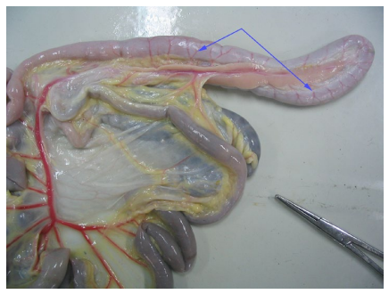
Small arterial vessels (see Figure 1) entering the duodenum are an offshoot of the right branch of the abdominal artery. The number of such arteries was 4.3 higher (p<0,05) in ST chickens. Still, we can’t claim that the increase in autonomic centers tonus enhances the duodenum blood supply. To receive a full picture it is necessary to explore the diameter of these blood vessels and intensity of blood flow in a specified area.

Figure 1: Intestinal vessels of chicken ST-NT, pour gelatin. The arrows show intestinal artery duodenum.

VMOJ-1-103 Fig 1


The next stage of our study reveals the peculiarities of relationship between the separate membranes of the duodenum wall and aggregate autonomic tonus.

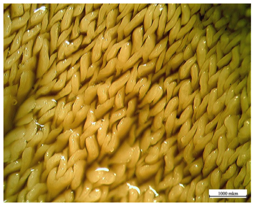
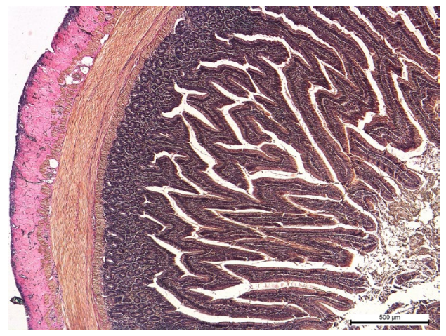
It is known that the functional characteristics of the intestinal mucosa are closely related to the number of fibers, their size and structure.31,32,33 Our study established that the increase in parasympathetic centers tonus in ST-NT chickens also increases the number of fibers per 1 sm2 of duodenum mucosa. In ST chickens the number turned out to be 61.5 villi (p<0,01) lower (see Figure 2). Combining this figure with the total mucosa area it was determined that the total number of villi was 5051.3 higher for ST-NT chickens.The same regularity is typical for the villus height. For ST chickens this figure was 143.7 micrometers (p<0,05) higher comparing to ST-NT chickens (see Figure 3). However, increase in sympathetic tonus exerts a stimulating influence on the depth of crypts which causes a 28.3 micrometers (p<0,05) higher value of this indicator in ST chickens.


Figure 2: Villi mucosa of ST chicken.

VMOJ-1-103 Fig 2


Figure 3: The structure of the wall of the duodenum ST-NT
chicken (stain Van Gieson).

VMOJ-1-103 Fig 3


Since the absolute values of villus height and crypt depth belong to different types of autonomic tonus, the relationship between these parameters is also different. For ST chickens it is 1.0 micrometers (or 19.2%) lower compared to ST-NT chickens.

Thus, the increase in parasympathetic centers tonus positively affects the increase in absorption surface of the duodenum mucous membrane rising the number and size of the villi. The indicated regularity should promote a better digestion and absorption of food nutrients, since it is widely known that the growth of these indicators is combined with a greater mass of the poultry.34,35,36

The thickness of the epithelium and mucosa muscle plate appeared to be the least dependent upon the tonic features of autonomic centers. Meanwhile, in different groups of poultry the epithelial layer thickness differed by only 0.1 micrometers with advantage in ST chickens. However, ST chickens had a somehow greater advantage (0.4 micrometers) in the thickness of the mucosa muscle plate.

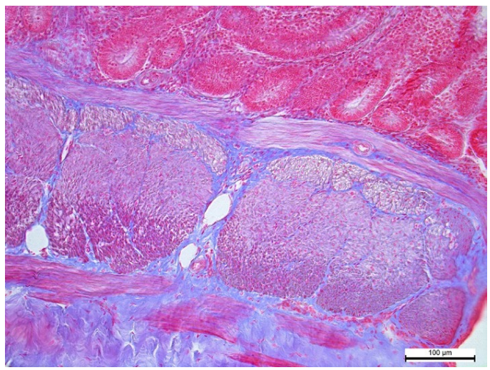
Describing the duodenum muscle membrane, first of all it should be noted that the thickness of its two layers becomes larger under a distinct sympathetic tonus in ST chickens. In the circular layer of the membrane differences between the poultry groups reach a value of 21 micrometers. Longitudinal muscle layer of the membrane is much thinner than the previous one (see Figure 4). That’s why its advantage in thickness is 4.9 micrometers (p<0,05) for ST-NT chickens.


Figure 4: The structure of the muscle layer of the duodenum STNT chicken (stain Pacini).

VMOJ-1-103 Fig 4


The represented morphological characteristics of the muscle membrane within the explored groups of poultry, of course, must be characterized by some functional manifestations. The main purpose of both layers is to provide a duodenum motor activity in the form of peristaltic, anti-peristaltic and segment contractions.37,38 Obviously, each type of autonomic tonus adjusts the very character and intensity of the duodenum motor activity, therefore affecting the intensity of feed mixing and its passage speed.

The amount of connective tissue within the duodenum wall structure is a direct evidence of its functional, adaptive and protective properties. Our research established that in the area of duodenum mucous membrane crypts of the ST chickens the relative area occupied by the connective tissue fibers is 0,21% (p<0,01) higher than the same indicator for ST-NT chickens. In the area of ​​the duodenum muscle membrane the percentage of the area occupied by connective tissue fibers increases in both poultry groups. Herewith, the advantage of ST chickens over ST-NT chickens still preserves but is less expressed and accounts for about 0,15% (p<0,05).

It should also be mentioned that for each type of autonomic tonus the amount of connective tissue fibers increases significantly around blood vessels and nerve nodes. It looks like some “connective tissue membranes” are formed around these structures.

CONCLUSION

Summarizing all the research material we can conclude that the increase in the sympathetic or parasympathetic tonus which is a characteristic feature of the selected groups of chickens causes the formation of structural changes complex in the duodenum wall. These morphological adjustments are inextricably linked with functional processes both in the intestine and the whole organism and are aimed at ensuring the optimal dissimilation and assimilation parameters in the organism of the poultry.

CONFLICTS OF INTEREST

The authors declare that they have no conflicts of interest.

1. Hughes RJ. Energy metabolism in chickens: Physiological limitations. Adelaide, SA, Australia: Rural Industries Research and Development Corporation; 2003: 1-62.

2. Karaca T, Yörük M. A morphological and histometrical study on distribution and heterogeneity of mast cells of chicken’s and quail’s digestive tract. YYU Vet Fak Derg. 2004; 15(1-2): 115-121. Web site. http://vfdergi.yyu.edu.tr/archive/2004/15_1-2/2004_15_(1-2)_115-121.pdf. Accessed March 28, 2016

3. Duke GE. Gastrointestinal physiology and nutrition in wild birds. Proc Nutr Soc. 1997; 56(3): 1049-1056. doi: 10.1079/PNS19970109

4. Nasrin M, Siddiqi MNH, Masum MA, Wares MA. Gross and histological studies of digestive tract of broilers during postnatal growth and development. J Bangladesh Agril Univ. 2012; 10(1): 69-77. doi: 10.3329/jbau.v10i1.12096

5. Shih BL, Chen Y-H, Hsu J-C. Morphological development of the small intestine in white Roman goslings. Afr J Biotechnol. 2013; 12(6): 611-617. doi: 10.5897/AJB11.1526

6. Kadhim KK, Bakar MZA, Noordin MM, Babjee MA, Saad MZ. Light and scanning electron microscopy of the small intestine of young malaysian village chicken and commercial broiler. Pertanika J Trop Agric Sci. 2014; 37(1): 51-64.

7. Mott CR, Siegel PB, Webb KE Jr, Wong EA. Gene expression of nutrient transporters in the small intestine of chickens from lines divergently selected for high or low juvenile body weight. Poultry Sci. 2008; 87(11): 2215-2224. doi: 10.3382/ps.2008-00101

8. Mahmud MA, Shaba P, Shehu SA, Danmaigoro A, Gana J, Abdussalam W. Gross morphological and morphometric studies on digestive tracts of three nigerian indigenous genotypes of chicken with special reference to sexual dimorphism. J. World’s Poult Res. 2015; 5(2): 32-41. Web site. http://agris.fao.org/agris-search/search.do?recordID=IR2015400050. Accessed March 28, 2016

9. Oliveira JE, Druyan S, Uni Z, Ashwell CM, Ferket P.R. Prehatch intestinal maturation of turkey embryos demonstrated through gene expression patterns. Poultry Sci. 2009; 88(12): 2600-2609. doi: 10.3382/ps.2008-00548

10. Fisinin VI, Suraj P. Gut immunity in birds: facts and reflections (review). Agricultural Вiology. 2013; 4: 3-25. Web site. http://cyberleninka.ru/article/n/gut-immunity-in-birds-facts-and-reflections-review. Accessed March 28, 2016

11.Horra MCDL, Cano M, Peral MJ, Calonge ML, Ilundain AA. Hormonal regulation of chicken intestinal NHE and SGLT-1 activities. Am J Physiol Regulatory Integrative Comp Physiol. 2001; 280: 655-660. Web site. http://ajpregu.physiology.org/content/280/3/R655.short. Accessed March 28, 2016

12. Muir WI, Bryden WL, Husband AJ. Immunity, vaccination and the avian intestinal tract. Dev Comp Immunol. 2000; 24(2-3): 325-342. doi: 10.1016/S0145-305X(99)00081-6

13. Rawdon B.B. Gastrointestinal hormones in birds: Morphological, chemical, and developmental aspects. J Exp Zool. 1984; 232(3): 659-670. doi: 10.1002/jez.1402320335

14. Vylitova M, Miksik I, Pacha J. Metabolism of corticosterone in mammalian and avian intestine. Gen Comp Endocrinol. 1998; 109(3): 315-324. doi: 10.1006/gcen.1997.7035

15. Stappenbeck TS, Hooper LV, Gordon JI. Developmental regulation of intestinal angiogenesis by indigenous microbes via Paneth cells. PNAS. 2002; 99(24): 15451-15455. doi: 10.1073/pnas.202604299

16. Dibner JJ, Richards JD. The digestive system: Challenges and opportunities. J Appl Poult Res. 2004; 13(1): 86-93. doi: 10.1093/japr/13.1.86

17. Adil S, Banday T, Ahmad Bhat G, Salahuddin M, Raquib M, Shanaz S. Response of broiler chicken to dietary supplementation of organic acids. JCEA. 2011; 12(3): 498-508 doi: 10.5513/JCEA01/12.3.947

18. Incharoen T. Histological adaptations of the gastrointestinal tract of broilers fed diets containing insoluble fiber from rice hull meal. Am J Anim Vet Sci. 2013; 8(2): 79-88. doi: 10.3844/ajavssp.2013.79.88

19. Revajová V, Slaminková Z, Grešáková Ľ, Levkut M. Duodenal morphology and immune responses of broiler chickens fed low doses of deoxynivalenol. Acta Vet Brno. 2013; 82: 337-342. doi: 10.2754/avb201382030337

20. Pelicano ERL, Souza PA, Souza HBA, Figueiredo DF, Amaral CMC. Morphometry and ultra-structure of the intestinal mucosa of broilers fed different additives. Rev Bras Cienc Avic. 2007; 9(3): 173-180. doi: 10.1590/S1516-635X2007000300006

21. Sittiya J, Yamauchi K. Growth performance and histological intestinal alterations of sanuki cochin chickens fed diets diluted with untreated whole-grain paddy rice. J Poult Sci. 2014; 51(1): 52-57. doi: 10.2141/jpsa.0130042

22. Bar-Shira E, Cohen I, Elad O, Friedman A. Role of goblet cells and mucin layer in protecting maternal IgA in precocious birds. Dev Comp Immunol. 2014; 44(1): 186-194. doi: 10.1016/j.dci.2013.12.010

23. Shao Y, Guo Y, Wang Z. β-1,3/1,6-Glucan alleviated intestinal mucosal barrier impairment of broiler chickens challenged with Salmonella enterica serovar Typhimurium. Poult Sci. 2013; 92(7): 1764-1773. doi: 10.3382/ps.2013-03029

24. Zhang, B., Shao Y, Liu D, Yin P, Guo Y, Yuan J. Zinc prevents Salmonella enterica serovar Typhimurium-induced loss of intestinal mucosal barrier function in broiler chickens. Avian Pathol. 2012; 41(4): 361-367. doi: 10.1080/03079457.2012.692155

25. Baevskij RM. Analiz variabel’nosti serdechnogo ritma: istorija i filosofija, teorija i praktika [Analysis of heart rate variability: the history and philosophy, theory and practice]. Klinicheskaja informatika i telemedicina [Clinical informatics and telemedicine]. 2004; 1: 54-64. (Rus.)

26. Baevskij RM, Kirilov OI, Kleckin SZ. Matematicheskij analiz izminenij serdechnogo ritma pri stresse [Mathematical analysis of changes in heart rate during stress]. Moscow. USSR: Nauka, 1984. (Rus.)

27. Hart rate variability. Standarts of measurements, physiological interpretation, and clinical use. Task force of the european of cardiology and the north american society of pacing and electrophysiology. Circulation. 1996; 93: 1043-1065. doi: 10.1161/01.CIR.93.5.1043

28. Mulisch M, Welsch U. Romeis – Mikroskopische Technik. Spektrum akademischer verlag; 2010. doi: 10.1007/978-3-8274-2254-5

29. Yovchev D, Dimitrov D, Penchev G. Age weight and morphometrical parameters of the bronze turkey’s (Meleagris meleagris gallopavo) intestines. Bulg J Agric Sci. 2013; 19(3): 611-614. Web site. http://www.agrojournal.org/19/03-36.pdf. Accessed March 28, 2016

30. Kadhim KK, Zuki ABZ, Noordin MM, Babjee SA, Khamas W. Growth evaluation of selected digestive organs from day one to four months post-hatch in two breeds of chicken known to differ greatly in growth rate. Journal of Animal and Veterinary Advances. 2010; 9(6): 995-1004. doi: 10.3923/javaa.2010.995.1004

31. Yamauchi Review on chicken intestinal villus histological alterations related with intestinal function. J Poult Sci. 2002; 39(4): 229-242. doi: 10.2141/jpsa.39.229

32. Mitjans M, Barniol G, Ferrer R. Mucosal surface area in chicken small intestine during development. Cell Tissue Res. 1997; 290(1): 71-78. doi: 10.1007/s004410050909

33. Pelicano ERL, Souza PA, Souza HBA, et al. Intestinal mucosa development in broiler chickens fed natural growth promoters. Brazilian Journal of Poultry Science. 2005; 7(4): 221-229. doi: 1590/S1516-635X2005000400005

34. Awad W, Ghareeb K, Böhm J. Intestinal structure and function of broiler chickens on diets supplemented with a synbiotic containing enterococcus faecium and oligosaccharides. Int J Mol Sci. 2008; 9(11): 2205-2216. doi: 10.3390/ijms9112205

35. Laudadio V, Passantino L, Perillo A, et al. Productive performance and histological features of intestinal mucosa of broiler chickens fed different dietary protein levels. Poultry Sci. 2012; 91(1): 265-270. doi: 10.3382/ps.2011-01675

36. Wali ON, Kadhim KK. Histomorphological comparison of proventriculus and small intestine of heavy and light line pre- and at hatching. Int J Anim Veter Adv. 2014; 6(1): 40-47. Web site. http://www.airitilibrary.com/Publication/alDetailedMesh?docid=20412908-201402-201507200026-201507200026-40-47. Accessed March 28, 2016

37. Clench MH, Mathias JR. Motility responses to fasting in the gastrointestinal tract of three avian species. The condor. 1995; 97(4): 1041-1047. doi: 10.2307/1369542

38. Hodgkiss JP. Intrinsic reflexes underlying peristalsis in the small intestine of the domestic fowl. J Physiol. 1986; 380(1): 311-328. doi: 10.1113/jphysiol.1986.sp016287

LATEST ARTICLES

Quality Assurance of General Purpose, Keratin Based and Dye lock Hair Shampoos

Saima Siddique*, Zahida Parveen, Zeeshan Ali and Sidra Mehmood

doi.

Blood Sample from the Patient

Hypertriglyceridemia-Induced Pancreatitis: A Case Report and Literature Review

Maarten Bulterys, Melvin Willems* and Agnes Meersman

doi.

From Neck Pain to a Life-Threatening Condition: A Case Report

Floris Vandewoude* and Sören Verstraete

doi.

LATEST ARTICLES

Original Research

2024 Aug

Saima Siddique*, Zahida Parveen, Zeeshan Ali and Sidra Mehmood

Cross Sectional Study, peer reviewed

2024 Jul

Amanuel P. Beta, Dereje Abera, Legese Belayneh and Isayas A. Kebede

Case Report, peer reviewed

2024 Jul

Syeda Rukh*, Sathyanarayana Machani and Milind Awale