Neuro

Open journal

ISSN 2377-1607

Immunomodulatory Effects of Mesenchymal Stem Cells on T- and B-Cells in a Quiescent State in a Chronic Experimental Model of Autoimmune Encephalomyelitis

Soraya Tabera, Arturo Mangas, Michel Geffard and Rafael Coveñas*

Rafael Coveñas, MSc, PhD

Universidad de Salamanca, Instituto de Neurociencias de Castilla y León (INCYL), Laboratorio 14, c/ Pintor Fernando Gallego, 1, 37007 Salamanca, Spain; Tel. 34-923-294400; ext 5315; Fax: 34-923-294549; E-mail: covenas@usal.esmangasam@usal.es

INTRODUCTION

Although, there are still many problems to be solved before cell therapy can be used reliably to repair mature central nervous system (CNS) lesions, one strategy that could be used in multiple sclerosis (MS) is the transplantation of stem cells. Several types of stem cells have been studied and characterized: embryonic stem cells, neural stem cells, haematopoietic stem cells, non-haematopoietic stem cells and bone marrow stromal cells.1,2,3,4,5,6,7 In this regard, neural precursor cells were the first candidates for cell-based therapy in neuroinflammation.8,9 However, the invasiveness associated with harvesting neural precursor cells, together with the reduced number of cells that can be obtained, may limit their clinical application. A potential alternative to neural precursor cells is mesenchymal stem cells (MSC). Recently, the actions exerted by adipose-derived mesenchymal stromal cells,10,11,12,13,14 placenta-derived mesenchymal stem cells,15,16 Wharton’s jelly mesenchymal stem cells,17 human decidua-derived mesenchymal stem cells18 and human umbilical cord blood mesenchymal stromal cells19 have been reported in experimental autoimmune encephalomyelitis (EAE) animal models.

MSC have been characterized by their multipotentiality and their capacity for immunomodulation, thus raising great hopes in regenerative medicine. These cells could be ideal candidates for application in human diseases, since they offer significant practical advantages: they are obtained from different adult tissues and can readily be cultured and expanded; they are multipotent and self-renewing. They have the capacity to differentiate into mesodermal-derived20 or into neural-like and glial-like cells21; they have been shown to have immunoregulatory properties10,12,13,14,17,18,22,23; they secrete factors that may stimulate endogenous neural stem cells in the CNS24,25; they can be safely injected autologously without the need for immunosuppression26; they decrease the number of infiltrating inflammatory cells, preserving axons and ameliorating demyelination,15,19,27,28 ameliorate neuroinflammation11,29 and exert a neurotrophic action.16 All these features make MSC suitable for therapy in autoimmune diseases. In fact, the beneficial effects of MSC have been reported in EAE models.25,30,31,32,33,34 Most of these studies were carried out in mice and in Lewis rats, and this means that to date the immunomodulatory effects of MSC in EAE Lewis 1A rats have not been studied. However, EAE Lewis 1A rats have been widely used to study the effects of new drug candidates for MS. Lewis 1A rats belong to the Major Histocompatibility Complex (MHC) congenic Lewis rat strain. The effects of MSC on T- and antigen-presenting cells have been studied in depth. Nevertheless, and contrary to their well-known effect on T-cells and antigen-presenting cells, the effect of MSC on B-cells remains unclear. B-cells are critical for myelin oligodendrocyte glycoprotein (MOG)-induced EAE, but are redundant in MOG (35-55)-induced EAE.35,36 This is important, because B-cells play a critical role in human MS.37 Moreover, MSC have been implicated in B-cell development in bone marrow, spleen and lymphoid follicles,38 exerting a negative control on B-cell lymphopoiesis. In addition, it has been reported that B-cell proliferation is inhibited by MSC,38 although this has not been confirmed by other authors.39 The distribution of MSC in bone marrow and secondary lymphoid organs allows an intimate interaction between both cell subsets, which contributes to normal lymph node development40 as well as to the support of tumor B-cells in follicular lymphomas.41 Therefore, the study of the effect of MSC on B-lymphocytes is necessary to increase our insight into autoimmune processes such as MS. In addition to the demonstration of the immunomodulatory effects of MSC on B- and T-lymphocytes in the EAE model, the effects of such cells on the abolition or not of EAE episodes must be studied. Another important point to make clear is the expression of α4 integrins. In this sense, murine bone marrow (BM)-MSC do not express α4 integrins,42 and one study on BM-MSC focusing on EAE lesions has provided conflicting results regarding the capacity of BM-MSC to migrate into inflammed CNS.25,30,31,32,43 In a mouse model of EAE, it has been reported the synergic effect (e.g., the expression of the brain-derived neurotrophic factor was increased) of BM-MSC when these cells were combined with fasudil44 (this combination reduced the severity of EAE in comparison with fasudil or BM-MSC alone); that rapamycin increased the immunomodulatory properties of BM-MSC,45 playing the latter combination an important role in neuroprotection, and that the combination of resveratrol and BM-MSC increased the immunomodulatory effects (pro-inflammatory cytokines were suppressed and anti-inflammatory ones were increased).46 Moreover, in a rat model of EAE, it has been demonstrated that the combination therapy of BM-MSC and EGb761 (a Ginkgo biloba extract) increased the neuroprotective effects, inhibited the secretion of pro-inflammatory cytokines and decreased the disease severity and the inflitrated cells.47 Finally, in a mouse model of EAE, it has been reported that MSC controlled the induction of T-cells with a regulatory phenotype and the inhibition of pro-inflammatory T-cells.48

In the light of the foregoing data, we tested the therapeutic potential of intravenously injected BM-MSC from healthy donors in a chronic EAE model (Lewis 1A rats). We studied in vitro the immunomodulatory effects of MSC on the B- and T-lymphocytes of both control and EAE animals and the effects of intravenously administered MSC on EAE episodes.

MATERIALS AND METHODS

Animals and Induction of EAE

Lewis 1A female rats (n=23) aged 10-11 weeks (weight around 190 g) obtained from CERJ Janvier (France) were used. The animals were kept under standardized lighting and temperature conditions and had free access to food and water. They remained for at least ten days in their cages before the experiments. The aminals were weighed and scored according to the scale described below in “Animal groups and clinical evaluation of EAE” 6 days/week throughout the experiments. The experimental design, protocols, and procedures were performed under the guidelines of the ethical and legal recommendations of Spanish and European law. The study was also approved by the experimental research commission of the University of Salamanca (Spain).

EAE rats were immunized with a solution containing 50 µg of MOG and complete Freund’s adjuvant (ACF), to which heat-inactivated Mycobacterium tuberculosis H37RA had been added. The rats were anaesthetized with isofluorane and the solution was injected intradermally at the base of the tail.

Animal Groups and Clinical Evaluation of EAE

Animals were classified in different groups: 1) Control animals (group 1; n=3); 2) EAE animals (group 2; n=5); 3) EAE animals to which MSC were administered before the induction of EAE (group 3; n=5); 4) EAE animals to which MSC were administered after the induction of EAE (group 4; n=5); and 5) EAE animals to which MSC were administered before and after the induction of the EAE (group 5; n=5).

The animals were weighed and scored according to a previously described scale49 6 days/week throughout the experiments. The neurological signs of EAE were assessed and scored using this scale: 0, no signs; 1, tail weakness or tail paralysis; 2, hind leg paraparesis or hemiparesis; 3, hind leg paralysis or hemiparalysis; 4, complete paralysis (tetraplegy). Any animal reaching a moribund state was immediately perfused (see below) in order to avoid suffering. At the end of the experiment, all the animals were deeply anaesthetized with urethane (1 g/kg, intraperitoneal) and perfused via the ascending aorta with 50-100 ml of cold physiologic saline (0.9% NaCl) and then with 500 ml of cold 4% paraformaldehyde in 0.1 M phosphate-buffer (pH 7.2).

Cell Isolation

MSC were isolated from the femora and tibias of wild-type female Lewis 1A rats (aged 10-11). MSC were harvested and placed in culture in DMEM containing 10% fetal bovine serum and 1% penicillin streptomycin. Cells were cultured at 37º C in a humidified atmosphere in the presence of 5% CO2. Cultures were incubated at a final concentration of 3 x 10⁶ nucleated cells per ml at 37° C in a 5% CO2 humidified incubator for 72 h. Non-adherent cells were aspirated on day 3 and the adherent population was cultured to achieve the maximum number of fibroblast colony-forming units prior to initial passage. Twice a week, adherent cells were fed by complete replacement of the medium. When the layer was confluent, cells were detached by treatment with trypsin and then subcultured at a concentration of 10,000 cells/cm2 until confluence. This process was repeated at least 3 times in order to obtain a sufficient number of cells for analysis. All cultures were used between the third and fifth passages.

Adipogenic, osteogenic and condrogenic differentiation was induced as previously described50 and recommended by the International Society of Cell Therapy (ISCT) consensus.51 Phenotypic characterization was performed using the following monoclonal antibody combinations: Fluorescein (FITC)/Phycoerythrin (PE)/Peridin chlorophil protein-Cyanine-5 (PerCP-Cy5)/Allo-phycocyanin (APC): CD45/CD106/CD90.1/MHC II. Data acquisition was performed with a FACScaliburTM flow cytometer and data analysis with the Paint-A-Gate program.

B- and T-lymphocytes were obtained from splenocytes from healthy donor rats using cell sorting. Cells were stained with CD45RA and CD3, following the manufacturer’s instructions, for B- or T-cells, respectively. For isolation, a FACSAria Cell Sorting flow cytometer was used. Positively selected cells contained >95% B- or T-cells, as assessed by flow cytometry.

Administration of MSC

Animals were deeply anaesthetized with isofluorane. MSC were administered intravenously in the tail vein. Depending on the animal group (3, 4 or 5), the rats were infused at the base of the tail with a solution of sterile phosphate-buffered saline (PBS) containing MSC. MSC were infused two weeks before the induction of EAE (groups 3 and 5); eighteen days after induction (groups 4 and 5); and five weeks after the induction of EAE (groups 4 and 5).

Cell Viability Assays

105 MSC were seeded in the plates and after 12 hours, 106 positively selected B- or T-lymphocytes were seeded in the 96-well culture plate. Cells were cultured for 3 days in Roswell Park Memorial Institute (RPMI) 1640 medium supplemented with 10% fetal bovine serum and 1% penicillin streptomycin. The effect of MSC on B-cell or T-cell growth was assessed by measuring the 3-(4.5-dimethylthiazol-2-yl)-2, 5-diphenyl tetrazolium bromide (MTT) dye absorbance of the cells. For this, 105 B-cells or T-cells/100 mL were plated in triplicate onto 96-well tissue culture dishes in culture medium with or without 104 MSC. MTT absorbance was assessed on the third day. Three wells were analyzed for each condition, and the results were presented as means±SD of triplicates.

For the detection of apoptosis, an AnnexinV-PE/7 amino-actinomycin (7-AAD) apoptosis detection kit was used. A minimum of 106 lymphocytes were washed and resuspended in binding buffer (1:10 diluted in PBS), maintaining a cell concentration of 1×10⁶ /mL. Annexin V-PE and 7-AAD, 5 mL each, were added for 15 minutes. In order to identify B- and T-lymphocytes, anti-CD45RA-FITC and anti-CD3-APC were also added. For each condition, 50,000 events were collected and analyzed. The samples were acquired using TrucountTM Tubes, which contain a calibrated number of fluorescent microbeads. The absolute count of annexin V-PE plus 7-AAD-negative cells was calculated using the following equation: (number of events in region containing annexin V-PE plus 7-AAD negative cells/number of events in the absolute count bead region) x (number of beads per-test/test volume). The Win MDI software was used for analysis.

Proliferation Assays

Studies were also performed on B-lymphocytes and T-lymphocytes cell cycles and DNA contents. For this, 5×10⁵ lymphocytes were cultured for 2 days. The cells were stained with 500 ml of solution B containing 0.5 g/L of RNAse; this solution was added for 10 minutes in the dark. Finally, 500 ml of solution C, containing 0.42 g/L of propidium iodide, was added to each tube and the cells were incubated in the dark for 15 minutes. After this period, measurements of DNA cell contents were performed on a FACScaliburTM flow cytometer. A minimum of 20,000 events were acquired. For the analysis of the distribution of the cells along the cell cycle phases, the model included in the ModFit LTTM software program was used after excluding cell debris and cell doublets in a FSC/FL2 area and a FL2 width/FL2 area dot plot, respectively.

Immunophenotipic Characterization

5×10⁴ MSC were seeded in the chamber of a 48-transwell plate and after 12 hours, 5×10⁵ positively selected B-lymphocytes were seeded in the chamber. The cells were cultured for 3 days in 1 mL of RPMI 1640 medium supplemented with 10% fetal bovine serum and 1% penicillin streptomycin.

T-cell activation was analyzed using the following monoclonal antibody combinations: FITC/PE/PerC-Cy5/APC: -/IL-4/-CD3, -/IFN-γ/-CD3. Standard intracellular cytokine staining was performed using a leukocyte activation cocktail—a ready-to-use polyclonal cell activation mixture with phorbol ester PMA (Phorbol 12-Myristate 13-Acetate), a calcium ionophore (ionomycin), and the protein transport inhibitor GolgiPlug™ (brefeldin A)—following the manufacturer’s instructions. After two washes with staining buffer, samples were first stained extracellularly with anti-CD3 before they were fixed and permeabilized for intracellular staining with phycoerythrin (PE)-conjugated anti-IL-14 or PE-conjugated anti-IFN-γ. Isotype-matched PE- and APC-conjugated monoclonal antibodies (mAbs) of irrelevant specificity were tested as negative controls. B-cell maturation was analyzed using the following monoclonal antibody combinations: FITC/PE/PerC-Cy5/APC: CD45RA/-CD86/-CD27. After staining for the antigens, cells were washed and resuspended in 0.5 ml of PBS until their acquisition in the flow cytometer. Data acquisition was performed on a FACSCaliburTM flow cytometer using the CellQuestTM software program, gated on the live population, and was analyzed using the Win MDI software. Analysis of both intracellular cytokines and the percentage of cells positive for surface antigens was performed on the gated population using the CellQuestTM software.

RESULTS

General Considerations

After analysing their morphology and phenotype, rat MSC can be said to fulfill the requirements established by the International Society of Cellular Therapy (ISCT).51 Moreover, rat MSC expressed the vascular cell adhesion molecule CD106 (VCAM) to a slight extent; this could explain the migratory effect of MSC. MSC promoted T- and B-lymphocyte survival in all cases studied and did not modify the T- or B-lymphocyte cell cycles in a quiescent state. The results also confirmed that MSC modulated EAE through the production of soluble cytokines. That is, the presence of MSC increased IFN-γ production and decreased IL-4 production in a different way. Thus, in vitro MSC decreased EAE by immunomodulating the T-helper 1 (Th1)/Th2 response. The presence of MSC modulated CD27/CD86 expression; they increased CD27/CD86 expression on the T-cells of healthy animals to a certain extent, and they significantly decreased CD27/CD86 expression in EAE animals.

Regarding the clinical evaluation of the EAE, it was observed that either MSC decreased the EAE clinical score or the animals did not develop EAE when those cells were infused prior to the induction of EAE. Finally, it is important to note that in order to confirm the results found in this study more animals must be used and a detailed statistical analysis must also be conducted. This procedure could confirm some of the data shown in the results section of this preliminary study.

Isolation, Differentiation and Characterization of MSC

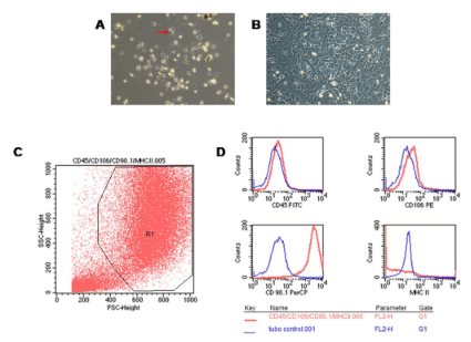
The isolation of BM-derived MSC was accomplished by culturing BM cells obtained from femora and tibias. MSC formed a heterogeneous population of cells that proliferated in vitro as plastic-adherent cells, had a fibroblast-like morphology, and formed colonies in vitro. After 2-3 passages, cells with a fibroblastic appearance reached confluence (Figure 1A, 1B). An enrichment in MSC was documented 1 month later by positive staining of cultured cells with anti-CD90.1 (Thy 1) and a slight expression of CD106 (V-CAM). However, they were negatively stained for CD45 (hematopoietic lineage marker) and MHC class II molecules (Figure 1C, 1D).


Figure 1. MSC Morphology at the Time of the First Passage (A), and at the Time of the Second Passage (B). MSC from Controls Proliferated and Acquired a
Spindle-Shaped Morphology (arrow). C: Immunophenotypic Assays of MSC from Healthy Donors. R1 Represents the Selection of the Percentages of Viable MSC. D: Differential Expression of the Markers Analyzed between Controls (blue line) and the MSC Phenotype (red line).

NOJ-4-128 Fig 1


Immunomodulatory Effects of MSC on T- and B-Cells

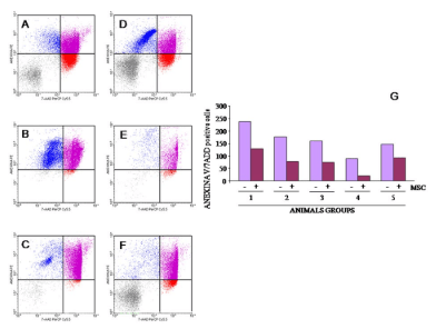
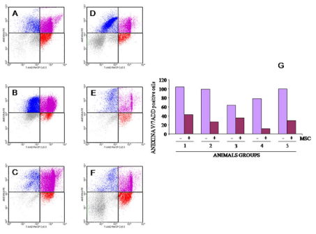
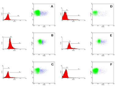
To investigate the immunomodulatory effects of purified MSCs, myelin-sensitized lymphocytes (obtained from EAE rats and healthy donor rats) were cultured with MSC at 1:10 concentration. For this purpose, purified T- or B-lymphocytes were cultured for 48 or 72 hours with or without MSC in 96- or 48-well plates. MSC increased the viability of T- and B-cells in all cases studied. As shown in Figures 2 and 3, respectively, the presence of MSC increased T-cell (Figure 2) and B cell viability (Figure 3). Similar results were obtained in the rest of the experimental groups analysed after 3 days of culture. These results are shown in Table 1. Analyses with MTT confirmed those findings (data not shown) (Figures 4 and 5).


Figure 2. T-Cell Viability Assessed by Annexin/7AAD Staining in the Absence (A to C) or Presence (D to F) of MSC in some Cases Analysed. CD45RA cells were Excluded and CD3 Cells Positively Selected. The First Two Dot Blots are from the Healthy Donor Animal Group, the Second two from the EAE Animal Group, and the Third Two are from EAE Animals to which MSC were Administered before and after the Induction of EAE. Percentage of
Annexin/7ADD-Positive Cells for Samples Cultured with (violet column) or without (maroon column) MSC. (1) Control animals (group 1); (2) EAE
Animals (group 2); (3) EAE Animals to which MSC were Administered before the Induction of EAE (group 3); (4) EAE Animals to which MSC were
Administered after the Induction of EAE (group 4); and (5) EAE Animals to which MSC were Administered before and after the Induction of EAE (Group 5) (G).

NOJ-4-128 Fig 2


Figure 3. B-Cell Viability Assesed by Annexin/7AAD Staining after 4 Days of Culture
Increased Significantly in the Presence of MSC. CD45RA Cells were Selected. B-Cell Viability Assessed by Annexin/7AAD Staining in the Absence (A to C) or Presence (D to F) of MSC in some Cases Analysed. The First Two Dot Blots are from the Healthy Donor Animal Group, the Second Two Dot Blots are from the EAE Animal Group, and the Third
Two Dot Blots are from the EAE Animals to which MSC were Administered before and after the Induction of EAE. G: Annexin/7ADD-Positive Cells for Samples Cultured with Violet Column) or without (Maroon Column) MSC: (1) Group 1; (2) Group 2; (3) Group 3; (4) Group 4; and (5) Group 5 of Experimental Animals.

NOJ-4-128 Fig 3


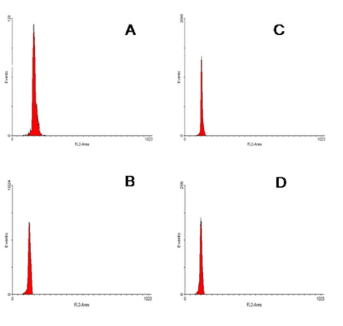
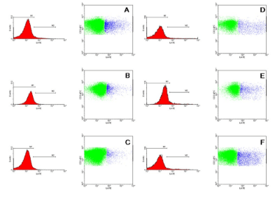
Figure 4: Cell Cycle of T-Cells Cultured in the Absence of MSC (A-B) or in the Presence of MSC (C-D). The First Two Dot Blots are from Group 1 of the Experimental Animals, and the Next Dot Blots
are from Group 5 of the Experimental Animals. No Changes in the T-Cell Cycle were Observed in any Case.

NOJ-4-128 Fig 4


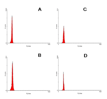
Figure 5: The same as in Figure 4 but Referring to the B-Cell Cycle.

NOJ-4-128 Fig 5


Table 1: Among these Cells the Number of Events Negative for both Annexin and 7-AAD was Calculated. A Calibrated Number of Microbeads was used.

1- MSC 1+ MSC 2- MSC 2+ MSC 3- MSC 3+ MSC 4- MSC 4+ MSC 5- MSC 5 + MSC
ANEXIN V+/7AAD+ 103.88 42.56 98.23 26.49 63.46 35.53 77.62 11.04 99.02

29.40

VIABLE

23.29 49.13 10.48 11.12 8.45 6.95 15.72 3.46 3.69

25.90

T- or B-cell culture induced a low proliferation, fewer than 5% of cells being in the S or G2M phases of the cell cycle. Culture with MSC does not modify the cell cycle of T (Figure 4) or B (Figure 5) cells in a quiescent state. Similar results were obtained in the rest of the experimental groups analysed after 3 days of culture, as shown in Table II.


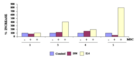
In order to analyse the production of soluble cytokines, the production of T-cell cytokines with or without MSC was studied. In the cases studied, MSC modulated the production of soluble cytokines in a different way. In control animals, MSC maintained or decreased the production of soluble IFN-γ, whereas in the EAE animals they increased its production (Figure 6). The opposite occurred with the production of IL-4 (Figure 7). Healthy T-lymphocytes secreted more IL-4 with MSC in the culture than EAE T-lymphocytes (Figure 8).


Figure 6: Percentage of IFN-g (Blue E)-Positive T-Cells after 3 Days of Culture without (A-C) MSC or with (D-F) MSC. Group 1 of the Experimental Animals (A, D), Group 3 of the Experimental Animals (B, E), and Group 5 of the Experimental Animals (C, F).

NOJ-4-128 Fig 6


Figure 7: Percentage of IL-4 (Blue Events)-Positive T-Cells after 3 Days of Culture without (A-C) MSC or with (D-F) MSC. Purified CD3-Positive Cells
were Analysed. The First Two Blots are from Group 1 of the Experimental Animals, the Second Two Blots are from Group 3 of the Experimental Animals, and the Third Two Blots are from Group 5 of the Experimental Animals.

NOJ-4-128 Fig 7

Figure 8: Increase in Positive Cells After the Culture of T-lymphocytes in the Absence of MSC (Violet Column) or in Presence of MSC for IFN-γ (Maroon Column) or IL-4 (Yellow Column), for the Different Cases Analysed. (1) Group 1; (3) Group 3; (4) Group 4; and (5) Group 5 of Experimental Animals.

NOJ-4-128 Fig 8


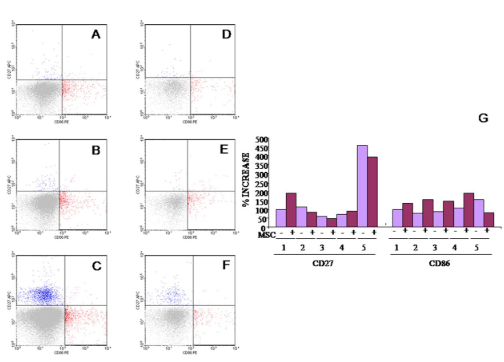
MSC modulated CD27/CD86 expression in B-cells. MSC increased the percentage of CD27-positive cells in healthy donors, whereas MSC significantly decreased CD27 expression in animals in which the clinical score was decreased due to MSC infusion (Figure 9). The same occurred with CD86 expression (Table 2).


Figure 9: Analysis of CD27/CD86-Positive B-Cells in the Absence of MSC (A-C) or in the Presence
of MSC (D-F) for 3 Days of Culture. The First Two Dot Blots are from Healthy Donor; the Next Ones
are from EAE Experimental Animals and the Last Ones are from EAE Animal to which MSC were
Administered before and after the Induction of EAE. G: Increase in Positive Cells after Culture of B Lymphocytes in the Absence of MSC (Violet Column) or in the Presence of MSC (Maroon Column) for CD27/CD86 Expression: (1) Group 1; (2) Group 2; (3) Group 3; (4) Group 4; and (5) Group 5 of
the Experimental Animals.

NOJ-4-128 Fig 9


Table 2: The Results Show, Respectively, the Different Phases of the Cell Cycle for T- or B-cells. In all the Cases not even 5% of the Cells were in S Phase and hence the Presence of MSC did not Modify the Cell Cycle in a Quiescent State.
T-Lymphocytes

1- MSC 1+ MSC 2- MSC 2+ MSC 3- MSC 3+ MSC 5- MSC 5+ MSC
G0/G1 88.25 96.9 96.02 98.9 92.4 97.22 97.25

96.64

S

1.71 1.78 3.29 2.05 0.15 0.41 0.01 0.03
G2/M 0 0 0 0 0 0 0

0

B-Lymphocytes

1- MSC 1+ MSC 2- MSC 2+ MSC 3- MSC 3+ MSC 5- MSC 5+ MSC
G0/G1 92.7 93.37 96.94 98.95 97.62 94.9 90.55

96.66

S

0.73 0.05 2.09 0 0.36 0.78 4.24 0.78
G2/M 0.26 0 0 0 0 0 0

0


Clinical Evaluation of EAE

Non-EAE immunized animals (group 1) showed a clinical score of 0. However, EAE-induced animals (groups 2-5) developed a mean clinical score of 2.2, with a standard deviation of 0.7 (the animals that did not develop the disease were not considered in the statistical data: one animal from group 3 and another animal from group 5). It is noteworthy that the animals infused with MSC prior to EAE immunization either failed to develop EAE or their EAE clinical scores were reduced. In contrast, the animals that received MSC after the induction of EAE developed a normal course of EAE.

In sum, rat MSC expressed the vascular cell adhesion molecule CD106 to a slight extent. MSC promoted T- or B-lymphocyte survival but did not modify the T- or B-lymphocyte cell cycles in the quiescent state. The results also confirmed that MSC modulated EAE though the production of soluble cytokines. In vitro, MSC decreased the EAE by immunomodulating the Th1/Th2 response. Moreover, MSC controlled CD27/CD86 expression in different ways. Animals infused with MSC prior to the EAE immunization did not develop EAE, or their EAE clinical scores were decreased, whereas animals that received MSC after the induction of EAE developed a normal EAE course.

DISCUSSION

MSC belong to a more recent field in the range of experimental therapies currently being developed to treat MS. While interest in the use of MSC was originally due to their potential capacity to differentiate into different cell lineages, recent work demonstrating their interesting immunological properties has led to a revised concept, envisaging their use for immunomodulatory purposes. These properties have already been exploited in the clinical setting for the treatment of severe autoimmune diseases. It has been reported that MSC inhibit monocyte-derived dendritic cell maturation as well as T-lymphocyte proliferation.52,53,54,55 MSC have been reported to inhibit the proliferation of T-cells through an MHC-independent mechanism,56 leading to the arrest of cell division.53 Although, the mechanisms involved in this inhibition of T-cell proliferation are still poorly understood, a veto-like activity has been reported,57 and a role for soluble molecules—including prostaglandin E2,54 transforming growth factor,58 and indoleamine 2,3-dioxygenase59—has been proposed. It has been reported that MSC increase the survival of unstimulated T-cells and inhibit the proliferation of activated T-cells.60 This is in accordance with previous studies indicating that MSC arrest T-cells in the G1 phase of the cell cycle53 and that this effect is mediated by the inhibition of cyclin D2 and the upregulation of p27kip1.

It is known that MSC inhibit B-cell proliferation and differentiation61; that MSC may induce both the expansion and differentiation of B-cells stimulated with an agonist of the Toll-like receptor (TLR) 9 in the absence of B cell receptor triggering62; and that MSC increase antibody secretion by human B-cells stimulated with lipopolysaccharide, cytomegalovirus, or varicellazoster virus.63 More recently, it has been described that MSC promote the survival and inhibit the proliferation and maturation of B-cells.64 These effects are mediated through the activation of MAPK pathways such as pErk 1/2 and p38. Here, it has been shown that MSC play a critical role in the immune regulation in EAE. Thus, we provide evidence that MSC given to Lewis 1A rats can, in vitro, support cell survival when T- or B-cells are in a quiescent state, leading to cell apoptosis. First, we observed that T- or B-cells cultured under non-stimulated conditions, a cell population physiologically prone to spontaneous cell death, are significantly rescued from apoptosis by the presence of MSC. Other authors have suggested that the protective effect of MSC mainly targets the “death receptor” pathway of apoptosis, through the downregulation of the Fas receptor and the Fas ligand on arrested T-cells.60 In this sense, and in the same manner, the increased viability induced by MSC in resting B-cells could explain the higher production of IgG upon weak stimulation, as shown in previous studies.63,64 However, we analysed the effect of MSC on T- or B-cell cycles and, contrary to the results showing that MSC can hamper T-cell proliferation through the inhibition of cell division and subsequent accumulation of cells in the G0 phase of the cell cycle,65 we did not find any difference between T- or B-cells cultured with or without MSC. This is also in part due to the absence of stimulating conditions and the low T- or B-cell proliferation capacity in the quiescent state.

However, there is a marked increase in the expression of TLR in MS brain lesions and cerebrospinal fluid mononuclear cells as well as in EAE brain lesions.66,67 TLR3 signals cause Th1 polarization with increased IFN-γ secretion concomitant with increased CD4 T-cell death.68 TLR signals are therefore potent modulators of microglial activation programs. The MSC-induced improvement was accompanied by changes in neural cell responses, with increased oligodendrocytes and decreased astrocytes in lesioned areas as well as changes in spleen cell responses.32 In active EAE, the predominant response is mediated through Th1 pro-inflammatory cells and the expression of their associated cytokines. In animals that received human BM-MSC, there was a significant reduction in pro-inflammatory cytokines, including IL-17, IFN-γ, IL-2, IL-12p70, and TNF-α, and a significant increase in anti-inflammatory cytokines, including IL-4 and IL-5. Moreover, it has been reported that MSC can suppress the T-cell proliferative response against TCR-dependent and -independent polyclonal stimuli.43 Such an effect was paralleled by a significant suppression of IFN-γ and TNF-α production by activated T-cells, supporting the notion of a profound inhibition of the Th1 response by MSC. Compatible with this, we observed that MSC regulated the balance of T lymphocytes between Th1/Th2 and modified the cytokines released during EAE. Thus, the presence of MSC increased the production of IFN-γ by T-cells in the EAE animals. However, this increase was not seen in the healthy animals or in the EAE animals belonging to experimental group 5 (animals to which MSC were administered before and after the induction of the EAE). Moreover, the production of IL-4 increased in all cases analysed. The most important aspect was the increase in the production of soluble IL-4 by T-cells in the EAE animals of group 5. This means that MS is a T-cell-mediated autoimmune disease, involving inflammatory demyelination of the CNS by CD4+ T-cells specific for myelin oligodendrocyte glycoprotein and other CNS autoantigens.69

In recent years, B-cells have emerged as a novel therapeutic target for treating MS, and clinical data with rituximab, as a B cell-depleting monoclonal antibody-based therapy, provide reciprocal conceptual support for a prominent role of B-cells in the pathogenesis of MS.70 Memory B-cells are significantly different from naïve B-cells, and the production of different effector cytokine profiles is a fundamental characteristic distinguishing naïve (CD27−) from memory (CD27+) human B-cells.71 Memory and naïve B-cells are considered to play different roles in immune regulation. However, the roles of memory and naïve B-cell subsets in MS have not yet been elucidated. Moreover, CD80 and CD86 are major costimulatory signals for T-cell activation, and variations in the expression of these proteins are likely to influence immune regulation in MS.72 Here, we examined the expression of CD86 and CD27 in B-cells by flow cytometry. In this regard, a high expression of CD27/CD86 was observed in the EAE animals of experimental group 5 versus healthy animals. In culture, this increment decreased in the presence of MSC. In the present study, we observed that subsets of memory and naïve B-cells differ between EAE animals and healthy control animals, and these differences could be exploited in the search for targets in MS therapies. Taken together, these results suggest that naïve and memory B-cell subsets play different active roles in the regulation of normal immune responses and indicate that abnormalities in these functions may contribute to MS. Moreover, the effect of MSC on B-cells antigen expression suggested that MSC could modulate the disease, which partly depended on the day on which the MSC were injected.

Finally, the clinical course of EAE was significantly ameliorated in the animals treated with MSC (intravenous administration). In this sense, it seems that the beneficial effects of MSC only appear if these cells are infused prior to the induction of EAE.

ACKNOWLEDGEMENTS

This work has been supported by Gemacbio (Saint Jean d’Illac, France), the Institut pour le Developpement de la Recherche en Pathologie Humaine et Therapeutique (IDRPHT, Talence, France), Red de Terapia Celular de Castilla y León (Spain), Consejería de Educación (Junta de Castilla y León, Spain) (SA099A08) and the INCYL-Federación de Cajas de Ahorro de Castilla y León (Spain). Arturo Mangas was supported by the Red de Terapia Celular de Castilla y León (Spain). The authors wish to thank to Professor Javier Yajeya (University of Salamanca, Spain) for his help in the studies carried out on the sciatic nerve motor conduction velocity. The authors also wish to thank Nicholas Skinner (University of Salamanca, Spain) for supervising the English text.

CONFLICTS OF INTEREST

The authors declare that they have no conflicts of interest.

1. Cao Q, Benton RL, Whittemore SR. Stem cell repair of central nervous system injury. J Neurosci Res. 2002; 68(5): 501-510. doi: 10.1002/jnr.10240

2. McDonald JW, Howard MJ. Repairing the damaged spinal cord: A summary of our early success with embryonic stem cell transplantation and remyelination. Prog Brain Res. 2002; 137: 299-309. doi: 10.1016/S0079-6123(02)37023-7

3. Campos LS. Neurospheres: Insights into neural stem cell biology. J Neurosci Res. 2004; 78: 761-769. doi: 10.1002/jnr.20333

4. Reubinoff BE, Itsykson P, Turetsky T, et al. Neural progenitors from human embryonic stem cells. Nat Biotech. 2001; 19: 1134-1140. doi: 10.1002/jnr.20333

5. Oyama Y, Craig RM, Traynor AE, et al. Autologous hematopoietic stem cell transplantation in patients with refractory Crohn’s disease. Gastroenterology. 2005; 128(3): 552-563. doi: 10.1053/j.gastro.2004.11.051

6. Mahmood A, Lu D, Wang L, Chopp M. Intracerebral transplantation of marrow stromal cells cultured with neurotrophic factors promotes functional recovery in adult rats subjected to traumatic brain injury. J Neurotrauma. 2002; 19(12): 1609-1617. doi: 10.1089/089771502762300265

7. Akiyama Y, Radtke C, Kocsis JD. Remyelination of the rat spinal cord by transplantation of identified bone marrow stromal cells. J Neurosci. 2002; 22: 6623-6630. doi: 10.1523%2FJNEUROSCI.22-15-06623.2002

8. Pluchino S, Quattrini A, Brambilla E, et al. Injection of adult neurospheres induces recovery in a chronic model of multiple sclerosis. Nature. 2003; 422: 688-694. doi: 10.1038/nature01552

9. Pluchino S, Zanotti L, Rossi B, et al. Neurosphere-derived multipotent precursors promote neuroprotection by an immunomodulatory mechanism. Nature. 2005; 436: 266-271. doi: 10.1038/nature03889

10. Anderson P, González-Rey E, O’Valle F, Martín F, Oliver FJ, Delagado M. Allogeneic adipose-derived mesenchymal cells ameliorate experimental autoimmune encephalomyelitis by regulating self-reactive T cell reponses and dendritic cell function. Stem Cells Int. 2017. doi: 10.1155/2017/2389753

11. Marín-Bañasco C, Benabdellah K, Melero-Jerez C, et al. Gene therapy with mesenchymal stem cells expressing IFN-β ameliorates neuroinflammation in experimental models of multiple sclerosis. Br J Pharmacol. 2017; 174: 238-253. doi: 10.1111/bph.13674

12. Mohammadzadeh A, Pourfathollah AA, Shahrokhi S, et al. Evaluation of AD-MSC (adipose-derived mesenchymal stem cells) as a vehicle for IFN-β delivery in experimental autoimmune encephalomyelitis. Clin Immunol. 2016; 169: 98-106. doi: 10.1016/j.clim.2016.06.015

13. Yousefi F, Ebtekar M, Soudi S, Soleimani M, Hashemi SM. In vivo immunomodulatory effects of adipose-derived mesenchymal stem cells conditioned medium in experimental autoimmune encephalomyelitis. Immunol Lett. 2016; 172: 94-105. doi: 10.1016/j.imlet.2016.02.016

14. Shalaby SM, Sabbah NA, Saber T, Abdel Hamid RA. Adipose-derived mesenchymal stem cells modulate the immune response in chronic experimental autoimmune encephalomyelitis model. IUBMB Life. 2016; 68: 106-115. doi: 10.1002/iub.1469

15. Jiang H, Zhang Y, Tian K, Wang B, Han S. Amelioration of experimental autoimmune encephalomyelitis through transplantation of placenta-derived mesenchymal stem cells. Sci Rep. 2017. doi: 10.1038/srep41837

16. Selim AO, Selim SA, Shalaby SM, Mosaad H, Saber T. Neuroprotective effects of placenta-derived mesenchymal stem cells in a rat model of experimental autoimmune encephalomyelitis. Cytotherapy. 2016; 18: 1100-1013. doi: 10.1016/j.jcyt.2016.06.002

17. Torkaman M, Ghollasi M, Mohammadnia-Afrouzi M, Salimi A, Amari A. The effect of transplanted human Wharton’s jelly mesenchymal stem cells treated with IFN-γ on experimental autoimmune encephalomyelitis. Cell Immunol. 2017; 311: 1-12. doi: 10.1016/j.cellimm.2016.09.012

18. Bravo B, Gallego MI, Flores AI, et al. Restrained Th17 response and myeloid cell infiltration into the central nervous sytem by human decidua-derived mesenchymal stem cells during experimental autoimmune encaphalomyelitis. Stem Cell Res Ther. 2016; 7: 43. doi: 10.1186/s13287-016-0304-5

19. Rafieemehr H, Kheyrandish M, Soleimani M. Neuroprotective effects of transplanted stromal cells-derived human umbilical cord blood neural progenitor cells in EAE. Iran J Allergy Asthma Immunol. 2015; 14(6): 596-604.

20. Prockop DJ. Marrow stromal cells as stem cells for nonhematopoietic tissues. Science. 1997; 276: 71-74. doi: 10.1126/science.276.5309.71

21. Bossolasco P, Cova L, Calzarossa C, et al. Neuro-glial differentiation of human bone marrow stem cells in vitro. Exp Neurol. 2005; 193: 312-325. doi: 10.1016/j.expneurol.2004.12.013

22. Krampera M, Pasini A, Pizzolo G, Cosmi L, Romagnani S. Annunziato F. Regenerative and immunomodulatory potential of mesenchymal stem cells. Curr Opin Pharmacol. 2006; 6: 435-441. doi: 10.1016/j.coph.2006.02.008

23. Spaggiari GM, Abdelrazik H, Becchetti F, Moretta L. MSCs inhibit monocyte-derived DC maturation and function by selectively interfering with the generation of immature DCs: Central role of MSC-derived prostaglandin E2. Blood. 2009; 113: 6576-6583. doi: 10.1182/blood-2009-02-203943

24. Chopp M, Li Y. Treatment of neural injury with marrow stromal cells. Lancet Neurol. 2002; 1: 92-100. doi: 10.1016/s1474-4422(02)00040-6

25. Zhang J, Li Y, Chen J, et al. Human bone marrow stromal cell treatment improves neurological functional recovery in EAE mice. Exp Neurol. 2005; 195: 16-26. doi: 10.1016/S1474-4422(02)00040-6

26. Bartholomew A, Polchert D, Szilagyi E, Douglas GW, Kenyon N. Mesenchymal stem cells in the induction of transplantation tolerance. Transplantation. 2009; 87: S55-S57. doi: 10.1097/TP.0b013e3181a287e6

27. Liao W, Pham V, Liu L, et al. Mesenchymal stem cells engineered to express selectin ligands and IL-10 exert enhanced therapeutic efficacy in murine experimental autoimmuneencephalomyelitis. Biomaterials. 2016; 77: 87-97. doi: 10.1016/j.biomaterials.2015.11.005

28. Gordon D, Pavlovska G, Uney JB, Wraith DC, Scolding NJ. Human mesenchymal stem cells infiltrate the spinal cord, reducede myelination, and localize to white matter lesions in experimental autoimmune encephalomyelitis. J Neuropathol Exp Neurol. 2010; 69: 1087-1095. doi: 10.1097/NEN.0b013e3181f97392

29. Hou Y, Heon Ryu C, Jun JA, Kim SM, Jeong CH, Jeun SS. Interferon β-secreting mesenchymal stem cells combined with minocycline attenuate experimental autoimmune encephalomyelitis. J Neuroimmunol. 2014; 274: 20-27. doi: 10.1016/j.jneuroim.2014.06.001

30. Gerdoni E, Gallo B, Casazza S, et al. Mesenchymal stem cells effectively modulate pathogenic immune response in experimental autoimmune encephalomyelitis. Ann Neurol. 2007; 61: 219-227. doi: 10.1016/j.jneuroim.2014.06.001

31. Kassis I, Grigoriadis N, Gowda-Kurkalli B, et al. Neuroprotection and immunomodulation with mesenchymal stem cells in chronic experimental autoimmune encephalomyelitis. Arch Neurol. 2008; 65: 753-761. doi: 10.1001/archneur.65.6.753

32. Bai L, Lennon DP, Eaton V, et al. Human bone marrow-derived mesenchymal stem cells induce Th2-polarized immune response and promote endogenous repair in animal models of multiple sclerosis. Glia. 2009; 57: 1192-1203. doi: 10.1002/glia.20841

33. Rafei M, Campeau PM, Aguilar-Mahecha A, et al. Mesenchymal stromal cells ameliorate experimental autoimmune encephalomyelitis by inhibiting CD4 Th17 T cells in a CC chemokine ligand 2-dependent manner. J Immunol. 2009; 182: 5994-6002. doi: 10.4049/jimmunol.0803962

34. Liu XJ, Zhang JF, Sun B, et al. Reciprocal effect of mesenchymal stem cell on experimental autoimmune encephalomyelitis is mediated by transforming growth factor-beta and interleukin-6. Clini Exp Immunol. 2009; 158: 37-44. doi: 10.1111/j.1365-2249.2009.03995.x

35. Iglesias A, Bauer J, Litzenburger T, Schubart A, Linington C. T- and B-cell responses to myelin oligodendrocyte glycoprotein in experimental autoimmune encephalomyelitis and multiple sclerosis. Glia. 2001; 36: 220-234. doi: 10.1002/glia.1111

36. Lyons JA, San M, Happ MP, Cross AH. B cells are critical to induction of experimental allergic encephalomyelitis by protein but not by a short encephalitogenic peptide. Eur J Immunol. 1999; 29: 3432-3439. doi: 10.1002/(SICI)1521-4141(199911)29:11<3432::AID-IMMU3432>3.0.CO;2-2

37. McLaughlin KA, Wucherpfennig KW. B cells and autoantibodies in the pathogenesis of multiple sclerosis and related inflammatory demyelinating diseases. Adv Immunol. 2008; 98: 121-149. doi: 10.1016/S0065-2776(08)00404-5

38. Shoham T, Parameswaran R, Shav-Tal Y, Barda-Saad M, Zipori D. The mesenchymal stroma negatively regulates B cell lymphopoiesis through the expression of activin A. Ann NY Acad Sci. 2003; 996: 245-260. doi: 10.1111/j.1749-6632.2003.tb03253.x

39. Krampera M, Cosmi L, Angeli R, et al. Role for interferon-gamma in the immunomodulatory activity of human bone marrow mesenchymal stem cells. Stem Cells. 2006; 24: 386-398. doi: 10.1634/stemcells.2005-0008

40. Nishikawa S, Honda K, Vieira P, Yoshida H. Organogenesis of peripheral lymphoid organs. Immunol Rev. 2003; 195: 72-80. doi: 10.1034/j.1600-065X.2003.00063.x

41. Amé-Thomas P, Maby-El HH, Monvoisin C, et al. Human mesenchymal stem cells isolated from bone marrow and lymphoid organs support tumor B-cell growth: Role of stromal cells in follicular lymphoma pathogenesis. Blood. 2007; 109: 693-702. doi: 10.1182/blood-2006-05-020800

42. De Ugarte DA, Alfonso Z, Zuk PA, et al. Differential expression of stem cell mobilization-associated molecules on multi-lineage cells from adipose tissue and bone marrow. Immunol Lett. 2003; 89: 267-270. doi: 10.1016/S0165-2478(03)00108-1

43. Zappia E, Casazza S, Pedemonte E, et al. Mesenchymal stem cells ameliorate experimental autoimmune encephalomyelitis inducing T-cell anergy. Blood. 2005; 106: 1755-1761. doi: 10.1182/blood-2005-04-1496

44. Yu JW, Li YN, Song GB, et al. Synergistic and superimposed effect of bone marrow-derived mesenchymal stem cells combined with fasudil in experimental autoimmune encephalomyelitis. J Mol Neurosci. 2016; 60: 486-497. doi: 10.1007/s12031-016-0819-3

45. Togha M, Jahanshahi M, Alizadeh L, et al. Rapamycin augments immunomodulatory properties of bone marrow-derived mesenchymal stem cells in experimental autoimmune encephalomyelitis. Mol Neurobiol. 2017; 54: 2445-2457. doi: 10.1007/s12035-016-9840-3

46. Wang D, Li SP, Fu JS, Bai L, Guo L. Resveratrol augments therapeutic efficiency of mouse bone marrow mesenchymal stem cell-based therapy in experimental autoimmune encephalomyelitis. Int J Dev Neurosci. 2016. doi: 10.1016/j.ijdevneu.2016.01.005

47. Hao F, Li A, Yu H, et al. Enhanced neuroprotective effects of combination therapy with bone marrow-derived mesenchymal stem cells and Ginkgo biloba extract (EGb761) in a rat model of experimental autoimmune encephalomyelitis. Neuroimmunomodulation. 2016; 23: 41-57. doi: 10.1159/000437429

48. Luz-Crawford P, Kurte M, Bravo-Alegría J, et al. Mesenchymal stem cells generate a CD4+CD25+Foxp3+ regulatory T cell population during the differentiation process of Th1 and Th17 cells. Stem Cell Res Ther. 2013; 4: 65. doi: 10.1186/scrt216

49. Weissert R, Wallstrom E, Storch MK, et al. MHC haplotype-dependent regulation of MOG-induced EAE in rats. J Clin Invest. 1998; 102: 1265-1273. doi: 10.1186/scrt216

50. Villarón EM, Almeida J, López-Holgado N, et al. Mesenchymal stem cells are present in peripheral blood and can engraft after allogeneic hematopoietic stem cell transplantation. Haematologica. 2004; 89(12): 1421-1427.

51. Dominici M, Le BK, Mueller I, et al. Minimal criteria for defining multipotent mesenchymal stromal cells. The International Society for Cellular Therapy position statement. Cytotherapy. 2006; 8: 315-317.

52. Rasmusson I, Ringden O, Sundberg B, Le Blanc K. Mesenchymal stem cells inhibit lymphocyte proliferation by mitogens and alloantigens by different mechanisms. Exp Cell Res. 2005; 305: 33-41. doi: 10.1016/j.yexcr.2004.12.013

53. Glennie S, Soeiro I, Dyson PJ, Lam EW, Dazzi F. Bone marrow mesenchymal stem cells induce division arrest anergy of activated T cells. Blood. 105: 2821-2827. doi: 10.1182/blood-2004-09-3696

54. Aggarwal S, Pittenger MF. Human mesenchymal stem cells modulate allogeneic immune cell responses. Blood. 2005; 105: 1815-1822. doi: 10.1182/blood-2004-04-1559

55. Jiang XX, Zhang Y, Liu B, et al. Human mesenchymal stem cells inhibit differentiation and function of monocyte-derived dendritic cells. Blood. 2005; 105: 4120-4126. doi: 10.1182/blood-2004-02-0586

56. Le BK, Tammik L, Sundberg B, Haynesworth SE, Ringden O. Mesenchymal stem cells inhibit and stimulate mixed lymphocyte cultures and mitogenic responses independently of the major histocompatibility complex. Scan J Immunol. 2003; 57: 11-20. doi: 10.1046/j.1365-3083.2003.01176.x

57. Potian JA, Aviv H, Ponzio NM, Harrison JS, Rameshwar P. Veto-like activity of mesenchymal stem cells: Functional discrimination between cellular responses to alloantigens and recall antigens. J Immunol. 2003; 171: 3426-3434. doi: 10.4049/jimmunol.171.7.3426

58. Di Nicola M, Carlo-Stella C, Magni M, et al. Human bone marrow stromal cells suppress T-lymphocyte proliferation induced by cellular or nonspecific mitogenic stimuli. Blood. 2002; 99: 3838-3843. doi: 10.1182/blood.V99.10.3838

59. Meisel R, Zibert A, Laryea M, Gobel U, Daubener W, Dilloo D. Human bone marrow stromal cells inhibit allogeneic T-cell responses by indoleamine 2,3-dioxygenase-mediated tryptophan degradation. Blood. 2004; 103: 4619-4621. doi: 10.1182/blood-2003-11-3909

60. Benvenuto F, Ferrari S, Gerdoni E, et al. Human mesenchymal stem cells promote survival of T cells in a quiescent state. Stem Cells. 2007; 25: 1753-1760. doi: 10.1634/stemcells.2007-0068

61. Corcione A, Benvenuto F, Ferretti E, et al. Human mesenchymal stem cells modulate B-cell functions. Blood. 2006; 107: 367-372. doi: 10.1182/blood-2005-07-2657

62. Traggiai E, Volpi S, Schena F, et al. Bone marrow-derived mesenchymal stem cells induce both polyclonal expansion and differentiation of B cells isolated from healthy donors and systemic lupus erythematosus patients. Stem Cells. 2008; 26: 562-569. doi: 10.1634/stemcells.2007-0528

63. Rasmusson I, Le BK, Sundberg B, Ringden O. Mesenchymal stem cells stimulate antibody secretion in human B cells. Scan J Immunol. 2007; 65: 336-343. doi: 10.1111/j.1365-3083.2007.01905.x

64. Tabera S, Pérez-Simón JA, Díez-Campelo M, et al. The effect of mesenchymal stem cells on the viability, proliferation and differentiation of B-lymphocytes. Haematologica. 2008; 93: 1301-1309. doi: 10.3324/haematol.12857

65. Krampera M, Glennie S, Dyson J, et al. Bone marrow mesenchymal stem cells inhibit the response of naive and memory antigen-specific T cells to their cognate peptide. Blood. 2003; 101: 3722-3729. doi: 10.1182/blood-2002-07-2104

66. Bsibsi M, Ravid R, Gveric D, van Noort JM. Broad expression of Toll-like receptors in the human central nervous system. J Neuropathol Exp Neurol. 2002; 61: 1013-1021. doi: 10.1093/jnen/61.11.1013

67. Andersson A, Covacu R, Sunnemark D, Danilov AI, Dal BA, Khademi M. Pivotal advance: HMGB1 expression in active lesions of human and experimental multiple sclerosis. J Leukoc Biol. 2008; 84(5): 1248-1255. doi: 10.1189/jlb.1207844

68. Jack CS, Arbour N, Blain M, Meier UC, Prat A, Antel JP. Th1 polarization of CD4+ T cells by Toll-like receptor 3-activated human microglia. J Neuropathol Exp Neurol. 2007; 66: 848-859. doi: 10.1097/nen.0b013e3181492a7

69. Williams KC, Ulvestad E, Hickey WF. Immunology of multiple sclerosis. J Clin Neurosci. 1994; 2: 229-245.

70. Hauser SL, Waubant E, Arnold DL, et al. B-cell depletion with rituximab in relapsing-remitting multiple sclerosis. New Eng J Med. 2008; 358: 676-688. doi: 10.1056/NEJMoa0706383

71. Duddy M, Niino M, Adatia F, et al. Distinct effector cytokine profiles of memory and naive human B cell subsets and implication in multiple sclerosis. J Immunol. 2007; 178: 6092-6099. doi: 10.4049/jimmunol.178.10.6092

72. Kuchroo VK, Das MP, Brown JA, et al. B7-1 and B7-2 costimulatory molecules activate differentially the Th1/Th2 developmental pathways: Application to autoimmune disease therapy. Cell. 1995; 80(5): 707-718. doi: 10.1016/0092-8674(95)90349-6

LATEST ARTICLES

Unraveling the Mysteries of Type-A Aortic Dissection Using POCUS/Echocardiography

Syeda Rukh*, Sathyanarayana Machani and Milind Awale

doi.

Blood Sample from the Patient

Hypertriglyceridemia-Induced Pancreatitis: A Case Report and Literature Review

Maarten Bulterys, Melvin Willems* and Agnes Meersman

doi.

From Neck Pain to a Life-Threatening Condition: A Case Report

Floris Vandewoude* and Sören Verstraete

doi.

LATEST ARTICLES