Advances in Food Technology and Nutritional Sciences

Open journal

ISSN 2377-8350

Green Tea Catechins as Neuroprotective Agents: Systematic Review of the Literature in Animal Pre-Clinical Trials

Natasha Katergaris, Lisa Dufficy, Paul D. Roach and Nenad Naumovski*

Nenad Naumovski, PhD

Assistant Professor of Food Science and Human Nutrition School of Public Health and Nutrition, Faculty of Health, University of Canberra, Canberra 2601, ACT, Australia; Tel. +61-02-6206-8719; Fax: +61-02-6201-5999; E-mail: nenad.naumovski@canberra.edu.au

INTRODUCTION

Mental disease represents one of the world’s leading disabilities affecting over 25% of people at some period during their lifetime. In 2004, it represented 13% of the global burden of disease and it is proposed to increase to 15% by the year 2020, which will place it as the second leading cause of disability in the world.1,2 Alzheimer’s Disease (AD) and cognitive impairment are on the increase and have been identified as contributing to the overall increase in mental disease, which indicates an increasing prevalence of disorders of later life rather than of childhood.3-5 The increasing incidence of these types of mental disease can also be viewed as being due to the rapid ageing of the world’s population and the fall in the birth rate, which has resulted in an overall lower prevalence of the types of mental disease associated predominately with adolescence.6

Much of the scientific research in the elderly has been devoted to studies on the implications of the cognitive decline and impairment that characterise the dementive-type illnesses. Dementia is a disease of the brain, and the term “dementia” is classified as an “umbrella” term for various types of diseases. These are characterised by the development of multiple cognitive impairments that can arise due to different causes such as direct physiological effects of a medical condition, persisting effects of substance abuse or multiple other aetiologies. Therefore, there are various types of dementia such as dementia of the Alzheimer’s type, vascular dementia and dementia due to various diseases such as HIV and Parkinson’s disease.7,8 It is also important to note that the aetiology of certain types of multiple cognitive deficits cannot be identified and thus, “Dementia Not Otherwise Specified” is another type of this disease.7

Studies have identified dementia of the Alzheimer’s type to be the most prevalent form of dementia.9,10 The AD dementia has therefore become the most well-known type of dementia chiefly associated with older age.7,11,12

The onset of AD is categorised by two subtypes according to age: early onset AD where symptoms of this type of dementia manifest themselves before 65 years of age and late onset AD with symptoms emerging after the age of 65.7 The use of 65 as the age cut-off point is completely arbitrary from a health point of view as it does not have any medical significance.13 Obviously however, it is related to the age that is normally associated with retirement from work.

The behavioural and cognitive onset of AD can be slow and gradual; it predominately manifests itself as a slow but progressive cognitive decline.7 Furthermore, there is no one universally accepted neuropathologic criteria to differentiate AD from healthy brain ageing primarily because AD is a complex neurodegenerative dementing illness.14-16 However, the fundamental neuropathologic features of AD, based on the post-mortem examination of patients’ brains, include reduced brain volume, enlarged ventricular spaces, region specific neurofibrillary tangles and neuritic amyloid plaques.17,18 The exact causative factor for these brain changes still remains unknown. However, the final resulting physiological outcome is death of neural cells which causes severe cognitive impairment and eventually death of the patient suffering from AD.11,13,15,16

It has been proposed that death of the neural cells is a result of a cascade of intracellular and extracellular events.16,17

There are several different factors which have been postulated to directly or indirectly play important roles in the initiation of these events: oxidative damage,19,20 genetic polymorphisms,21,22 gene mutations23 and abnormal levels of β-amyloid and tau proteins.24 Of these, the deposition of β-amyloid protein is seen as the primary event in the pathogenesis of AD. The other changes such as neurofibrillary tangles, synaptic degeneration and neuronal cell death are proposed to arise only as a consequence of the deposition of this protein in the brain.25

In addition, neurodegeneration is a common feature of AD with the main reasons described to be the combination of multi-factorial events such as neuro-inflammation, glutamate rich excitotoxicity and depletion of antioxidants.26 Hence, in recent years there has been developing evidence proposing that dietary polyphenols can potentially counteract and suppress the neuronal injury.26-29

Polyphenols are ubiquitous compounds classified as wide and complex group of plant secondary metabolites and to date there have been over 8000 compounds identified. Additionally, these compounds are exceptionally diverse in their structures, ranging from very simple molecules (phenolic acids) to more complex and highly polymerized structures (proanthocyanidins).30-36 The polyphenols are generally divided into two groups based on their molecular weight, low (500-3000 Da) and high molecular weight (>3000 Da).37 In addition, these compounds can also be classified into different groups based on the number of phenolic rings that they contain and on the basis of the structural elements by which these rings are connected to one another. They include such compounds as phenolic acids, flavonoids, stilbenes and lignans (Figure 1).

Figure 1: Brief representation of the polyphenols classification with focus on green tea catechins.

AFTNSOJ-1-108Fig1


One of the foods rich in bioactive compounds, particularly flavonoids, is green tea (Camellia sinensis). In recent years, there has been a significant upward trend in the consumption of green tea and taking into consideration that overall tea (black and green) is one of the most consumed beverages in the world, it is not surprising that this trend was closely followed by academic research interest.

The most abundant polyphenolic compounds in green tea are the catechins, which account for nearly 30% of the dry tea leaf weight.38 In the recent literature, the Green Tea Catechins (GTC) have been related to a variety of different beneficial health effects particularly with respect to their potential for preventing and treating different cancers,39,40 cardiovascular diseases,41-43 inflammatory diseases44-47 and some of the neurodegenerative diseases45,48,49 in humans. Accounting for at least half of the total GTC,38 the most predominant catechin found in green tea is Epigallocatechin gallate (EGCG), which has been ascribed numerous beneficial properties including antioxidant,50 antiinflammatory,51,52 anti-microbial53,54 and anticancer effects.55-57

Although current trend in the scientific writing of systematic reviews is chiefly reserved for trials with human subjects from various cohorts, the use of well-designed pre-clinical animal trials is gaining value. This is primarily due to the increased number of well-developed gene-knockout animal models that can be used to mimic certain diseases commonly occurring in humans. Additionally, some of the illnesses such as AD can only be successfully diagnosed post-mortem. Therefore, keeping in mind that findings from animal models are not necessarily directly transferrable to humans, these studies still provide valuable insight into the potential mechanisms of action that may be applied to clinical research in humans. Therefore, the aim of this systematic review is to look into what is known about the relationship between GTC and the leading biomarkers associated with the development of AD.

METHODS

Animals

The animals used as models in the pre-clinical animal studies covered in this systematic review were rodents in particular rats (JCL: Wistar) and mice (C57/BL; Tg2576; APPsw; non-transgenic).

Search Strategy

The standardized criteria for conducting and reporting systematic reviews of observational studies based on the PRISMA 2009 guidelines58 were used. The search strategies were applied for articles published since the year 2000 using the following electronic databases: PubMed (http://www.ncbi.nlm.nih. gov/pubmed/), the Cochrane Library (http://www.thecochranelibrary.com/view/0/index.html), and Scopus (http://www.elsevier. com/online-tools/scopus). Titles and abstracts were scanned for relevance and appropriate articles were selected.

The search used terms relating to the topic of this systematic review; “Green tea polyphenols” and “Alzheimer’s Disease” were used in the primary search followed by “Beta Amyloid”, “Neuroprotection” and “Catechins” in the secondary search. In addition, reference list checks of selected articles with relevance to the topic were also performed.

Selection Criteria

For the purpose of this systematic review, articles were selected only if they were entirely published in English, in peer-reviewed journals and were identified as pre-clinical animal trials. They were included if GTC were delivered as a part of a supplement, injection, oral consumption in tea, water, food or similar delivery methods. Furthermore, studies selected for review had sample sizes of over 5 animals (at the end of the study) of any gender and had identified looking at whether or not GTC affected one or all of the biomarkers in relation to the development and onset of AD.

Primary Outcomes

The primary outcomes were cognitive learning and abnormal levels of β-amyloid and tau proteins in brain homogenates.

Secondary Outcomes

The secondary outcomes were oxidative stress identification by the presence of Lipid peroxide (LPO) by-products and generation of Reactive Oxygen Species (ROS).

Materials

Studies used green tea extracts, GTC or pure EGCG in the intervention and the control groups were always administered with water containing no additional compounds or in a case of injections, animals were injected Intraperitonealy (IP) with sterile saline or not injected at all.

RESULTS

Characteristics of the Studies

Preliminary searches of the selected databases identified 4635 records with the terms “Green tea polyphenols” and “Alzheimer’s disease” alone (Figure 2) but the secondary search reduced this to 500 articles. These 500 abstracts were screened, of which only 3 studies (1 in rats and 2 in mice) were included in the review because they fitted all the eligibility criteria.59-61 These 3 articles were published after the year 2000; included animals in the pre-clinical setting treated orally or injected with a GTC supplement or an individual purified catechin and also included control groups.

Figure 2: Flow chart of the publication selection process.

AFTNSOJ-1-108Fig2


GTC or EGCG Supplementation Doses

The 3 articles included in this review utilized either a GTC preparation or pure EGCG as the treatment method in animals. In the study with rats,59 the animals were orally treated with an aqueous GTC extract, referred to as Polyphenon E (Mitusi Norin CO. Ltd, Tokyo, Japan), which had an initial concentration of total catechins of 0.5% (w/v). The extract was diluted daily in the animals’ drinking water to provide the following concentrations of the individual catechins: EGCG (63%); EC (11%); EGC (6%) and ECG (6%). The control group received drinking water with no catechins in it.

One of the 2 studies in mice60 also used a GTC powdered extract (Plant extract GmbH & Co, KG, Vestenbergsgreuth, Germany) in which the catechin concentration was said to vary between 12 and 17% (w/w). In this study, some mice were IP injected with 0.5, 1 or 5 mg/kg of the GTC extract in saline (twice on the first day of the experiment with a 6 hr interval) followed by once for the next 4 days. Control mice were injected with saline. In the same paper, other mice received oral treatment with pure EGCG at two concentration levels (2 and 10 mg/kg/day) for 14 days.

In the other mice study,61 some of the animals (Tg2576 females) were orally administered with pure EGCG (50 mg/kg) in water or water only (control) on a daily basis for 6 months. Other mice (female APPsw) were treated by IP administration of EGCG (20 mg/kg) or vehicle only (control) daily for 60 days.

Cognitive Testing

Rats were tested for learning-related cognitive ability with the use of the eight-arm radial maze with a particular focus on the reference memory error and working memory error.59 The Radial Arm Water Maze (RAWM) was also used to assess the experimental mice for working memory.61

Induction of Cognitive Deficits/Mimicking of Ad Symptoms in Animals

One of the mice studies included “Swedish” mutant amyloid precursor protein overexpressing (APPsw, Tg) mice,61 while the other study injected the mice IP with N-methyl-4-phenyl-1,2,3,6-tetrahydropyridine (MPTP) at levels of 24 mg/kg/ day in order to replicate the clinical AD symptoms.60 However, in the Wistar rat study, the animals were infused with Aβ1-40 (4.9- 5.5 mmol/l) into the exposed left ventricle after the injection of AlCl3 (0.5 µg) was performed in the right ventricle.59

Tissue Analysis/Testing

In the mice studies, the brain homogenate tissues were analyzed using Western blots and immunohistochemistry techniques with a specific focus on soluble Aβ1-40 61 and tyrosine hydroxylase.60 In the rat study,59 plasma and brain homogenates were tested for LPO concentrations using the Thiobarbituric acid-reactive substances (TBARS) assay while plasma was also analyzed for total antioxidant activity using the Ferric Reducing Ability of Plasma (FRAP) assay and for plasma triglycerides and total cholesterol using commercially available enzyme kits.

Study Outcomes

One of the studies by Rezai-Zadeh, et al.61 was done to determine if the oral administration of ECGC would cause a reduction in plaque build-up in the APPsw mice. It was found that the ECGC (50 mg/kg) treated mice had significantly reduced (p<0.05) plaque build-up compared to controls (Table 1). This was supported by the micrographs of the brain sections where it was identified that β-amyloid stained antibody sections were substantially reduced in the cingulate and entorhinal cortex, as well as in the hippocampus by 54%, 51% and 43%, respectively. This was further supported by analysis of the anterior quarter brain homogenates, which indicated that the oral consumption of EGCG significantly (p><0.05) decreased both the soluble and insoluble forms of β-amyloid plaques.61 In addition, the Tau protein formation induced by the deposition of β-amyloid plaques was also suppressed in the EGCG treated mice.>< 0.05) plaque build-up compared to controls (Table 1). This was supported by the micrographs of the brain sections where it was identified that β-amyloid stained antibody sections were substantially reduced in the cingulate and entorhinal cortex, as well as in the hippocampus by 54%, 51% and 43%, respectively. This was further supported by analysis of the anterior quarter brain homogenates, which indicated that the oral consumption of EGCG significantly (p<0.05) decreased both the soluble and insoluble forms of β-amyloid plaques.61 In addition, the Tau protein formation induced by the deposition of β-amyloid plaques was also suppressed in the EGCG treated mice><0.05) decreased both the soluble and insoluble forms of β-amyloid plaques.61 In addition, the Tau protein formation induced by the deposition of β-amyloid plaques was also suppressed in the EGCG treated mice.

The second study by Rezai-Zadeh, et al.61 was done to examine whether EGCG had potential cognitive benefits, either by IP injection or by oral intake (Table 1). The results identified that the animals, after two months of IP injections with EGCG, had an improved working memory compared to the controls (p<0.001). This was supported during the final stage of the RAWM testing where the EGCG treated transgenic animals showed improvements between the trials while the control nontransgenic mice showed no working memory improvements. A similar finding was also identified for animals that consumed the EGCG orally (50 mg/kg); the transgenic mice were not weakened in working memory compared to the non-transgenic control animals. >< 0.001). This was supported during the final stage of the RAWM testing where the EGCG treated transgenic animals showed improvements between the trials while the control nontransgenic mice showed no working memory improvements. A similar finding was also identified for animals that consumed the EGCG orally (50 mg/kg); the transgenic mice were not weakened in working memory compared to the non-transgenic control animals.

Table 1: Summary of GTC effects in animal pre-clinical trials.

Author (Year) Animals, sample size, age Aim Intervention Results
Rezai-Zadeh, et al. (2008) Female “Swedish” mutant amyloid precursor protein overexpressing (APPsw) (All n=10; C:n=5; T:n=5) Female Tg2576 (All n=10; C:n=5; T:n=5) Female Non-transgenic mice (n=5) Age at start: 8 mth Anti-amyloidgenic effects of orally administered EGCG in APPsw mice Oral EGCG (50mg/kg) or control (water) treated for 6 mth. Treatment resulted in a significant reduction in plaque buildup (p>0.05) Treatment decreased soluble and insoluble forms of Aβ1-40,42, increased ADAM10 maturation and increased sAAP-α release (p<0.001) Provision of comparable attenuation of amyloid pathology to that of IP injections.><0.001) Provision of comparable attenuation of amyloid pathology to that of IP injections
Effect of IP injection EGCG treatment on Tau protein physiology in APPsw mice Treated 2 months with IP injection of pure EGCG (20mg/kg) or orally consumed EGCG (50mg/kg). Control group did not receive any treatment. Both treatments (oral and IP injection) decreased Tau hyperphosphorylation (p<0.001)
Cognitive benefits after oral administration of EGCG in APPsw mice Animals provided with EGCG treatment had substantially improved working memory performance, comparable to nontransgenic non-treated mice (p<0.001).
Haque, et al. (2008) Male JCL:Wistar rats (All=49; C:n=25; T:n=24) Age at start: 5 wk. Long-term administration of GTC and effect on oxidative stress and cognitive impairment in Aβ1-40 infused AD rat model. Treatment group was administered GTC combination [EGCG (63%), EC (11%), EGC (6%) and ECG (6%)] in water for 26 wk. Treatment prevented β-amyloid associated impairment in cognitive learning (p<0.0001). Treated rats showed significant decreased number of reference and working memory errors (p><0.05). Treated rats also had lower LPO (TBARS) in hippocampus and plasma and ROS in hippocampus and cortex and higher FRAP in plasma. ><0.05). Treated rats also had lower LPO (TBARS) in hippocampus and plasma and ROS in hippocampus and cortex and higher FRAP in plasma.
Levites, et al. (2001) Male C57/BL mice (All; n=144) The effects of GTC extract in MPTP injected animals Mice were IP injected twice on the first day with GTC extract (0.5mg/ kg), followed by injection of MPTP (24mg/kg) and GTC extract (0.5mg/ kg) for the subsequent four days. For EGCG studies, mice were orally administered EGCG (2 or 10mg/kg/ day) for 10 days and then EGCG (2 or 10mg/kg/day) and MPTP (24mg/ kg/day) for the following 4 days. Control mice received only saline or GTC extract The injected GTC extract and oral EGCG showed a protective effect against MPTPinduced decreases in dopamine and tyrosine hydroxylase protein levels (p<0.05).
The effect of EGCG on MPTP-induced neuro-degeneration The injected GTC extract also prevented the MPTP-induced decrease in dopamine neurons in the brains. (p<0.02).
The prevention of the MPTP-induced increase in the antioxidant enzymes SOD and Catalase by EGCG Oral EGCG at 2mg/kg/day, but not at 10mg/kg/day, prevented the MPTP-induced increase in SOD and Catalase.


The study by Haque, et al.59 in JCL: Wistar rats was focused on the effects of GTC on three measures: AD precursors/indicators, plasma and brain tissue (hippocampal and cerebral cortex) oxidative stress and outcomes of the eight arm radial maze test (working and reference memory errors) (Table 1). The plasma TBARS concentrations were significantly (p=0.02) higher in the control rats infused with the vehicle only and in rats infused with Aβ1-40, whether they were pre-administered with Polyphenon E or not, compared to the animals infused with Polyphenon E only. However, the plasma FRAP was significantly (p< 0.0001) higher in the Polyphenon E pre-administered groups, compared to controls, whether they were infused with Aβ1-40 or not. Furthermore, the plasma cholesterol levels were significantly (p<0.05) lower in the polyphenon E treated Aβ1-40 infused group compared to the rest of the experimental groups although the plasma triglycerides were not significantly(p>0.05) different amongst the groups.

The hippocampal TBARS concentrations were significantly (p<0.0001) lower as were the ROS levels (p=0.0029) in the Aβ1-40 infused rats pre-administered with Polyphenon E. The cerebral cortex TBARS concentrations were unaffected (p=0.2) amongst the groups but the Polyphenon E only group (no Aβ1-40 infused) had significantly (p<0.05) lower concentrations of ROS compared to the other groups.

The subset analysis of the number of radial maze error scores also indicated that Polyphenon E has significant effects in both the vehicle only and Aβ1-40 infused rats. The Polyphenon E pre-treated groups had significantly (p<0.0001) lower error scores than either the Aβ1-40 infused or vehicle treated animals.

The study by Levites, et al.60 investigated the neuroprotective properties of a GTC extract and pure EGCG in the MPTP mice model of dopaminergic neurodegeneration similar to that found in sufferers of Parkinson’s disease (Table 1). In this study, it was identified that pre-treatment of the mice with either GTC at two doses (0.5 and 1 mg/kg) or with pure EGCG at two doses (2 and 10 mg/kg) prevented the neuronal losses caused by the MPTP treatment and concomitantly prevented the depletion of dopamine and tyrosine hydroxylase in the substantia nigra of the mice’s brains. It was identified that the GTC extract provided significant protection against the MPTP induced decrease in the melanin containing dopamine neurons at the GTC doses of 0.5 and 1 mg/kg although the higher dose of 5 mg/kg GTC was not effective. In addition, the pure EGCG at 2 and 10 mg/kg also considerably prevented the decrease in striatal dopamine neurons induced by the MPTP.

Furthermore, the activities of the SOD and catalase enzymes were assessed in the striatum of the mice treated with EGCG, MPTP or both of these compounds. It was observed that the MPTP increased the activity of both of these enzymes while pre-treatment with EGCG prevented these effects at 2 mg/kg but not at 10 mg/kg.

DISCUSSION

The recent advances in drug development as well as more identifiable characteristics of neurodegeneration have set a path for the utilization of drugs that exhibit free radical scavenging and iron-chelating properties. These compounds can consequently serve as potential treatment of neurodegeneration and act as vehicle mediated “backbones” for human trials on suppressing the development of AD and Parkinson’s disease alike. The utilization of pure EGCG and more complex GTC mixtures in the prevention of neurodegeneration has been associated with the catechol-like structures as it is well established that catecholcontaining structures are potential antioxidants and free-radical scavengers.62,63

The studies outlined in this review have exemplified some of the putative neuroprotective properties that GTC extracts and pure EGCG have on the suppression of neurodegeneration and the preservation of learning and behavioral patterns commonly affected by AD. The studies reviewed identified the benefits of GTC on either the suppression of development or the maintenance of symptoms associated with AD.

The study by Rezai-Zadeh, et al.61 identified that the oral administration of EGCG diminished amyloidosis in an ADinduced animal model, which is readily identified as one of the hallmarks in the development of AD; EGCG reduced the formation of β-amyloid plaque in the primary neuronal cells.61,64 Previous studies have predicted and observed that EGCG promotes the cleavage of Amyloid Precursor Protein (APP) into solubleAPP associated with an elevation of α-secretase cleavage, the main pathway for APP processing.65 Therefore, the findings by Rezai-Zadeh and colleagues61 indicate that EGCG may potentially reduce the formation of the toxic sarkosyl soluble phospho-tau isoforms that are considered to be one of the features of AD.64 In this mouse study, both delivery methods of IP injection and oral consumption of EGCG also independently promoted benefits for cognition and learning by reducing the number of maze errors in the AD-induced animal model.61

Furthermore, in the same article,61 pure EGCG administration to amyloid precursor protein overexpressing (APPsw, Tg) transgenic mice, successfully reduced the development of the neuro-toxic portion of the Tau protein strands.66,67 The oral administration of EGCG for a six-month period to the AD transgenic mice also reduced β-amyloid deposition and its build up in important cognitive areas of the brain, improving working memory to near perfect results. Therefore, the findings of this study also supported the potential of EGCG to provide cognitive benefits in the transgenic mice models of AD as measured by RAWMS. Finally, these findings strengthen the possibility that the consumption of EGCG can be seen as a feasible therapeutic approach against AD and other similar cognitive impairments, which result from neuro-degeneration.

The study by Haque, et al.59 demonstrated that the long term pre-administration of PolyphenonE could prevent the development of β-amyloid-induced spatial cognitive learning difficulties in an AD rat model. In this model, the infusion of Aβ1-40 into the rat’s hippocampus induced a deficit in long-term potentiation and also in working memory. The β-amyloid infusion also increased hippocampal ROS concentrations, suggesting that ROS generation may lead to impaired learning cognitive functions.

The findings indicated that the neuroprotection and prevention of cognitive learning impairments seen with the GTC extract may have been due to its ability to decrease the β-amyloid induced oxidative stress.59 The pre-administration of PolyphenonE decreased the LPO (TBARS) and ROS concentrations, induced by β-amyloid infusion, in both the brain and plasma of the rats; the Polyphenon E also increased the FRAP in plasma. These results suggested that there was a significant and independent antioxidant effect of the GTC extract, which could potentially be harnessed to prevent cognitive damage in AD. This increase in antioxidant activity was proposed by the authors to be the main enhancing influence on the memory-related learning observed.59 These findings were also consistent with other observations that the oral consumption of GTC and other antioxidants is associated with the activation of antioxidative enzymes in various mouse models.68-70

In one part of the study in mice by Levites, et al.60 they utilized MPTP as an initiator of neuro-degeneration caused by depletion of dopamine and tyrosine hydroxylase and showed that a GTC extract could prevent the MPTP-induced decreases in dopamine and tyrosine hydrolase. In a further study using a mice model of cerebral ischemia, pure EGCG injected immediately after ischemia, resulted in less memory impairment and reduced hippocampal neuron damage. Levites Y, et al.60 suggested that the EGCG’s neuroprotective properties may also include the regulation of antioxidant enzymes.

In conclusion, the papers reviewed in this systematicreview indicate that GTC and/or pure EGCG possess neuroprotective properties, which may be useful for preventing the development, or for the maintenance, of AD. Each of the papers reviewed also show effects on one or more of the current biomarkers linked to the development of AD. Although the findings of these articles are associated with pre-clinical animal models, they never the less provide a “stepping stone” for the utilization of GTC or EGCG as nootropic nutraceuticals for the potential suppression of neurodegenerative diseases such as AD.

ACKNOWLEDGMENTS

All authors included in this manuscript are employees of their respective universities or students in the postgraduate courses.

CONFLICTS OF INTEREST: None.

1. WHO. The World health report 2001 – Mental Health: New Understanding, New Hope.
Website: http://www.who.int/whr/2001/en/. 2001; Accessed 2015.

2. Collins PY, Patel V, Joestl SS, March D, Insel TR, Daar AS et al. Grand challenges in global mental health. Nature. 2011; 475(7354): 27-30.
doi: 10.1038/475027a

3. Dixon J, Ferdinand M, D’Amico F, Knapp M. Exploring the cost-effectiveness of a one-off screen for dementia (for people aged 75 years in England and Wales). Int J Geriatr Psychiatry. 2015; 30(5): 446-452. doi: 10.1002/gps.4158

4. Giebel CM, Sutcliffe C, Challis D. Activities of daily living and quality of life across different stages of dementia: A UK study. Aging Ment Health. 2015; 19(1): 63-71. doi: 10.1080/13607863.2014.915920

5. Russ TC, Gatz M, Pedersen NL, et al. Geographical variation in dementia: examining the role of environmental factors in Sweden and Scotland. Epidemiology. 2015; 26(2): 263-270. doi: 10.1097/EDE.0000000000000230

6. Murray CJL, Lopez AD. A Comprehensive assessment of mortality and disability from diseases, injuries, and risk factors in 1990 and projected to 2020. Global Burden of Disease and Injury Series. 1996, Harvard School of Public Health on behalf of the World Health Organization and the World Bank. 43.

7. APA. Diagnostic and Statistical Manual of Mental Disorders. 5th ed. Arlington, VA, USA: American Psychiatric Association; 2013.
doi: 10.1176/appi.books.9780890425596

8. Pansari K, Gupta A, Thomas P. Alzheimer’s disease and vascular factors: Facts and theories. International Journal of Clinical Practice. 2002; 56(3): 197-203.

9. Lopez OL, Kuller LH, Fitzpatrick A, Ives D, Becker JT, Beauchamp N. Evaluation of dementia in the cardiovascular health cognition study. Neuroepidemiology. 2003; 22(1): 1-12. doi: 10.1159/000067110

10. Holstein J, Chatellier G, Piette F, Moulias R. Prevalence of associated diseases in different types of dementia among elderly institutionalized patients: Analysis of 3447 records. J Am Geriatr Soc. 1994; 43(4): 972-977.  doi: 10.1111/j.1532-5415.1994.tb06590.x

11. Jackson GA, Dementia: Alzheimer’s disease. Nurs Times. 2002; 98(45): 32-34.

12. Larson EB, Shadlen MF, Wang L, et al. Survival after initial diagnosis of Alzheimer disease. Ann Intern Med. 2004; 140(7): 501-509.
doi: 10.7326/0003-4819-140-7-200404060-00008

13. Schwartz, S, Abnormal Psychology. Mauntain View. California, USA: Mayfield Publishing Company; 2000.

14. McKeel DW Jr, Price JL, Miller JP, et al. Neuropathologic criteria for diagnosing Alzheimer disease in persons with pure dementia of Alzheimer type. J Neuropathol Exp Neurol. 2004; 63(10): 1028-1037. doi: 10.1093/jnen/63.10.1028

15. Zea-Sevilla MA, Fernández-Blázquez MA, Calero M, Bermejo- elasco P, Rábano A. Combined Alzheimer and cerebrovascular staging explains advanced dementia cognition. Alzheimers Dement. 2015; pii: S1552- 260(15)00059-X. doi: 10.1016/j.jalz.2015.01.004

16. Kumar A, Singh A, Ekavali. A review on Alzheimer’s disease pathophysiology and its management: an update. Pharmacol Rep. 2015; 67(2): 195-203. doi: 10.1016/j.pharep.2014.09.004

17. Clark CM, Karlawish JH. Alzheimer disease: current concepts and emerging diagnostic and therapeutic strategies. Ann Intern Med. 2003; 138(5): 400-410. doi: 10.7326/0003-4819- 138-5-200303040-00010

18. Miller JW, Green R, Mungas DM, Reed BR, Jagust WJ. Homocysteine, vitamin B-6, and vascular disease in AD patients. Neurology. 2002; 58(10): 1471-1475. doi: 10.1212/wnl.58.10.1471

19. Zhu X, Raina AK, Lee HG, Casadesus G, Smith MA, Perry G. Oxidative stress signalling in Alzheimer’s disease. Brain Res. 2004; 1000(1-2): 32-39. doi: 10.1016/j.brainres.2004.01.012

20. Barnham KJ, Masters CL, Bush AI. Neurodegenerative diseases and oxidative stress. Nat Rev Drug Discov. 2004; 3(3): 205-214.
doi: 10.1038/nrd1330

21. Mace S, Cousin E, Ricard S, et al. ABCA2 is a strong genetic risk factor for early-onset Alzheimer’s disease. Neurobiol Dis. 2005; 18(1): 119-125. doi: 10.1016/j.nbd.2004.09.011

22. Kan R, Wang B, Zhang C, et al. Genetic association of BACE1 gene polymorphism C786G with late-onset Alzheimer’s disease in chinese. J Mol Neurosci. 2005; 25(2): 127-132. doi: 10.1385/JMN:25:2:127

23. Zekanowski C, Styczyńska M, Pepłońska B, et al. Mutations in presenilin 1, presenilin 2 and amyloid precursor protein genes in patients with early-onset Alzheimer’s disease in Poland. Exp Neurol. 2003; 184(2): 991-996. doi: 10.1016/S0014-4886-(03)00384-4

24. Sobow T, Flirski M, Liberski PP. Amyloid-beta and tau proteins as biochemical markers of Alzheimer’s disease. Acta Neurobiol Exp (Wars). 2004; 64(1): 53-70.

25. Kowalska A. The beta-amyloid cascade hypothesis: a sequence of events leading to neurodegeneration in Alzheimer’s disease. Neurol Neurochir Pol. 2004; 38(5): 405-411.

26. Vauzour D, Vafeiadou K, Rodriguez-Mateos A, Rendeiro C, Spencer JP. The neuroprotective potential of flavonoids: a multiplicity of effects. Genes Nutr. 2008; 3(3-4): 115-126. doi: 10.1007/s12263-008-0091-4

27. Vauzour D. Dietary polyphenols as modulators of brain functions: biological actions and molecular mechanisms underpinning their beneficial effects. Oxid Med Cell Longev; 2012. 2012:914273. doi: 10.1155/2012/914273

28. Pavlica S, Gebhardt R. Protective effects of ellagic and chlorogenic acids against oxidative stress in PC12 cells. Free Radic Res. 2005; 39(12): 1377-1390. doi: 10.1080/09670260500197660

29. Wang Q, Sun AY, Simonyi A, et al. Neuroprotective mechanisms of curcumin against cerebral ischemia-induced neuronal apoptosis and behavioral deficits. J Neurosci Res. 2005; 82(1):138-148. doi: 10.1002/jnr.20610

30. Borriello A, Cucciolla V, Della Ragione F, Galletti P. Dietary polyphenols: focus on resveratrol, a promising agent in the prevention of cardiovascular diseases and control of glucose homeostasis. Nutr Metab Cardiovasc Dis, 2010; 20(8): 618-625. doi: 10.1016/j.numecd.2010.07.004

31. Dai J, Mumper RJ. Plant phenolics: extraction, analysis and their antioxidant and anticancer properties. Molecules. 2010; 15(10): 7313-7352. doi: 10.3390/molecules15107313

32. Franz C, Chizzola R, Novak J, Sponza S. Botanical species being used for manufacturing plant food supplements (PFS) and related products in the EU member states and selected third countries. Food Funct. 2011; 2(12): 720-730. doi: 10.1039/c1fo10130g

33. Recio MC, AndujarI, Rios JL. Anti-inflammatory agents from plants: progress and potential. Curr Med Chem. 2012; 19(14): 2088-2103. doi: 10.2174/092986712800229069

34. Schaffer S, Asseburg H, Kuntz S, Muller WE, Eckert GP. Effects of polyphenols on brain ageing and Alzheimer’s disease: focus on mitochondria. Mol Neurobiol. 2012; 46(1): 161-178. doi: 10.1007/s12035-012-8282-9

35. Lorrain, B, et al. Evolution of analysis of polyhenols from grapes, wines, and extracts. Molecules. 2013; 18(1): 1076-1100. doi: 10.3390/molecules18011076

36. Naumovski N. Bioactive Composition of Plants and Plant Foods, in Plant Bioactive Compounds for Pancreatic Cancer Prevention and Treatment. In Scarlett CJ, Vuong QV. Nova Publishers: New York. 2015; 322.

37. Eschenauer G, Sweet BV. Pharmacology and therapeutic uses of theanine. Am J Health Syst Pharm. 2006. 63(1): 26, 28-30.
doi: 10.2146/ajhp050148

38. Vuong QV, Golding JB, Nguyen M, Roach PD. Extraction and isolation of catechins from tea. J Sep Sci. 2010; 33(21):3415-3428.
doi: 10.1002/jssc.201000438

39. Khan MA, Hussain A, Sundaram MK, et al. (-)-Epigallocatechin-3-gallate reverses the expression of various tumor-suppressor genes by inhibiting DNA methyltransferases and histone deacetylases in human cervical cancer cells. Oncol Rep. 2015; 33(4): 1976-1984.
doi: 10.3892/or.2015.3802

40. Shan HM, Shi Y, Quan J. Identification of green tea catechins as potent inhibitors of the polo-box domain of polo-like kinase 1. ChemMedChem. 2015; 10(1): 158-163. doi: 10.1002/cmdc.201402284

41. Nabavi SM, Daglia M, Moghaddam AH, Nabavi SF, Curti V. Tea consumption and risk of ischemic stroke: a brief review of the literature. Curr Pharm Biotechnol. 2014; 15(4): 298-303. doi: 10.2174/1389201015666140617100945

42. Li M, Hagerman AE. Role of the flavan-3-ol and galloyl moieties in the interaction of (-)-epigallocatechin gallate withserum  albumin. J Agric Food Chem. 2014; 62(17): 3768-3775. doi: 10.1021/jf500246m

43. Koutelidakis AE, Rallidis L, Koniari K, et al. Effect of green tea on postprandial antioxidant capacity, serum lipids, C-reactive protein and glucose levels in patients with coronary artery disease. Eur J Nutr. 2014; 53(2): 479-486. doi: 10.1007/s00394-013-0548-0

44. Hagiwara M, Matsushita K. Epigallocatechin gallate suppresses LPS endocytosis and nitric oxide production by reducing Rab5-caveolin-1 interaction. Biomed Res. 2014; 35(2): 145-151. doi: 10.2220/biomedres.35.145

45. Cai J, Jing D, Shi M, et al. Epigallocatechin gallate (EGCG) attenuates infrasound-induced neuronal impairment by inhibiting microglia-mediated inflammation. J Nutr Biochem. 2014; 25(7): 716-725. doi: 10.1016/j.jnutbio.2014.02.012

46. Yang EJ, Lee J, Lee SY, et al. EGCG attenuates autoimmune arthritis by inhibition of STAT3 and HIF-1alpha with Th17/Treg control. PLoS One. 2014; 9(2): e86062. doi: 10.1371/journal. pone.0086062

47. Liu D, Zhang X, Jiang L, Guo Y, Zheng C. Epigallocatechin3-gallate (EGCG) attenuates concanavalin A-induced hepatic injury in mice. Acta Histochem. 2014; 116(4): 654-662. doi: 10.1016/j.acthis.2013.12.002

48. Apetz N, Munch G, Govindaraghavan S, Gyengesi E. Natural compounds and plant extracts as therapeutics against chronic inflammation in Alzheimer’s disease–a translational perspective. CNS Neurol Disord Drug Targets. 2014; 13(7): 1175-1191.
doi: 10.2174/1871527313666140917110635

49. Nguyen P, Derreumaux P. Understanding amyloid fibril nucleation and abeta oligomer/drug interactions from computer simulations. Acc Chem Res. 2014; 47(2): 603-611. doi: 10.1021/ar4002075

50. Lee HS, Jun JH, Jung EH, Koo BA, Kim YS. Epigalloccatechin-3-gallate inhibits ocular neovascularization and vascular permeability in human retinal pigment epithelial and human retinal microvascular endothelial cells via suppression of MMP-9 and VEGF activation. Molecules. 2014; 19(8): 12150-12172. doi: 10.3390/molecules190812150

51. Betts JW, Wareham DW. In vitro activity of curcumin in combination with epigallocatechin gallate (EGCG) versus multidrug-resistant Acinetobacter baumannii. BMC Microbiol. 2014; 14: 172. doi: 10.1186/1471-2180-14-172

52. Zhao J, Suyama A,Tanaka M, Matsui T. Ferulic acid enhances the vasorelaxant effect of epigallocatechin gallate in tumor necrosis factor-alpha-induced inflammatory rat aorta. J Nutr Biochem. 2014; 25(7): 807-814. doi: 10.1016/j.jnutbio.2014.03.013

53. Reygaert WC. The antimicrobial possibilities of green tea. Front Microbiol. 2014; 5: 434. doi: 10.3389/fmicb.2014.00434

54. Gundimeda U, McNeill TH, Fan TK, et al. Green tea catechins potentiate the neuritogenic action of brain-derived neurotrophic factor: role of 67-kDa laminin receptor and hydrogen peroxide. Biochem Biophys Res Commun. 2014; 445(1): 218-224. doi: 10.1016/j.bbrc.2014.01.166

55. Saiko P, Steinmann MT, Schuster H, et al. Epigallocatechin gallate, ellagic acid, and rosmarinic acid perturb dNTP pools and inhibit de novo DNA synthesis and proliferation of human HL-60 promyelocytic leukemia cells: Synergism with arabinofuranosylcytosine. Phytomedicine. 2015; 22(1): 213-222. doi: 10.1016/j.phymed.2014.11.017

56. Wei R, Zhu G, Jia N, Yang W. Epigallocatechin-3-gallate sensitizes human 786-O renal cell carcinoma cells to TRAILinduced apoptosis. Cell Biochem Biophys. 2014. doi: 10.1007/s12013-014-0428-0

57. Eom DW, Lee JH, Kim YJ, et al. Synergistic effect of curcumin on epigallocatechin gallate-induced anticancer action in PC3 prostate cancer cells. BMB Rep. 2014; pii: 2990. doi: 10.5483%2FBMBRep.2015.48.8.216

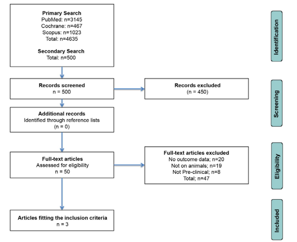
58. Moher D, Liberati A, Tetzlaff J, Altman DG; PRISMA Group. Preferred reporting items for systematic reviews and meta-analyses: The PRISMA statement. Int J Surg. 2010; 8(5): 336-341. doi: 10.7326/0003-4819-151-4-200908180-00135

59. Haque AM, Hashimoto M, Katakura M, Hara Y, Shido O. Green tea catechins prevent cognitive deficits caused by Abeta1-40 in rats. J Nutr Biochem. 2008; 19(9): 619-626. doi: 10.1016/j.jnutbio.2007.08.008.

60. Levites Y, Weinreb O, Maor G, Youdim MB, Mandel S. Green tea polyphenol (-)-epigallocatechin-3-gallate prevents Nmethyl-4-phenyl-1,2,3,6-tetrahydropyridine-induced dopaminergic neurodegeneration. J Neurochem. 2001; 78(5): 1073-1082. doi: 10.1046/j.1471-4159.2001.00490.x

61. Rezai-Zadeh K, Arendash GW, Hou H, et al. Green tea epigallocatechin-3-gallate (EGCG) reduces beta-amyloid mediated cognitive impairment and modulates tau pathology in Alzheimer transgenic mice. Brain Res. 2008; 1214: 177-187. doi: 10.1016/j. brainres.2008.02.107

62. Saeed NM, El-Naga RN, El-Bakly WM, Abdel-Rahman HM, El-Demerdash E. Epigallocatechin-3-gallate pretreatment attenuates doxorubicin-induced cardiotoxicity in rats: A mechanistic study. Biochem Pharmacol. 2015; pii: S0006-2952-(15)00110-0. doi: 10.1016/j.bcp.2015.02.006

63. Joshi R, Rana A, Gulati A. Studies on quality of orthodox teas made from anthocyanin-rich tea clones growing in Kangra valley, India. Food Chem. 2015; 176: 357-366. doi: 10.1016/j.foodchem.2014.12.067

64. Obregon DF, Rezai-Zadeh K, Bai Y, et al. ADAM10 activation is required for green tea (-)-epigallocatechin-3-gallateinduced alpha-secretase cleavage of amyloid precursor protein.J Biol Chem. 2006; 281(24): 16419-16427. doi: 10.1074/jbc.M600617200

65. Lichtenthaler SF, Haass C. Amyloid at the cutting edge: Activation of alpha-secretase prevents amyloidogenesis in an Alzheimer disease mouse model. J Clin Invest. 2004; 113(10):1384-1387. doi: 10.1172/JCI21746

66. Dickey CA, Kamal A, Lundgren K, et al. The high-affinity HSP90-CHIP complex recognizes and selectively degrades phosphorylated tau client proteins. J Clin Invest. 2007; 117(3):648-658. doi: 10.1172/JCI29715

67. Kosik KS, Shimura H. Phosphorylated tau and the neurodegenerative foldopathies. Biochim Biophys Acta. 2005; 1739(2-3):298-310.
doi: 10.1016/j.bbadis.2004.10.011

68. Khan SG, Katiyar SK, Agarwal R, Mukhtar H. Enhancement of antioxidant and phase II enzymes by oral feeding of green tea polyphenols in drinking water to SKH-1 hairless mice: Possible role in cancer chemoprevention. Cancer Res. 1992; 52(14):4050-4052.

69. McGahon BM, Martin DS, Horrobin DF, Lynch MA. Agerelated changes in LTP and antioxidant defenses are reversed by an alpha-lipoic acid-enriched diet. Neurobiol Aging. 1999; 20(6): 655-664. doi: 10.1016/S0197-4580(99)00050-0

70. Murray CA, Lynch MA. Dietary supplementation with vitamin E reverses the age-related deficit in long term potentiation in dentate gyrus. J Biol Chem. 1998; 273(20): 12161-12168. doi: 10.1074/jbc.273.20.12161

LATEST ARTICLES

Practical Pointers for Drug Development and Medical Affairs

Gerald L. Klein*, Roger E. Morgan, Shabnam Vaezzadeh, Burak Pakkal and Pavle Vukojevic

doi.

10.17140/CTPOJ-7-125

Prevalence and Risk Factors of Subclinical Mastitis of Goats in Banadir Region, Somalia

Omar M. Salah*, Yasin H. Sh-Hassan, Moktar O. S. Mohamed, Mohamed A. Yusuf and Abas S. A. Jimale

doi.10.17140/VMOJ-9-184

Use of Black Soldier Fly (Hermetia illucens) Prepupae Reared on Organic Waste

Maggot Debridement Therapy: A Natural Solution for Wound Healing

Isayas A. Kebede*, Haben F. Gebremeskel and Gelan D. Dahesa,

doi.10.17140/VMOJ-9-183

Figure 11. Risk Map for the Introduction of Ruminant Diseases at Borders

Ovine Network in Morocco: Epizootics Spread Prevention and Identification of the At-Risk Areas for “Peste des Petits Ruminants” and “Foot and Mouth Disease”

Yassir Lezaar*, Mehdi Boumalik, Youssef Lhor, Moha El-Ayachi, Abelilah Araba and Mohammed Bouslikhane

doi.

10.17140/EPOJ-8-131

The Impact of Family Dynamics on Palliative Care at the End-of-Life

Neil A. Nijhawan*, Rasha Mustafa and Aqeela Sheikh

doi.10.17140/PMHCOJ-10-154

Long-Term Follow-Up After Laparoscopic Radical Prostatectomy for Localized and Locally Advanced Prostate Cancer

Shrenik J. Shah*, Abhishek Jha, Chirag Davara, Rushi Mistry and Kapil Kachhadiya

doi.

10.17140/UAOJ-7-147

Treatment and Control Methods of Bovine Mastitis: A Review

Isayas A. Kebede* and Gelan D. Dahesa

doi.10.17140/VMOJ-9-182

LATEST ARTICLES

Prevalence (%) of Types of Anthropometric Failure among Previous and Present Studied Tribal Children

Original Research, peer reviewed

2024 Apr

Biswajit Mahapatra and Kaushik Bose*

Opinion

2024 Apr

Gerald L. Klein*, Roger E. Morgan, Shabnam Vaezzadeh, Burak Pakkal and Pavle Vukojevic

Original Research

2024 Mar

Omar M. Salah*, Yasin H. Sh-Hassan, Moktar O. S. Mohamed, Mohamed A. Yusuf and Abas S. A. Jimale