Advances in Food Technology and Nutritional Sciences

Open journal

ISSN 2377-8350

Giant Iatrogenic Lumbar Pseudomeningocele: A Case Report and Literature Review

Abolfazl Rahimizadeh*, Housain Soufiani, Shahrzad Rahimizadeh, Naser Asgari and Mahan Amirzadeh

Received: April 4th, 2021; Revised: June 28th, 2021; Accepted: June 28th, 2021; Published: June 29th, 2021

INTRODUCTION

Post-discectomy pseudomeningocele was reported first in 1947 by Swanson and Fincher.1 Three-years later in 1950, Winkler et al2 reported two additional cases of a pseudomeningocele after a lumbar discectomy. In 1968, Miller et al3 reported 10 new cases and classified pseudomeningoceles into congenital, iatrogenic and traumatic. Congenital pseudomeningoceles have been described in the patients with Marfan disease and neurofibromatosis mostly in the thoracic and lumbosacral regions respectively.4,5

Majority of the traumatic pseudomeningoceles develop subsequent to blunt traumatic events and are mostly seen in the cervical spine, but seldom in the lumbar region or with penetrating injuries.6,7,8

Iatrogenic pseudomeningoceles may occur in the lumbar, cervical or thoracic regions in decreasing frequency.9-13 In the lumbar region, they are mostly seen following the laminectomy for lumbar disc herniation or canal stenosis.

The cause of these cystic cerebrospinal fluid-containing lesions are incidental dural tears.9-13 The growth of the iatrogenic lumbar pseudomeningoceles is limited but in rare occasions it may continue to grow, till the pressure of the cyst’s contents and the surrounding tissues reach to equilibrium.9-13 Most of the pseudomeningoceles remain relatively minute (below 5 cm in size) and a small number will grow above 5 cm in size which are classified as large. Very rarely this pathologic sac may grow beyond 8 cm being classified as giant subtype.14-16

In 1963, Miller et al3 were the first to report 3 cases with giant iatrogenic pseudomeningocele and since then several cases with detailed information have been published so far.17-27 Herein, we present a middle age woman with giant pseudomeningocele which had developed after L4-L5 discectomy. With consideration of the previously published cases of giant pseudomeningocele with detailed information, the current case will be the 21st in the literature. The detailed information of 21 cases with iatrogenic giant lumbar pseudomeningoceles including the current case are presented separately (Table 1).

Table 1. Data on the Surgical Hardware Used in the Course of Surgical Treatment of Patients with Fracture-Dislocations of the PH

Author

Year Age Sex Lumbar Procedure Clinical Picture

Outcome

Miller et al3

1963

F

40

Low lumbar laminectomy Low back and sciatic pain Good
Miller et al3

1963

M

35

L5- S1 laminectomy S1 radiculopathy Good
Miller et al3

1963

M

31

L4-L5 laminectomy Headache & LBP relieved by lying down Good
Rinaldi et al17 1969 50

M

L3-L4 hemilaminectomy Radiculopathy Good
Rinaldi et al17

1969

42 F L5-S1 discectomy LBP, bilateral S1 radiculopathy Good
Rinaldi et al18

1970

49

M

Subsequent to discectomy for recurrent L4-L5 disc LBP, Unilateral, radiculopathy Good
Schumacher et al19

1988

39

M

L3-L4 hemilaminectomy+ discectomy Radiculopathy Good
Lee et al20

1992

18

M

Right L4-L5 discectomy+ PLIF Persistent LBP, Right radiculopathy Good
Lee et al20

1992

25

F

Right L4-L5 hemilaminectomy Palpable mass, L5 Paresthesia Good


Pavlou et al21

2005

59

F

L4-L5 discectomy Weakness of dorsiflexion Good
Hamilton et al22

2009

51

M

3 time surgery for L3-L4 spondylolisthesis Retroperitoneal mass on left ureter Good
Weng et al23

2010

26

F

L4-L5 laminectomy discectomy Not described Good
Liu et al24 2011 40

F

L4-L5 laminectomy& Screw-rod fixation Radiculopthyy & Postural headache Good
Alvarez et al25

2018

39

M

L4-L5 interlaminar-Laminectomy + discectomy LBP & radiculopathy Good
Alvarez et al25

2018

92

M

L3-L4 & L4-L5 laminectomy LBP, Right leg radiculopathy Good
Eneke et al15

2018

64

F

L4 &L5 laminectomy, L4-L5 discectomy LBP, Radiculopathy, N claudication Fair
Hamdan et al26

2018

26

F

L5-S1 discectomy Episodes of black out while lying Good
Rahimizadeh et al14

2019

30

F

L5-S1 interlaminar laminectomy+ discectomy LBP & left S1 radiculopathy Good
Rahimizadeh et al14

2019

52

F

Multilevel laminectomy Neurogenic claudication Good
Jah

2021

38

M

L4-L5Inter- laminectomy, discectomy LBP & subcutaneous lump Good
Current case

2021

49

F

L4-L5Inter- laminectomy, discectomy LBP & Left L5 radiculopathy, Lump Good
CASE PRESENTATION

A 49-year-old female who had undergone a lumbar laminectomy for a central L4-L5 disc herniation eight months earlier was referred to our facility after the appearance of a large subcutaneous midline lumbar mass which was associated with severe low back pain for a duration of 3-months (Figure 1). Neurological examination of motor and sensory nerves was normal. With the diagnosis of a pseudomeningocele, magnetic resonance imaging (MRI) was performed and displayed a very large pseudomeningocele at the site of the previous surgery. The pseudomeningocele was extended from L2 to S4, with a total length of 13 cm (Figure 2).

Figure 1. A Photograph of the Lumbar Region; Shows a Giant Subcutaneous Lump

ORTOJ-6-121Fig1

Figure 2. (a) T2 Weighted Sagittal MRI, Shows a Giant Pseudomeningocele Extending from L2 to S4, (b) T2 Weighted Axial Images Showing the Narrow Stalk of the Pseudomeningocele (white arrows)

ORTOJ-6-121Fig2a

ORTOJ-6-121Fig2b

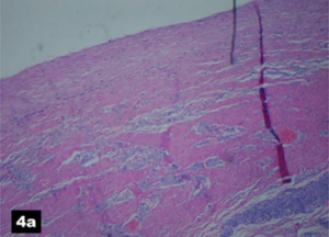
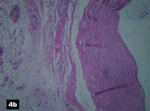
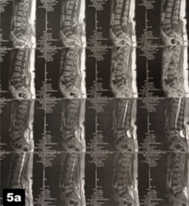
At surgery, the abnormally thick wall of the sac was widely opened. After evacuation of cerebrospinal fluid and at the depth of the cavity; a small breach with slow flow of cerebral spinal fluid (CSF) was found (Figure 3). The defect was subsequently enlarged and closed tightly with interrupted silk sutures. This was subsequently covered with free fat graft being reinforced with suturing of the lower part of the pseudomeningocele’s fibrotic wall. This was followed by a placement of a drain and closure of the wound in three layers. The pathological result of the wall of the pseudomeningocele was composed of connective tissue, mainly fibroblasts being aggregate with foamy macrophages (Figure 4). The patient recovered well-during the 4-days hospital stay and all her complaints had ceased at the time of the one month follow-up encounter. The patient had complete relief of her back pain and was doing well 5-months post-operatively. Successful excision and closure of the pseudomeningocele was confirmed in a MRI taken at six months follow-up (Figure 5).

Figure 3. (a) An Intraoperative Photograph Showing a Breach is Demonstrated at the Bottom of the Surgical Scene. (b) An Intraoperative Photograph showing a Pediatric Nasogastric Tube
Inserted intrathecal via the Breach Only to Show its Communication with Thecal Sac, so was Later Removed. (c) After Closure of the Breach, Reinforced with a Second Layer Composed of the Fibrotic Cyst’s Wall

An Intraoperative Photograph Showing a Breach is Demonstrated at the Bottom


Figure 4. Histopathologic Result of the Cyst’s Wall (a) Note an External Layer being Composed of Fibroblasts. This Layer is Aggregated with Foamy Macrophages. No Epithelial Lining is Seen. (b) Histopathologic Result of the Cyst’s Wall with Higher MagnificationORTOJ-6-121Fig4a

ORTOJ-6-121Fig4b

Figure 5. Post-Operative Sagittal and Axial Lumbar Spine MRI at 6-months Follow-up, (a & b) both T1 and T2-Weighted Sagittal and Axial Images after Excision of the Pseudomeningocele, the Small Hyperintense Mass is Probably Free Fat Graf

ORTOJ-6-121Fig5a

ORTOJ-6-121Fig5b

DISCUSSION

Lumbar pseudomeningoceles are uncommon complication of lumbar disc surgeries which develop as a consequence of an incidental or unrepairable dural tear. The true incidence of post-discectomy lumbar pseudomeningocele is difficult to ascertain because many remain asymptomatic.16 However, it is estimated to be between 0.1 and 2%.14,19

A small dural tear with concomitant arachnoid layer perforation results in a gradual extradural accumulation of the cerebrospinal fluids.14,16,28 According to one theory, the small dural tears leads to a higher probability of pseudomeningocele formation with respect to ball-valve mechanism.28,29 With subsequent reactive fibrosis, the fluid will be enveloped resulting in a cystic mass with false walls or pseudomeningoceles.16-27

Unnoticed dural tear with intact arachnoid and a ball valve mechanism will result in the development of a true cyst lined with arachnoid.29 This type of iatrogenic cyst is called true meningocele where surrounding connective tissue might reinforce the arachnoid capsule with time.14,16,28

Higher frequency of the pseudomeningoceles in the lumbar region in comparison to thoracic and cervical region is due to the relatively higher CSF pressure in the caudal thecal sac and the more frequency of lumbar disc surgeries.

The size of the lumbar pseudomeningoceles in majority remain minute, but in a small number of the cases, in a time frame which varies from a few months to a year after laminectomy, the pathology continue to grow till it reaches to an equilibrium. In rare occasions, a pseudomeningocele might grow beyond 8 cm.

Weng et al23 have attributed the giant size of a pseudomeningocele to high body mass index. However, we believe that intra-operative extensive dissection of the paravertebral muscles and fatty degeneration of these muscles might be the other predisposing factors.

Clinically, most of the lumbar pseudomeningoceles remain small and asymptomatic, this is in contrast to symptomatic ones which are relatively large. Large pseudomeningoceles might present a large subcutaneous lump. Nonetheless, in symptomatic cases, low back pain (LBP) which characteristically tends to be aggravated with straining and Valsalva maneuver is the most frequent clinical feature.30 If a rootlet is extruded through the breach and trapped within, radiculopathy may coexist.16,31-33 Such manifestation is clinically quite similar to a recurrent lumbar disc herniation.16,31-33 Rarely, in those with anterior dural breach, some rootlets might be trapped in corresponding collapsed intervertebral disc space.34 Occasionally, lower limbs motor dysfunction and incontinence and even cauda equina syndrome might occur.35 Headache as well as syncope and hypotension may be caused by compression of the subcutaneous lump of the pseudomeningocele. Positional headache has been also described in an iatrogenic lumbar pseudomeningocele.36 Positional syncope is another rare presentation of pseudomeningoceles which has been reported in a case report.

The patient’s symptom disappeared following the excision of the pseudomeningocele.37 Headache due to chronic subdural hematoma is another rare complication of the pseudomeningoceles.38 Clinical features of meningitis have been reported in an infected pseudomeningocele.39,40

An abdominal mass due to retroperitoneal growth of a pseudomeningocele is another rare presentation of the scenario.41 Hydronephrosis secondary to ureteral obstruction caused by a retroperitoneal growth of a giant pseudomeningocele has been reported in a rare case report.22

On plain radiographs, erosion of the surrounding bones might be seen in long standing cases.42 Ossification of the cyst’s wall is an infrequent scenario.43-47

The degree and extent of ossification can be best demonstrated in reconstructed computed tomography (CT) images.47

Computed tomography myelography can detect the small pseudomeningoceles, even in those tiny ones that grow intraosseously.48

Magnetic resonance imaging remains the most useful diagnostic tool for the demonstration of a pseudomeningocele and its short fistulous tract. A pseudomeningocele displays low signal intensity in T1-weighted and high signal intensity in T2-weighted MRI images.49 Furthermore, the measurement of the length of the pseudomeningoceles and their classification to minute, large and giant became possible with the aid of MRI.16-27 Generally, this specific CSF-containing mass is located posterior to the dural sac; although in rare instances it might grow into the intervertebral disc space and even progress into retroperitoneal space.22,50

Owing the high chances of spontaneous regression, conservative treatment is recommended for asymptomatic cases.51 Spontaneous regression may occur within 3-months to a few years following the diagnosis.51 According to Solomon et al51 healing of the dural defect with the gradual resorption of cerebrospinal fluid is the possible mechanism for the spontaneous resolution of pseudomeningoceles.16,52 Surgery for a symptomatic lumbar pseudomeningocele starts with widely opening the cyst and closure of the dural breach itself.14-20 For closure of the breach, both interrupted suture and titanium U shape clips can be used.14-16,52,53 In the case of a radiculopathy where entrapment of a rootlet is responsible; reduction of the rootlet into the thecal sac through the breach is the key to adequate treatment.16,31-33

Following the closure of the breach, lumbar myofascial flap was introduced by Misra et al.54 Myofascial flap which can be achieved with advancement of lumbar paravertebral muscles has been advocated in those with large dead space.

CONCLUSION

Incidental dural tears with CSF leakage during lumbar laminectomy should be properly addressed. Lumbar pseudomeningocele should be suspected in patients with a delayed reappearance of lower back pain or radiculopathy within a few months to several years after the initial laminectomy. Appropriate surgical intervention should be decided upon and undertaken once the diagnosis is reached. There remains no difference in the management of large and giant pseudomeningoceles.

CONSENT

The authors have received written informed consent from the patient.

CONFLICTS OF INTEREST

The authors declare that they have no conflicts of interest.

1. Swanson HS, Fincher EF. Extradural arachnoidal cysts of traumatic origin. J Neurosurg. 1947; 4: 530-538. doi: 10.3171/ jns.1947.4.6.0530

2. Winkler H, Powers JA. Meningocele following hemilaminectomy; Report of 2 cases. N C Med J. 1950; 11: 292-294.

3. Miller PR, Elder FW. Meningeal pseudocysts (meningocele spurius) following laminectomy. Report of ten cases. J Bone Joint Surg. 1968; 50A: 268-276. doi: 10.2106/00004623-196850020-00005

4. Stone JG, Bergmann LL, Takamori R, Donovan DJ. Giant pseudomeningocele causing urinary obstruction in a patient with Marfan syndrome. J Neurosurg Spine. 2015; 23: 77-80. doi: 10.3171/2014.11. SPINE131086

5. Dobran M, Iacoangeli M, Ruscelli P, Costanza MD, Nasi D, Scerrati1 M. A giant lumbar pseudomeningocele in a patient with neurofibromatosis Type 1: A case report. Case Rep Med. 2017; 2017: 4681526. doi: 10.1155/2017/4681526

6. Rahimizadeh A, Ehtesham S, Yazdi TT, Rahimizadeh S. Remote paraparesis due to a traumatic extradural arachnoid cyst developing 2 Years after brachial plexus root avulsion injury: Case report and review of the literature. J Brachial Plex Peripher Nerve Inj. 2015; 10: e43-e49. doi: 10.1055/s-0035-1558426

7. Hader WJ, Fairholm D. Giant intraspinal pseudomeningoceles cause delayed neurological dysfunction after brachial plexus injury: Report of three cases. Neurosurgery. 2000; 46(5): 1245-1249. doi: 10.1097/00006123-200005000-00044

8. Rahimizadeh A, Javadi SA. Symptomatic intraspinal lumbosacral pseudomeningocele, A late consequence of root avulsion injury secondary to a gunshot wound. North American Spine society Journal (NASS J). 2020; 3: 100025. doi: 10.1016/j.xnsj.2020.100025

9. Barazi SA, D’Urso PI, Thomas NW. Pseudomeningocele after anterior cervical discectomy and fusion: Case report. Cent Eur Neurosurg. 2012; 73: P050. doi: 10.1055/s-0032-1316252

10. Rahimizadeh A, Soufiani H, Rahimizadeh S. Remote cervical pseudomeningocele following anterior cervical corpectomy and fusion: Report of a case and review of the literature. Int J Spine. Surg. 2016; 10: 36. doi: 10.14444/3036

11. Macky M, Lo S, Bydon M, Kaloostian P, Bydon A. Post-surgical thoracic pseudomeningocele causing spinal cord compression. J Clin Neurosci. 2014; 21: 367-372. doi: 10.1016/j.jocn.2013.05.004

12. Filho AAP, de David G, Pereira Filho GA, Brasil AVB. Symptomatic thoracic spinal cord compression caused postsurgical pseudomeningocele. Arq Neuropsiquiatr. 2007; 65: 279-282. doi: 10.1590/s0004-282×2007000200017

13. Pagni CA, Cassinari V, Bernasconi V. Meningocele spurious following hemilaminectomy in a case of lumbar discal hernia. J Neurosurg. 1961; 18: 709-710. doi: 10.3171/jns.1961.18.5.0709

14. Rahimizadeh A, Kaghazchi M, Rahimizadeh A. Post-laminectomy lumbar pseudomeningocele: Report of three cases and review of the literature. World Spinal Column J( WScJ). 2014; 4: 103-108.

15. Eneke O, Dannaway J, Tait M, New CH. Giant lumbar pseudomeningocele after revision lumbar laminectomy: A case report and review of the literature. Spinal Cord Ser Cases. 2018; 4: 82. doi: 10.1038/s41394-018-0118-z

16. Rahimizadeh A, Mohsenikabir N, Asgari N. Iatrogenic lumbar giant pseudomeningocele: A report of two cases. Surg Neurol Int. 2019; 10: 213. doi: 10.25259/SNI_478_2019

17. Rinaldi I, Peach WF. Postoperative lumbar meningocele: Report of two cases. J Neurosurg. 1969; 30: 504-507. doi: 10.3171/jns.1969.30.4.0504

18. Rinaldi I, Hodges TO. Iatrogenic lumbar meningocele: Report of three cases. J Neurol Neurosurg Psychiatry. 1970; 33: 484-492. doi: 10.1136/jnnp.33.4.484

19. Schumacher H-W, Wassman H, Podlinski C. Pseudomeningocele of the lumbar spine. Surg Neurol. 1988; 29: 77-78. doi: 10.1016/0090-3019(88)90127-9

20. Lee KS, Hardy IM II. Postlaminectomy lumbar pseudomeningocele: Report of four cases. Neurosurgery. 1992; 30: 111-114. doi: 10.1227/00006123-199201000-00020

21. Pavlou G, Bucur SD, van Hille PT. Entrapped spinal nerve roots in a pseudomeningocoele as a complication of previous spinal surgery. Acta Neurochir (Wien). 2006; 148: 215-220. doi: 10.1007/ s00701-005-0696-y

22. Hamilton RG, Brown SW, Goetz LL, Miner M. Lumbar pseudomeningocele causing hydronephrosis. J Spinal Cord Med. 2009; 32(1): 95-98. doi: 10.1080/10790268.2009.11760758

23. Weng YJ, Cheng CC, Li YY, Huang TJ, Hsu RWW. Manage ment of giant pseudomeningoceles after spinal surgery. BMC Musculoskelet Disord. 2010; 11: 53. doi: 10.1186/1471-2474-11-53

24. Liu C, Cai HX, S Fan SW, Liu YJ. Postoperative pseudomeningocele in a 40-year-old man. Ir J Med Sci. 2011; 180: 925-927. doi: 10.1007/s11845-010-0598-8

25. Alvarez CM, Urakov TM, Vanni S. Repair of giant postlaminectomy pseudomeningocele with fast-resorbing polymer mesh: technical report of 2 case. J Neurosurg Spine. 2018; 28: 341-344. doi: 10.3171/2017.6.SPINE161292

26. Hamdan A, Saxena A, Rao G Ivanov M. Compression of a giant pseudomeningocele causing transient anoxic seizures: A case report. Acta Neurochirug. 2018; 160: 479-485. doi: 10.1007/s00701- 017-3446-z

27. Hawk MW, Kim KD. Review of spinal pseudomeningoceles and cerebrospinal fluid fistulas. Neurosurg Focus. 2000; 9(1): e5. doi: 10.3171/foc.2000.9.1.5

28. Cobb C, Ehni G. Herniation of the spinal cord into an iatrogenic meningocele. Case report. J Neurosurg. 1973; 39(4): 533-536. doi: 10.3171/jns.1973.39.4.0533

29. Rahimizadeh A, Kaghazchi M, Shariati M, Abdolkhani E. Spinal extradural arachnoid cysts. Coluna/Columna. 2013; 12(2): 112- 118. doi: 10.1590/S1808-18512013000200004

30. Aldrete JA, Ghaly R. Postlaminectomy pseudomeningocele. An unsuspected cause of low back pain. Reg Anesth. 1995; 20: 75-79. doi: 10.1136/rapm-00115550-199520010-00013

31. Hadani M, Findler G, Knoler N, Tadmor R, Sahar A, Shacked I. Entrapped lumbar nerve root in pseudomeningocele after laminectomy: Report of three cases. Neurosurgey. 1986; 19: 405-497. doi: 10.1227/00006123-198609000-00011

32. O’Connor D, Maskery N, Griffiths WE. Pseudomeningocele nerve root entrapment after lumbar discectomy. Spine (Phila Pa 1976). 1998; 23: 1501-1502. doi: 10.1097/00007632-199807010- 00014

33. Kamali R, Beni ZN, Beni AN, Forouzandeh M. Postlaminectomy lumbar pseudomeningocele with nerve root entrapment: A case report with review of literature. Eur J Orthop Surg Traumatol. 2012; 22 (Suppl 1): S57-S61. doi: 10.1007/s00590-011-0934-3

34. Toppich HG, Feldmann H, Sandvoss G, Meyer F. Intervertebral space nerve root entrapment after lumbar disc surgery. Spine (Phila Pa 1976). 1994; 19: 249-250. doi: 10.1097/00007632- 199401001-00021

35. Asha MJ, George KJ, Choksey M. Pseudomeningocele presenting with cauda equina syndrome: Is ball-valve theory the answer. Br J Neurosurgery. 2011; 25: 766-768. doi: 10.3109/02688697.2011.578768

36. Marlin AE, Epstein F, Rovit R. Positional headache and syncope associated with a pseudomeningocele. Arch Neurol. 1980; 37: 736-737. doi: 10.1001/archneur.1980.00500600084022

37. De luca GC, Garza I, Lanzino G, Watson JC. An unexpected cause of orthostatic headache: Delayed postlaminectomy pseudomeningocele. Neurology. 2010; 74: 1553. doi: 10.1212/ WNL.0b013e3181dd4319

38. Kuhn J, Hofman B, Knitelious HO, Coenen HH, Bewermyer H. Bilateral subdural hematoma and lumbar pseudomeningocele due to chronic leakage of liquor cerebrospinalis after lumbar discectomy with application of Adcon-L gel. J Neurol Neurosurg Psychiat. 2005; 76(103): 1031-1033. doi: 10.1136/jnnp.2004.046276

39. Koo J, Adamson R, Wagner FC, Jr, Hrdy DB. A new cause of chronic meningitis, infected lumbar pseudomeningocele. Am J Med. 1989; 86(1): 103-104. doi: 10.1016/0002-9343(89)90238-6

40. Nairus JG, Richman RJ, Douglas RA. Retroperitoneal pseudo-meningocele complicated by meningitis following a lumbar burst fracture. A case report. Spine (Phila Pa 1976). 1995; 21: 1090- 1093. doi: 10.1097/00007632-199605010-00020

41. Kolawole TM, Patel PJ, Naim-Ur-Rahaman. Post-surgical anterior pseudomeningocele presenting as an abdominal mass. Comput Radiol. 1987; 11: 237-240. doi: 10.1016/0730-4862(87)90004-7

42. Lau KK, Stebnyckyj M, McKenzie A. Post-laminectomy pseudomeningocele: An unusual cause of bone erosion. Australas Radiol. 1992; 36: 262-264. doi: 10.1111/j.1440-1673.1992.tb03166.x

43. Tsuji H, Handa N, Handa O, Tajima G, Mori T. Postlaminectomy ossified extradural pseudocyst. Case report. J Neurosurg. 1990; 73: 785-787. doi: 10.3171/jns.1990.73.5.0785

44. Al-Erdus SA, Mukari SAM, Ganesan D, Rimali N. Ossified lumbar pseudomeningocele: Imaging findings. Spine J. 2011; 11: 796-797. doi: 10.1016/j.spinee.2011.05.013

45. Akhaddar A, Boulahourd O, Boucetta M. Nerve root herniation into a calcified pseudomeningocele after lumbar laminectomy. Spine J. 2012; 12: 273. doi: 10.1016/j.spinee.2012.02.008

46. Lau KK, Stebnyckyj M, McKenzie A. Post-laminectomy pseudomeningocele: An unusual cause of bone erosion. Australas Radiol. 1992; 36: 262-264. doi: 10.1111/j.1440-1673.1992.tb03166.x

47. Teplick JG, Peyster RG, Teplick SK, Goodman LR, Haskin ME. CT identification of postlaminectomy pseudomeningocele. AJR Am J Roentgenol. 1983; 140: 1203-1206. doi: 10.2214/ajr.140.6.1203

48. Paolini S, Ciapetta P, Piattella MC. Intraspinous postlaminectomy pseudomeningocele. Eur Spine J. 2003; 12: 325-327. doi: 10.1007/s00586-002-0482-y

49. Murayama S, Numaguchi Y, Whitecloud TS, Brent CR. Mag netic resonance imaging of postsurgical pseudomeningocele. Comput Med Imaging Graph. 1989; 13: 335-339. doi: 10.1016/0895- 6111(89)90211-5

50. Buy X, Alberti N, Pointillart V, Loiseau H, Palussière J. Intravertebral pseudomeningocele: An unusual complication after disc surgery. Spine J. 2014; 14(11): e1-e4. doi: 10.1016/j.spinee.2014.08.011

51. Solomon P, Sekharappa V, Krishnan V, David KS. Spontaneous resolution of postoperative lumbar pseudomeningoceles: A report of four cases. Indian J Orthop. 2013; 47: 417-421. doi: 10.4103/0019- 5413.114937

52. Gupta R, Narayan S. Post-operative pseudomeningocele after spine surgery: Rare cause of failed back syndrome. Iran J Neurosurg. 2016; 2(1): 15-18. doi: 10.18869/acadpub.irjns.2.1.15

53. Nicolletti GF, Umana GE, Florio FG A, Scalia G. Repair of spinal pseudomeningocele in a delayed postsurgical cerebrospinal fluid leak using titanium U clips: Technical note. Interdisciplinary Neurosurgery. 2020; 21: 1000742. doi: 10.1016/j.inat.2020.100742

54. Misra SN, Morgan HW, Sedler R. Lumbar myofacial flap for pseudomeningocele repair. Neurosurg Focus. 2003; 15(3): E13. doi: 10.3171/foc.2003.15.3.13

LATEST ARTICLES

Use of Black Soldier Fly (Hermetia illucens) Prepupae Reared on Organic Waste

review

2024 Mar

Isayas A. Kebede*, Haben F. Gebremeskel and Gelan D. Dahesa,
Figure 11. Risk Map for the Introduction of Ruminant Diseases at Borders

Original Research, peer reviewed

2024 Mar

Yassir Lezaar*, Mehdi Boumalik, Youssef Lhor, Moha El-Ayachi, Abelilah Araba and Mohammed Bouslikhane

Commentary, End-of-Life, Family Dynamics, palliative Care, peer reviewed

2024 Mar

Neil A. Nijhawan*, Rasha Mustafa and Aqeela Sheikh

Retrospective Study

2024 Mar

Shrenik J. Shah*, Abhishek Jha, Chirag Davara, Rushi Mistry and Kapil Kachhadiya

Attention, Disaster, Mindfulness, Original Research, peer reviewed

2024 Mar

Christina Hagen*, Lars Lien, Edvard Hauff and Trond Heir

review

2024 Mar

Isayas A. Kebede* and Gelan D. Dahesa

review

2024 Feb

Bekiyad S. Daro*; Yared A. Deneke, Jiregna G. Negasa and Ishetu N. Dibaba