Cancer Studies and Molecular Medicine

Open journal

ISSN 2377-1518

From Elegance to Complexity: The Intricate Dance of Myometrial Stem Cells Transforming into Leiomyoma Stem Cells: Unraveling the Symphony of Genetic and Epigenetic Refinement

Somayeh Vafaei*

Somayeh Vafaei, PhD

Department of Obstetrics and Gynecology, University of Chicago, Chicago, IL, USA; E-mail: somayehv@uchicago.edu

INTRODUCTION

The most prevalent tumor among women is uterine leiomyoma (LM, or fibroid), which affects around 70-80% of women globally. It disproportionately negatively impacts African American women, commencing at a younger age and with a higher incidence overall.1,2 Even though it does not have a malignant source, LM can result in severe morbidities, such as excessive uterine hemorrhage, repeated miscarriages, and pelvic pain. These manifestations might potentially be used to misdiagnose or cover up malignant tumors.3 Women who no longer wish to bear children are most commonly treated with hysterectomy. This procedure is linked to morbidity, as well as a significant financial burden on the healthcare delivery system.4 The abundant accumulation of extracellular matrix (ECM), which plays a crucial role in the size and stiffness of benign smooth muscle tumors, has been linked to these tumors. This deposition of ECM is connected with these tumors.5 Notwithstanding their widespread occurrence, little is understood about the tumors’ specific pathophysiology. Normal tissue and pathological development cannot occur without stem cells. The involvement of stem cells in LM has been established by a number of investigations, and these cells are necessary for multiplication, regeneration, and the development of tumors.6

The endometrium, myometrium, and perimetrium are the three layers of tissue that make up the human uterus.7 The layer of smooth muscle of uterus known as the myometrium is frequently distinguished by its capacity to regenerate and remodel both during and following pregnancy.8 Myometrial stem cells, based on these remarkable characteristics, may exist and strictly control myometrial development.9 Likewise, tumor-initiating cells are a group of cells inside a cancerous cell community that maintain their capacity to sustain malignancies through asymmetric proliferation. Single somatic stem cell (SSCs) in the myometrium may have been changed and transformed, leading to the formation of myometrial fibroids, monoclonal tumors of the myometrium, which then spread and grow in a steroid-regulated manner.10 Uterine LM are thought to be the result of a myometrial stem cell turning into a mutant stem cell (through acquired cytogenetic abnormalities), which then multiples to produce a LM.11 It is believed that numerous factors including hypoxic niche, changed epigenome, and aberrant estrogen signaling, contribute to the transition of a normal stem cell into a LMforming stem cell.8

Numerous genetic and epigenetic variables associated with an elevated risk of LM have been discovered through genome-wide association investigations and analysis of LM tumors.12 An epigenetic change in phenotype does not occur because of an altered deoxyribonucleic acid (DNA) sequence; rather, it is a consequence of alterations in gene expression. There are many factors that influence stem cell regulation and disease advancement through epigenetic modifications, including DNA methylation.13 DNA methylation, which occurs within cancer stem cells, has the effect of silencing the genes necessary for differentiation, which in turn leads to aberrant clonal growth.14 Cancer stem-like cells can be “reset” to a differentiated phenotype by DNA-hypomethylating drugs in numerous tumor forms, which makes them more sensitive to chemotherapeutic approaches.15,16

Therefore, gaining knowledge of these processes may present significant prospects for the enhancement of diagnostic and prognostic techniques and the development of innovative clinical strategies. Genetic and epigenetic modifications in myometrial stem cells resulting in LM stem cell type are discussed in this review.

CELLULAR ORIGIN OF LEIOMYOMA

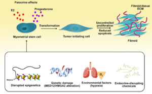
Considering that LM (leiomyoma) is a monoclonal tumor, the aberrant conversion of myometrial stem cells into a tumor-initiating cell type is responsible for the development of the tumor (Figure 1). Myometrial and leiomyoma tissues can be used to extract cells exhibiting stem or progenitor properties.17 It is fascinating to note that these cells appear to possess modest concentrations of sex steroid hormone receptors; however, they need these steroids for tumor progression. This leads one to speculate that an extra paracrine process, mediated by mature adjacent cells expressing these receptors, is indispensable for these cells’ transformation.18 Progenitor cells can become cancerous when exposed to early-life endocrine-disrupting chemicals like xenoestrogens.19

Figure 1. Cellular Origin of Uterine Leiomyoma

CSMMOJ-8-136Fig1

Some environmental and epigenetic/genetic factors may cause the conversion of myometrial stem cells into tumor initiating cells with uncontrolled proliferation and decreased apoptosis rate, may eventually lead to a leiomyoma tumor.

Furthermore, mediator complex subunit 12 (MED12) mutations are found in progenitor cells from the LM but not the myometrium, implying that one genetic hit leads to myometrial stem cell conversion into a tumor-initiating cell and develop these tumors.10,20 There have also been reports of mutations in myometrial cells which alter the transcription of the high mobility group A2 (HMGA2) gene. These mutated myometrial cells led to develop LM-like tissue after xenotransplantation into immunocompromised mice.21 It is unknown whether mutations in MED12 and HMGA2 cause myometrial stem cells to develop into LM stemprogenitor cells or whether they simply sustain LM stem-progenitor cells that are already present, despite the fact that abnormalities in both genes appear to be mutually unique.7 From the cellular perspective, LM establishment and development may be affected by environmental and epigenetic factors, including hypoxia in the uterus or abnormal methylation.22-24 In conclusion, these myometrial stem/progenitor cells provide fresh and robust prospective targets for treatment or prophylactic measures, although an additional definition of the cellular origin of LM is required. It is likely that forthcoming therapies focus on the growth acceleration phase of fibroid advancement since it appears that the conversion of myometrial stem cells into pre-fibroids is a common, if not universal process.

MYOMETRIAL STEM CELLS

The ability of the female reproductive tract to regenerate and modify is indicative of the presence of endometrial and myometrial stem cell networks. The previous study demonstrated the isolation of side population (SP) cells from non-gravid human myometrium to validate the existence of stem cells and their potential role. Transcription of the ATP-binding cassette, subfamily G, member 2 (ABCG2) protein is necessary to remove intracellular DNA-binding dyes in human SP cells.25 Human myoSP cells did not multiply in vitro in a medium with a normoxic oxygen concentration (20% O2 ), but they proliferated effectively in vitro when the oxygen tension was set at 2%.25 In vitro, tissue-specific stem cells can survive and thrive in low-oxygen circumstances.26 Based on these findings, it appears that such stem cells are well adapted for proliferation and survival in a hypoxic condition (in vivo). For multiplication and spontaneous conversion into smooth muscle cells, myoSP cells demand a hypoxic condition. Human myoSP cells may also have a role in the growth of the uterus.17 Shynlova et al demonstrated that mechanical stretching of the uterine wall during pregnancy might lead to hypoxia in the myometrium in the rat by employing a biochemical probe detecting hypoxia. They also found hypoxia-activated caspases in the rat myometrium. Hypoxia is well-known to control trophoblast and placenta growth, differentiation, and function.27 It has been shown that hypoxia encourages the multiplication of hypothesized endometrial stem/progenitor cells such as myoSP.28 Hence, because of the impact that oxygen tension has on the trophoblast, uterus, and placenta, it may be one of the most important factors in determining whether or not a pregnancy will be successful. As a result, it is possible that mechanical stretching during pregnancy causes hypoxia and promotes the growth of human myoSP. Consequently, human myoSP may play a role in the expansion and remodeling of the uterus that occurs during pregnancy.

LEIOMYOMA STEM CELLS

More than 80% of African-American women and 70% of Caucasian women suffer from uterine leiomyomas by the age of 50.29 In around 15-30% of cases, LM result in excessive uterine hemorrhage, anemia, recurrent miscarriages, pelvic discomfort, preterm labor, and urinary incontinence.30 Hysterectomy is performed most frequently because of these symptoms.31 One percent of tumor cells are leiomyoma SP (LMSP) types, which have features of tumor-initiating stem cells.32 Tumor development is stimulated by the proliferation of LMSP cells in vivo. This development is hormone-dependent, despite the fact that these cells express significantly lower numbers of estrogen/progesterone receptors (ER, PR) than matured myometrial or leiomyoma cells.33 It is generally accepted that LM are forms of monoclonal tumors.34 In an ovarian steroid-dependent way, it has been postulated that each LM arises from a single converted myometrium-specific stem cell.35 Nevertheless, this theory has yet to be verified conclusively. Multipotent tissue-specific stem cells are essential for the in vivo proliferation of human LM tissue reliant on estrogen and progesterone.15,32 Matured myometrial or LM cells with increased numbers of steroid receptors and interactions are necessary for their proliferation. A paracrine mechanism proposed to explain how steroid hormones affect LM stem cells through matured myometrial cells (tumor commencement) or matured LM cells (growth maintenance) has been used to support this hypothesis. It is believed that the self-renewal of the LM stem cells is supported by a paracrine contact with the ER-1-and/or PR-positive niche cells that are located in the surrounding area. Downregulation of genes necessary for progesterone-induced LM stem cell development is the outcome of PR gene hypermethylation. By modulating the ten-eleven translocation (TET) enzymes, PR knockdown was linked to an enhancement in global DNA methylation.36

Uterine LM are quite common, yet little is known about their cellular or molecular origins. Epigenetic processes, including DNA methylation, micro ribonucleic acid (miRNA) regulated modifications, and histone modification, according to recent research findings may be involved in LM.37,38 LM development has been linked to genetic mutations, including those causing a deficiency in the fumarate hydratase (FH).39

GENETIC ALTERNATION

Sequencing of exosome in uterine LM from 18 Finnish patients found MED12 recurrent somatic mutations.40 Additional investigation of a total of 225 tumors originating from 80 individuals indicated that an astounding 70% of uterine LM exhibit MED12 mutations, rendering it the most commonly mutated gene. Exon 2 and the intron 1-exon 2 junctions, where all mutations were found, are evolutionarily conserved regions of the gene. Numerous investigations encompassing numerous ethnic groups have confirmed this outcome.41-43 Tumorigenesis may be facilitated by the encoded region’s abnormal functioning. It was shown that LM size was inversely associated with MED12 mutations in the MED12 mutation studies.44,45 Particular genetic alterations, such as MED12, are present in most LM, which demonstrates that the transition of healthy myocytes into aberrant myocytes is essential at some time during the origin of the LM development. The MED12 gene connects regulatory factors in the promoters of gene to the RNA polymerase II initiation complex and encodes a mediator complex, that comprises 26 subunits and modulates transcription and also elongation.40

Other genes, particularly those participating in the cell cycle and tumor suppression, including cell cycle associated protein 1 (CAPRIN1), Decorin (DCN), and aryl hydrocarbon receptor (AHR)12 as well as particular mitochondrial genes, are said to contain specific point mutations in LM.46 Owing to translo cations, duplications, and deletions of the chromosomes 6, 7, 12, and 14, chromosomal defects have been documented in LM. Whole-genome sequencing supported these findings, indicating a single origin for the rearrangements found in LM.47 To categorize patients experiencing LM according to their molecular and clinical characteristics, several types of cytogenetic mutations are being considered benchmarks. In cytogenetically aberrant fibroids, the tumor size tends to increase,14 whereas deletions in several areas, like chromosome 1p, may be related to prognosis and histological variations of LM.48

Despite the fact that chromosomal abnormalities can occur anywhere in the genome in LM, certain genes are commonly compromised by these sorts of incidents, resulting in interrupted biological mechanisms responsible for the development of tumors. It has been shown that upregulation of the HMGA2 gene results from rearrangements in the region 12q14-15 that are present in fibroids.49 The DNA-repairing gene RAD51B is one of the preferential translocation partners of HMGA2, so, changes in the transcription of this protein, that is participated in transcription regulation, have been linked to the advancement of LM through various processes, such as neovascularization,17ER-mediated cell multiplication,10 and homologous recombination DNA repair.50 Rearrangements that target region 6p21 can also affect another family member known as HMGA1, which is disrupted in LM. Both HMGA1 and HMGA2 may accomplish a physiological function that is analogous to one another, which would describe why the effects of disrupting either gene are comparable.51 In contrast, despite the fact that deletions in the 7q22 area have been related to LM, the impacted target gene has not been found definitively. Despite the fact that these deletions change the expression of several genes, including lipoma HMGIC fusion partner (LHFPL3), Laminin Subunit Beta 1 (LAMB1), or human polybromo-1 (HPB1),10,20 however, cut like homeobox 1 (CUX1) is the most widely regarded target gene. Both up- and downregulation of CUX1 can be the consequence of 7q deletions, demonstrating that this gene may play either a tumor suppressor or an oncogenic role, which may lead to the progression of LM in a manner analogous to that of RAD51B.52 There has also been a link found between Alport syndrome and the establishment of smooth muscle tumors known as diffuse leiomyomatosis, which includes uterine LM, and loss of collagen IV genes including COL4A5 and COL4A6, due to X chromosomal deletions.53 Some of the key mutated genes in LM development have been summarized in Table 1.

Table 1. Key Mutated Genes Identified in Leiomyoma Progression

Genes

Type of tissue

Reference

Mediator Complex Subunit 12 (MED12) Uterine leiomyoma, Leiomyosarcoma

44,79,80

High Mobility Group (HMGA1/HMGA2) Uterine myometrium, Leiomyosarcoma

47,79

Collagen Alpha-5/Collagen Alpha-6/COL4A5/COL4A6 Uterine leiomyoma, Oesophagus leiomyoma

47,81

Fumarate Hydratase (FH) Fumarate Hydratase (FH)

47,82

EPIGENETIC ALTERNATIONS

In humans, the development of LM is significantly influenced by epigenetic processes, which are characterized by the fact that they alter the expression of genes.3,54 Regulation via DNA methylation, miRNAs, histone modifications, and long-noncoding RNAs (lncRNAs) are some of the primary epigenetic processes. Foxo1, TERT and WNT4 are examples of genes affected by abnormalities in the DNA methylation process, which is necessary for healthy development.25 They are linked to an increased level of methylation in tumor suppressor genes and/or a decreased level of methylation in oncogenes, suggesting that they may have a role in the development of tumors.55 Particularly, LM, in comparison to normal myometrium, is linked to modifications of DNA methylation, upregulation of ER1 messenger RNA (mRNA), and DNA methyltransferases in cancerous specimens.56,57 Upregulation of HMGA2 has been shown to be attributed to hypomethylation in the HMGA2 gene recently; however, this association does not necessarily seem to be dependent on metastasis.58 Receptor activator of nuclear factor kappa-Β ligand (RANKL) gene expression is affected by progesterone/PR-mediated DNA methylation and MED12 mutation, which together form a complex regulatory network. This network is critical for activating stem cell multiplication and the emergence of LM.59,60 These data indicate that by changing the normal myometrial mRNA expression profile, DNA methylation may have a role in uterine LM pathogenesis. Additional research utilizing epigenetic modulators including 50-Aza-Cytidine may contribute to the development of innovative medicines that inhibit the formation of tumors by reducing the number of stem cells in the affected area via various demethylation processes.61 Aside from histone tail methylation and acetylation, other epigenetic changes have been related to uterine LM, including enhancer of zeste homolog 2 (EZH2) and histone deacetylases (HDACs).3 Histone acetylation is exclusively involved in the regulation of gene activation, in contrast to histone methylation, which can either activate or suppress gene expression.62 EZH2 methylation, in particular, is responsible for the silencing of gene function in LM33; even though HDACs are essential in the control of the tumor suppressor gene krüppel like factor-11 (KLF11), its expression is reduced throughout these benign tumors.63 In conclusion, it is essential to highlight the considerable part played by miRNAs belonging to families let-7, miR-93, miR-21, miR-200, and miR-106b in the epigenetic processes that contribute to proliferation, inflammatory processes, neovascularization, and the production of extracellular matrix constituents. As a result, by targeting of important signaling pathways, including Wnt/β-catenin and Wnt/MAPK, they contribute to the formation of the LM.64,65 Recently, it was discovered that, compared to the myometrium, the transcription of long noncoding RNAs (RNA transcripts with more than 200 nucleotides) like Xinactive specific transcript (XIST) was changed in LM.66 Innovative therapeutic approaches which target uterine LM can be identified by better understanding the role of epigenetic controls in the development of LM and their involvement in tumorigenesis (possibly related to a hypoestrogenism phenotype in myometrial advancement.67

Stem Cells Regulation by Epigenetic Changes in Leiomyoma

SSCs are undifferentiated cells involving many processes in the body. To replace dying cells, heal injured organs, and then develop into tissue-specific cell types, these cells can divide and proliferate. The SSCs are responsible for creating the dynamic environment necessary for cellular and tissue homeostasis. Therefore, tumor stem cells have the ability to maintain and further develop the tumor, in addition to their potential for self-renewal. The development process, stem cells maintenance, and the differentiation of cells all require a large number of chromatin regulators. By regulating the transcriptional availability of various sections of genome packing or opening various portions of chromatin, epigenetic processes enable genetically identical cells to acquire distinct phenotypes in a stable manner. There is a possibility that distinct components of stem cell phenotype require a specific combination of transcriptional elements and chromatin remodeling factors.68 Increasing evidences show that the function of stem cells and the pathogenesis of cancer are significantly stimulated by polycomb repressive complex (PRC1) and PRC2 as well as HDAC1- and HDAC2-containing complexes (NuRD, Sin3, and CoREST).69,70 Furthermore, a growing amount of evidence indicates that during multipotent stem cell differentiation, particular CpG methylation sites are modified.71 In hematopoietic stem cells, 5-mC and 5-hmC and active/repressive histone coding marks related to DNA Methyltransferase 3 Alpha (DNMT3α) transcription have been mapped genome-wide recently, revealing a new epigenetic environment.

Leiomyomas of the uterine lining are monoclonal cancers originating from the smooth muscle cells of uterine.72 It has been determined that familial uterine LM syndromes are connected with a small number of genetic abnormalities that are passed down through germ cells. For instance, although hereditary leiomyomatosis is related to germline mutations in FH, there is no documented evidence indicating the role of epigenetic modifications in these patients yet.73 Multiple lines of evidence imply that each LM arises from the transformation of a single SSC despite the fact that the cellular origin of uterine LM is mainly unidentified.11,74 MED12 mutations have been found to be present in stem cells originating from LM, further indicating that at least one genetic hit is needed to convert myometrial stem cells.

Employing 5-bromo-2’-deoxyuridine, which makes it possible to identify label-retaining cells, and the side population approach, respectively, researchers in 2007 were able to determine for the first time that presumed SSCs occur in mouse and nonpregnant human myometrium.75 There is a population of myometrial cells that may be extracted from human myometrium called the side population. These cells have properties that are comparable to stem cells. In contrast to the primary community of myometrial cells, After transplantation of side population of myometrial cells to the uteri of severely immunodeficient mice, they effectively develop functional human myometrial organs.76 Afterward, the side population formed from LM possessing stem cell properties was identified and shown to be pivotal for cell multiplication and tumor development.15 WNT/β-catenin pathway is stimulated in a paracrine manner in LM stem cells, which leads to the stimulation of tumor growth, despite the fact that LM have a reduced side population cells percentage in comparison to healthy myometrium. Numerous dimensions of tumorigenesis, tissue homeostasis, and embryonic development, are regulated by WNT/β-catenin signaling.76 To shed new light on how aberrantly controlled epigenetic components contribute to the pathogenesis of LM and to offer a prospective clinical strategy for targeting LM stem cells, optimized methods in the identification and separation of myometrium- and LM-derived SSCs are needed.

POSSIBLE THERAPEUTIC STRATEGIES

Epigenetic mechanisms, as opposed to irreversible genetic alterations, have the possibility of being reversed, and as a result, they are prospective therapeutic options for the treatment of cancer. It is possible that epigenetic medicines could target CpG-rich areas, which are present in around 70% of identified gene promoters. The particular TET inhibitors and activators, as well as their modes of action, ought to be identified and characterized in upcoming investigations. It is possible to prevent epigenetic alterations in other organs of the organism by administering the drug directly to the modified tissue and the area surrounding it. The therapy induces hypermethylation at particular loci and hypomethylation at other loci; therefore, one of the problems is targeting specific genes. In addition, it is still completely unclear what the biological importance of TET proteins is, as well as the precise involvement of their enzymatic and non-enzymatic functions. Therefore, there is a requirement for additional in-depth study on chemicals that can affect the function of the TET enzyme to identify alternative epigenetic modulators having curative properties on LM that decrease the growth of tumors.77,78

CONCLUSIONS AND PERSPECTIVES

DNA methylation, histone modifications, and miRNAs dysregulation are a consequence of aberrant genetic and epigenetic alterations, marking pivotal points in the journey of tumorigenesis. While significant strides have been made in unraveling the epigenetic intricacies associated with tumor development, the understanding of the processes and implications of epigenetics in LM remains in its infancy.

Recent advancements in epigenomic exploration, including methodologies such as mapping the distribution of 5-methylcytosine (5 mC) and its oxidized derivatives throughout the entire genome, have provided a deeper comprehension of the epigenome’s role within the realm of tumor biology and somatic cell reprogramming. Noteworthy revelations have emerged, indicating the presence of stem cell-like side populations in both myometrium and LM. This discovery holds promise for elucidating the formation of these stem cells and unraveling the intricate interplay of genetic and epigenetic events governing these processes.

Innovative techniques, such as single-cell sequencing and genome editing, particularly employing CRISPR-Cas technologies, have become instrumental in advancing our understanding of LM. These cutting-ed ge methodologies open avenues to dissect the intricate mechanisms underpinning tumor development. Utilizing such methodologies in LM investigations holds the potential to unravel the complexities of how tumors evolve, providing valuable insights for effective therapeutic interventions.

The identification of stem cell-like populations within LM, coupled with a refined understanding of aberrant signaling and genetic and epigenetic regulations, paves the way for the development of targeted curative strategies. A holistic approach, leveraging advanced methodologies and a comprehensive understanding of LM stem cells, is crucial for the formulation of therapeutic interventions that not only mitigate the severity and extent of uterine LM but also minimize associated complications. In the ongoing pursuit of unraveling the mysteries of LM, the integration of these diverse approaches promises to shape a more nuanced and effective landscape for addressing this benign yet clinically significant condition.

1. Kjerulff KH, Langenberg P, Seidman JD, Stolley PD, Guzinski GM. Uterine leiomyomas. Racial differences in severity, symptoms and age at diagnosis. J Reprod Med. 1996; 41(7): 483-490.

2. Huyck KL, Panhuysen CIM, Cuenco KT, et al. The impact of race as a risk factor for symptom severity and age at diagnosis of uterine leiomyomata among affected sisters. Am J Obstet Gynecol. 2008; 198(2): 168.e1-168.e9. doi: 10.1016/j.ajog.2007.05.038

3. Rezk A, Kahn J, Singh M. Fertility sparing management. In: Uterine Fibroids. Treasure Island (FL), USA: StatPearls Publishing; 2022. https://www.ncbi.nlm.nih.gov/books/NBK574504/. Accessed November 11, 2023.

4. Yang Q, Mas A, Diamond MP, Al-Hendy A. The mechanism and function of epigenetics in uterine leiomyoma Development. Reprod Sci. 2016; 23(2): 163-175. doi: 10.1177/1933719115584449

5. Jamaluddin MFB, Nahar P, Tanwar PS. Proteomic characterization of the extracellular matrix of human uterine fibroids. Endocrinology. 2018; 159(7): 2656-2669. doi: 10.1210/en.2018-00151

6. Ratajczak MZ, Bujko K, Mack A, Kucia M, Ratajczak J. Cancer from the perspective of stem cells and misappropriated tissue regeneration mechanisms. Leukemia. 2018; 32(12): 2519-2526. doi: 10.1038/s41375-018-0294-7

7. Chaudhry SR, Liman MNP; Peterson DC. Anatomy, Abdomen and Pelvis: Stomach. Treasure Island (FL), USA: StatPearls Publishing. 2021. Webstie. https://www.ncbi.nlm.nih.gov/books/ NBK482334/. Accessed November 11, 2023.

8. Brakta S, Mas A, Al-Hendy A. The ontogeny of myometrial stem cells in OCT4-GFP transgenic mouse model. Stem Cell Res Ther. 2018; 9(1): 333. doi: 10.1186/s13287-018-1079-7

9. Bozorgmehr M, Gurung S, Darzi S, et al. Endometrial and menstrual blood mesenchymal stem/stromal cells: Biological properties and clinical application. Front Cell Dev Biol. 2020; 8: 497. doi: 10.3389/fcell.2020.00497

10. Ono M, Bulun SE, Maruyama T. Tissue-specific stem cells in the myometrium and tumor-initiating cells in leiomyoma. Bio Reprod. 2014; 91(6): 149. doi: 10.1095/biolreprod.114.123794

11. Mas A, Cervello I, Gil-Sanchis C, Simón C. Current understanding of somatic stem cells in leiomyoma formation. Fertil Steril. 2014; 102(3): 613-620. doi: 10.1016/j.fertnstert.2014.04.051

12. Al-Hendy A, Myers ER, Stewart E. Uterine fibroids: Burden and unmet medical need. Semin Reprod Med. 2017; 35(6): 473-480. doi: 10.1055/s-0037-1607264

13. Sheaffer KL, Kim R, Aoki R, et al. DNA methylation is required for the control of stem cell differentiation in the small intestine. Genes Dev. 2014; 28(6): 652-664. doi: 10.1101/gad.230318.113

14. Mazloumi Z, Farahzadi R, Rafat A, et al. Effect of aberrant DNA methylation on cancer stem cell properties. Exp Mol Pathol. 2022; 125: 104757. doi: 10.1016/j.yexmp.2022.104757

15. Wang Y, Cardenas H, Fang F, et al. Epigenetic targeting of ovarian cancer stem cells. Cancer Res. 2014; 74(17): 4922-4936. doi: 10.1158/0008-5472.CAN-14-1022

16. Liu S, Yin P, Xu J, et al. Targeting DNA methylation depletes uterine leiomyoma stem cell-enriched population by stimulating their differentiation. Endocrinology. 2020; 161(10): bqaa143. doi: 10.1210/endocr/bqaa143

17. Mas A, Cervelló I, Gil-Sanchis C, et al. Identification and characterization of the human leiomyoma side population as putative tumor-initiating cells. Fertil Steril. 2012; 98(3): 741-751.e6. doi: 10.1016/j.fertnstert.2012.04.044

18. Moravek MB, Yin P, Ono M, et al. Ovarian steroids, stem cells and uterine leiomyoma: Therapeutic implications. Hum Reprod Update. 2015; 21(1): 1-12. doi: 10.1093/humupd/dmu048

19. Mas A, Stone L, O’Connor PM, et al. Developmental exposure to endocrine disruptors expands murine myometrial stem cell compartment as a prerequisite to leiomyoma tumorigenesis. Stem Cells. 2017; 35(3): 666-678. doi: 10.1002/stem.2519

20. Ono M, Qiang W, Serna VA, et al. Role of stem cells in human uterine leiomyoma growth. PLoS One. 2012; 7(5): e36935. doi: 10.1371/journal.pone.0036935

21. Mas A, Cervelló I, Fernández-Álvarez A, et al. Overexpression of the truncated form of High Mobility Group A proteins (HMGA2) in human myometrial cells induces leiomyoma-like tissue formation. Mol Hum Reprod. 2015; 21(4): 330-338. doi: 10.1093/molehr/gau114

22. Zhou S, Yi T, Shen K, Zhang B, Huang F, Zhao X. Hypoxia: The driving force of uterine myometrial stem cells differentiation into leiomyoma cells. Med Hypotheses. 2011; 77(6): 985-986. doi: 10.1016/j.mehy.2011.08.026

23. Maruyama T, Ono M, Yoshimura Y. Somatic stem cells in the myometrium and in myomas. Semin Reprod Med. 2013; 31(01): 77- 81. doi: 10.1055/s-0032-1331801

24. Liu B, Chen G, He Q, et al. An HMGA2-p62-ERα axis regulates uterine leiomyomas proliferation. FASEB J. 2020; 34(8): 10966-10983. doi: 10.1096/fj.202000520R

25. Ono M, Maruyama T, Masuda H, et al. Side population in human uterine myometrium displays phenotypic and functional characteristics of myometrial stem cells. Proc Natl Acad Sci U S A. 2007; 104(47): 18700-18705. doi: 10.1073/pnas.0704472104

26. Mohyeldin A, Garzón-Muvdi T, Quiñones-Hinojosa A. Oxygen in stem cell biology: A critical component of the stem cell niche. Cell Stem Cell. 2010; 7(2): 150-161. doi: 10.1016/j. stem.2010.07.007

27. Red-Horse K, Zhou Y, Genbacev O, et al. Trophoblast differentiation during embryo implantation and formation of the maternal-fetal interface. J Clin Invest. 2004; 114(6): 744-754. doi: 10.1172/JCI22991

28. Cervello I, Gil-Sanchis C, Mas A, et al. Human endometrial side population cells exhibit genotypic, phenotypic and functional features of somatic stem cells. PLoS One. 2010; 5(6): e10964. doi: 10.1371/journal.pone.0010964

29. Baird DD, Dunson DB, Hill MC, Cousins D, Schectman JM. High cumulative incidence of uterine leiomyoma in black and white women: Ultrasound evidence. Am J Obstet Gynecol. 2003; 188(1): 100-107. doi: 10.1067/mob.2003.99

30. Okolo S. Incidence, aetiology and epidemiology of uterine fibroids. Best Pract Res Clin Obstet Gynaecol. 2008; 22(4): 571-588. doi: 10.1016/j.bpobgyn.2008.04.002

31. Wallach EE, Vlahos NF. Uterine myomas: An overview of development, clinical features, and management. Obstet Gynecol. 2004; 104(2): 393-406. doi: 10.1097/01.AOG.0000136079.62513.39

32. Ono M, Qiang W, Serna VN, et al. Role of stem cells in human uterine leiomyoma growth. PLoS One. 2012; 7(5): e36935. doi: 10.1371/journal.pone.0036935

33. Ono M, Yin P, Navarro A, et al. Paracrine activation of WNT/β-catenin pathway in uterine leiomyoma stem cells promotes tumor growth. Proc Natl Acad Sci U S A. 2013. 110(42): 17053-17058. doi: 10.1073/pnas.1313650110

34. Zhang P, Zhang C, Hao J, et al. Use of X-chromosome inactivation pattern to determine the clonal origins of uterine leiomyoma and leiomyosarcoma. Hum Pathol. 2006; 37(10): 1350-1356. doi: 10.1016/j.humpath.2006.05.005

35. Linder D, Gartler SM. Glucose-6-phosphate dehydrogenase mosaicism: utilization as a cell marker in the study of leiomyomas. Science. 1965; 150(3692): 67-69. doi: 10.1126/science.150.3692.67

36. Liu S, Yin P, Xu J, et al. Progesterone receptor-DNA methylation crosstalk regulates depletion of uterine leiomyoma stem cells: A potential therapeutic target. Stem Cell Reports. 2021; 16(9): 2099-2106. doi: 10.1016/j.stemcr.2021.07.013

37. Navarro A, Yin P, Monsivais D, et al. Genome-wide DNA methylation indicates silencing of tumor suppressor genes in uterine leiomyoma. PLoS One. 2012; 7(3): e33284. doi: 10.1371/journal.pone.0033284

38. Georgieva B, Milev I, Minkov I, Dimitrova I, Bradford AP, Baev V. Characterization of the uterine leiomyoma microRNAome by deep sequencing. Genomics. 2012; 99(5): 275-281. doi: 10.1016/j.ygeno.2012.03.003

39. Tomlinson IP, Alam NA, Rowan AJ, et al. Germline mutations in FH predispose to dominantly inherited uterine fibroids, skin leiomyomata and papillary renal cell cancer. Nat Genet. 2002; 30(4): 406-410. doi: 10.1038/ng849

40. Mäkinen N, Mehine M, Tolvanen J, et al. MED12, the mediator complex subunit 12 gene, is mutated at high frequency in uterine leiomyomas. Science. 2011; 334(6053): 252-255. doi: 10.1126/science.1208930

41. de Graaff MA, Cleton-Jansen A-M, Szuhai K, Bovée JVMG. Mediator complex subunit 12 exon 2 mutation analysis in different subtypes of smooth muscle tumors confirms genetic heterogeneity. Hum Pathol. 2013; 44(8): 1597-1604. doi: 10.1016/j. humpath.2013.01.006

42. Rieker RJ, Agaimy A, Moskalev EA, et al. Mutation status of the mediator complex subunit 12 (MED12) in uterine leiomyomas and concurrent/metachronous multifocal peritoneal smooth muscle nodules (leiomyomatosis peritonealis disseminata). Pathology, 2013. 45(4): 388-392. doi: 10.1097/PAT.0b013e328360bf97

43. Ravegnini G, Mariño-Enriquez A, Slater J, et al. MED12 mutations in leiomyosarcoma and extrauterine leiomyoma. Mod Pathol. 2013; 26(5): 743-749. doi: 10.1038/modpathol.2012.203

44. Heinonen H-R, Sarvilinna NS, Sjöberg J, et al. MED12 mutation frequency in unselected sporadic uterine leiomyomas. Fertil Steril. 2014; 102(4): 1137-1142. doi: 10.1016/j.fertnstert.2014.06.040

45. Markowski DN, Bartnitzke S, Löning T, Drieschner N, Helmke BM, Bullerdiek J. MED12 mutations in uterine fibroids—their relationship to cytogenetic subgroups. Int J Cancer. 2012; 131(7): 1528-1536. doi: 10.1002/ijc.27424

46. Shaik NA, Lone WG, Khan IA, et al. Detection of somatic mutations and germline polymorphisms in mitochondrial DNA of uterine fibroids patients. Genet Test Mol Biomarkers. 2011; 15(7- 8): 537-541. doi: 10.1089/gtmb.2010.0255

47. Mehine M, Kaasinen E, Mäkinen N, et al. Characterization of uterine leiomyomas by whole-genome sequencing. N Engl J Med. 2013; 369(1): 43-53. doi: 10.1056/NEJMoa1302736

48. Hodge JC, Pearce KE, Clayton AC, Taran FA, Stewart EA. Uterine cellular leiomyomata with chromosome 1p deletions represent a distinct entity. Am J Obstet Gynecol. 2014; 210(6): 572.e1- 572.e7. doi: 10.1016/j.ajog.2014.01.011

49. Klemke M, Meyer A, Maliheh Hashemi Nezhad, et al. Overexpression of HMGA2 in uterine leiomyomas points to its general role for the pathogenesis of the disease. Genes Chromosomes Cancer. 2009. 48(2): 171-8. doi: 10.1002/gcc.20627

50. Mehine M, Kaasinen E, Heinonen H-R, et al. Integrated data analysis reveals uterine leiomyoma subtypes with distinct driver pathways and biomarkers. Proc Natl Acad Sci U S A. 2016; 113(5): 1315-1320. doi: 10.1073/pnas.1518752113

51. Nezhad MH, Drieschner N, Helms S, et al. 6p21 rearrangements in uterine leiomyomas targeting HMGA1. Cancer Genetics and Cytogenet. 2010; 203(2): 247-252. doi: 10.1016/j.cancergencyto.2010.08.005

52. Schoenmakers, E.F.P.M., et al. Identification of CUX1 as the recurrent chromosomal band 7q22 target gene in human uterine leiomyoma. Genes, Chromosomes Cancer. 2013. 52(1): 11-23. doi: 10.1002/gcc.22001

53. Nozu K, Minamikawa S, Yamada S, et al. Characterization of contiguous gene deletions in COL4A6 and COL4A5 in Alport syndrome-diffuse leiomyomatosis. J Human Genet. 2017; 62(7): 733-735. doi: 10.1038/jhg.2017.28

54. Laganà AS, Vergara D, Favilli A, et al. Epigenetic and genetic landscape of uterine leiomyomas: A current view over a common gynecological disease. Arch Gynecol Obstet. 2017; 296(5): 855-867. doi: 10.1007/s00404-017-4515-5

55. Miozzo, M., V. Vaira, and S.M. Sirchia, Epigenetic alterations in cancer and personalized cancer treatment. Future Oncology, 2015. 11(2): 333-348. doi: 10.2217/fon.14.237

56. Styer AK, Rueda BR. The epidemiology and genetics of uterine leiomyoma. Best Pract Res Clin Obstet Gynaecol. 2016. 34: 3-12. doi: 10.1016/j.bpobgyn.2015.11.018

57. Maekawa, R., Shun Sato, Yoshiaki Yamagata, et al. Genomewide DNA methylation analysis reveals a potential mechanism for the pathogenesis and development of uterine leiomyomas. PLoS One. 2013; 8(6): e66632. doi: 10.1371/journal.pone.0066632

58. George JW, Fan H, Johnson B, et al. Integrated epigenome, exome, and transcriptome analyses reveal molecular subtypes and homeotic transformation in uterine fibroids. Cell Reports. 2019; 29(12): 4069-4085.e6. doi: 10.1016/j.celrep.2019.11.077

59. Liu S, Yin P, Kujawa SA, Coon JS, Okeigwe I, Bulun SE. Progesterone receptor integrates the effects of mutated MED12 and altered DNA methylation to stimulate RANKL expression and stem cell proliferation in uterine leiomyoma. Oncogene. 2019; 8(15): 2722-2735. doi: 10.1038/s41388-018-0612-6

60. Ali M, Ciebiera M, Vafaei S, et al. Progesterone signaling and uterine fibroid pathogenesis; Molecular mechanisms and potential therapeutics. Cells. 2023; 12(8): 1117. doi: 10.3390/cells12081117

61. Vaiman D. Towards an epigenetic treatment of leiomyomas? Endocrinology. 2020; 161(12): bqaa172. doi: 10.1210/endocr/ bqaa172

62. Albert M, Helin K. Histone methyltransferases in cancer. Semin Cell Dev Biol. 2010; 21(2): 209-220. doi: 10.1016/j. semcdb.2009.10.007

63. Wei L-H, Torng P-L, Hsiao S-M, Jeng Y-M, Chen M-W, Chen C-A. Histone deacetylase 6 regulates estrogen receptor α in uterine leiomyoma. Reprod Sci. 2011; 18(8): 755-762. doi: 10.1177/1933719111398147

64. Segars JH, Al-Hendy A. Uterine leiomyoma: New perspectives on an old disease. Semin Reprod Med. 2017; 35(06): 471-472. doi: 10.1055/s-0037-1606569

65. Borahay MA, Al-Hendy A, Kilic GS, Boehning D. Signaling pathways in leiomyoma: Understanding pathobiology and implications for therapy. Mol Med. 2015; 21(1): 242-256. doi: 10.2119/molmed.2014.00053

66. Chuang TD, Rehan A, Khorram O, Functional role of the long noncoding RNA X-inactive specific transcript in leiomyoma pathogenesis. Fertil Steril. 2021; 115(1): 238-247. doi: 10.1016/j.fertnstert.2020.07.024

67. Ciarmela P, Petraglia F. New epigenetic mechanism involved in leiomyoma formation. Fertili Steril. 2021; 115(1): 94-95. doi: 10.1016/j.fertnstert.2020.09.143

68. Rinaldi L, Benitah SA. Epigenetic regulation of adult stem cell function. FEBS J. 2015; 282(9): 1589-1604. doi: 10.1111/ febs.12946

69. Mazzarella L, Jørgensen HF, Soza-Ried J, et al. Embryonic stem cell–derived hemangioblasts remain epigenetically plastic and require PRC1 to prevent neural gene expression. Blood. 2011; 117(1): 83-87. doi: 10.1182/blood-2010-03-273128

70. Laugesen A, Helin K. Chromatin repressive complexes in stem cells, development, and cancer. Cell Stem Cell. 2014; 14(6): 735-751. doi: 10.1016/j.stem.2014.05.006

71. Ezura, Y., et al. Methylation status of CpG islands in the promoter regions of signature genes during chondrogenesis of human synovium–derived mesenchymal stem cells. Arthritis Rheum. 2009; 60(5): 1416-1426. doi: 10.1002/art.24472

72. Bulun SE. Uterine fibroids. N Engl J Med. 2013; 369(14): 1344- 1355. doi: 10.1056/NEJMra1209993

73. Linehan WM, Rouault TA. Molecular pathways: Fumarate hydratase-deficient kidney cancer—targeting the Warburg effect in cancer. Clin Cancer Res. 2013; 19(13): 3345-3352. doi: 10.1158/1078-0432.CCR-13-0304

74. Maruyama T, Ono M, Yoshimura Y. Somatic stem cells in the myometrium and in myomas. Semin Reprod Med. 2013; 31(1): 77- 81. doi: 10.1055/s-0032-1331801

75. Szotek PP, Chang HL, Zhang L, et al. Adult mouse myometrial label-retaining cells divide in response to gonadotropin stimulation. Stem Cells. 2007; 25(5): 1317-1325. doi: 10.1634/ stemcells.2006-0204

76. Ono M, Yin P, Navarro A, et al. Inhibition of canonical WNT signaling attenuates human leiomyoma cell growth. Fertil Steril. 2014; 101(5): 1441-1449. e1. doi: 10.1016/j.fertnstert.2014.01.017

77. Włodarczyk M, Nowicka G, Ciebiera M, Ali M, Yang Q, AlHendy A. Epigenetic regulation in uterine fibroids—the role of ten-eleven translocation enzymes and their potential therapeutic application. Int J Mol Sci. 2022; 23(5): 2720. doi: 10.3390/ ijms23052720

78. Ali M, Bariani MV, Vafaei S, et al. Prevention of uterine fibroids: Molecular mechanisms and potential clinical application. J Endometr Uterine Disord. 2023; 1: 100018. doi: 10.1016/j. jeud.2023.100018

79. Bertsch E, Qiang W, Zhang Q, et al. MED12 and HMGA2 mutations: Two independent genetic events in uterine leiomyoma and leiomyosarcoma. Mod Pathol. 2014; 27(8): 1144-1153. doi: 10.1038/modpathol.2013.243

80. Schwetye KE, Pfeifer JD, Duncavage EJ. MED12 exon 2 mutations in uterine and extrauterine smooth muscle tumors. Hum Pathol. 2014; 45(1): 65-70. doi: 10.1016/j.humpath.2013.08.005

81. Sá MJN, Fieremans N, de Brouwer APM, et al. Deletion of the 5’ exons of COL4A6 is not needed for the development of diffuse leiomyomatosis in patients with Alport syndrome. J Med Genet. 2013; 50(11): 745-753. doi: 10.1136/jmedgenet-2013-101670

82. Vanharanta S, Pollard PJ, Lehtonen HJ, et al. Distinct expression profile in fumarate-hydratase-deficient uterine fibroids. Hum Mol Genet. 2006; 15(1): 97-103. doi: 10.1093/hmg/ddi431

LATEST ARTICLES

Chest X-ray Showed a Hazy Left Upper Lung Infiltrate

A Noteworthy Case of Myasthenic Crisis Induced by Levofloxacin

Ada Young*, Ramya Ramesh and Milind Awale

doi.

The Right Thigh Anterior Compartment was Swollen, and the Skin was Ulcerated due to the Traditional Cautery

Primary Skeletal Muscle Lymphoma: A Case Report and Literature Review

Solomon Bishaw*, Addisu Alemu and Abel Tefera

doi.

An Unusual Presentation of Encephalitis in a Patient with Lyme Neuroborreliosis

Maithily Patel*, Jazmin Jatana, Ramya Ramesh and Milind Awale

doi.

Practical Pointers for Drug Development and Medical Affairs

Gerald L. Klein*, Roger E. Morgan, Shabnam Vaezzadeh, Burak Pakkal and Pavle Vukojevic

doi.

10.17140/CTPOJ-7-125

Prevalence and Risk Factors of Subclinical Mastitis of Goats in Banadir Region, Somalia

Omar M. Salah*, Yasin H. Sh-Hassan, Moktar O. S. Mohamed, Mohamed A. Yusuf and Abas S. A. Jimale

doi.10.17140/VMOJ-9-184

LATEST ARTICLES