INTRODUCTION
An abscess is the commonest swelling observed on the skin surface of an animal body, sometimes caused by microbial infection.1 It’s a circumscribed inflammatory lesion that consists of a purulent exudate ‘the pus’ enclosed by a ‘limiting membrane’ the pyogenic membrane.2 Pus is the end result of a war established between the invasive pyogenic microorganisms and living tissues. It’s consisting of dead and living polymorph nuclear leucocytes, cellular debris, pyogenic microorganisms, lymph, and serum.3,4 Symptom might occur superficially on the skin or internally in tissues or organs any place within the body usually in the middle of the swelling.5
A superficial abscess is one of the common types of abscess that seems outwardly as a swollen, lumpy mass beneath the surface of the skin.1,2 Common predilection seats of superficial abscesses in line with the manner of the entrance of infection in some domestic animal salivary gland region, mandibular region, sub-conjunctival, cheek region, ears, neck, thoracic limbs, the thoracic wall behind the elbow, ventral abdominal wall, umbilicus, girdle limbs, gluteal region, perineal region, tail and female genital organ lips.6 Causes of abscess formation in domestic animals are variable and embrace a breach on the surface of the skin or mucosa and entrance of pyogenic microorganisms through it, infected foreign bodies migrating from the lumen of the alimentary tract, the non-sterilized needle used for injection, pierced wounds elicited by infected foreign bodies and blood and lymph-borne infection.6,7
Pain, heat, swelling, tenderness, and redness are the typical symptoms and signs of cutaneous and subcutaneous abscesses.
Whereas, for deep abscesses, local pain, tenderness, and systemic symptoms, especially fever, anorexia, and weight loss, are common findings.8 Diagnosis and differential diagnosis of body surface swellings (hematomas, abscesses, hernias, neoplasms, bursitis, and cysts) depend mainly on anamnesis and clinical examination such as palpation, exploratory puncture, and aspiration3 and that of deep abscesses often require imaging/ultrasonography examination since the local pain, skin thickness and critical body location may hinder the clinical decision.4,8,9 Abscesses can be treated in several ways, depending on the type of abscess and its size. However, the routine management and treatment of abscess in livestock involve maturation of the abscess if not, shaving of hair and washing the site with an antiseptic solution, drainage of abscess, and disrupting of a pyogenic membrane of the abscess cavity by iodine tincture accompanied by a systemic antibiotic.2 Therefore, this case report briefly describes the surgical en bloc excisions of chronic abscesses in the bull.
CASE DESCRIPTION
Case History and Examination
A nine-years-old male Zebu cattle weighing 300 kg was presented to the surgery unit of the Veterinary Teaching Hospital, College of Veterinary Medicine and Agriculture, Addis Ababa University with a history of swelling on the check, and a gradual reduction of feed intake and water consumption. The owner also added that the swelling was small in size at starting time before four-years of admitted days. As the owner said that he was trying to incise the swelling by using a blade at home for three years and the white viscous fluids were oozing out. Following his incision, bleeding occurred and he was unable to stop the bleeding so he took the bull to Hidi Veterinary Clinic. Then, the clinician was trying to stop the bleeding and prescribed the drugs for consecutive three-days. But the swelling was not disappeared and get enlargement gradually. The swelling was subcutaneously located at the body of the mandible and checked (Figure 1A). Up on clinical examination, the mass was cold, hard, and not painful when palpated. To differentiate it from other types of lesions causing swelling like a tumor, hematoma, cyst, etc., aspiration of the content from the swollen mass was performed using a 16-gauge sterile needle that revealed nothing was aspirated. A sample was taken from the swelling and Corynebacterium pseudotuberculosis was isolated through laboratory findings by gram staining procedures and culturing on the blood agar. The clinical parameters such as heart rate, respiratory rate, body temperature; capillary refill time, body weight, and age were recorded. Measurement of vital signs and all were within range of physiological limit. Based on clinical examination and laboratory finding the case was diagnosed as caseous lymphadenitis and was managed as en bloc excisions.
Pre-operative Preparation of the Bull
After physical restraining and stabilizing of the animal in the crush the area surrounding the swelling was prepared aseptically by washing with soap and tap water. The hair was shaved from the skin that covers the swelling and surrounding area (Figure 1B). Then, the area was again washed by using an antiseptic solution (savlon) to remove any fall of hair. Finally, the shaved area was scrubbed with 1% povidone-iodine in a circular manner starting from the center to prefer, and made ready for aseptic abscess management.
Anesthesia and Animal Control
Bull was controlled in a standing position by using physical and chemical methods by intramuscular injection of xylazine hydrochloride @ 0.01 mg/kg of body weight. Field block anesthesia was used by making walls of infiltration of 2% lidocaine hydrochloride enclosing the swollen parts. The swollen and surrounding area was given a final scrub with 1% povidone-iodine in a circular manner starting from the center to prefer prior to proceeding to en bloc excision of chronic abscess as a result of caseous lymphadenitis.
Surgical Procedure and Techniques
Two elliptical skin incisions were made on the base of the swelling and advanced gently with blunt dissection by using a mayo scissor (Figure 1C). After careful handling and blunt dissection of adjacent tissues, the swelling mass that was enclosed with the capsule was surgically excised (Figure 1D). Bleeding during the procedure was managed by using blunt dissection. In addition, applying sterile gauze, using different straight and curved hemostatic forceps was applied. Then, the cavity was irrigated by using 1% povidone-iodine (Figure 1E) and the skin flap was trimmed in order to achieve a cosmetic wound appearance while suturing the wound edge. Before skin closure, obliteration of dead space was made through continuous apposing sutures of subcutaneous tissue including the underlying tissues in a continuous interlocking suture pattern with natural absorbable suture material (catgut No. 2–0) to prevent accumulation of serosanguinous fluid. Finally, the skin was closed with a vertical suture pattern using non-absorbable suture material (silk No. 2) (Figure 1F). Lastly, the area was properly cleaned and dressed in a 1% povidone-iodine solution, and the peripheral area of the incised swollen was sprayed with cyclo spray to prevent the entrance of bacteria from the skin.
Figure 1. Chronic Abscess and its Surgical Correction through En Bloc Excision in Bull
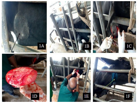
(A) Clinical presentation of the case (B) On progression of shaving of hair from surgical site
(C) Elliptical incision and blunt dissection on progression (D) Excised mass
(E) Irrigation of cavity (F) Wound appearance after completion of skin suture.
Post-operative Care and Outcome
The wound was regularly cleaned by using savlon and dressed with 1% povidone-iodine solution for five days daily. The bull was administered short-acting oxy-tetracycline (10 mg/kg) I.M for three days. Diclofenac sulfate was administered at a dose rate of 2 mg/ kg intramuscularly daily for three successive days for pain management. The wound spray (cyclo spray) was applied to the wound for five days daily. After continuous daily follow-up for five-days, the follow-up was continuous through phone communication for two weeks. Then, the sutures were removed after 15-days (Figure 2A) and the wound was successfully recovered without any other complications (Figure 2B).
Figure 2. Post-operative Care and Follow-up
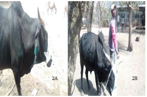
(A) Healing of the wound and removal of suture on 15th days of follow up (B) Close observation of an animal after one months at home and wound on satisfactory result.
DISCUSSION
Abscess is the most common swelling observed on the skin surface of animal body that result from a microbial invasion of the dermis and its supporting structures, leading to the formation of a collection of pus.7,10 Bacterial infection is the most common cause of abscess in an animal. According to the clinical and bacteriological study of subcutaneous abscesses caused by gram-positive bacteria in cows and sheep conducted by AL-Tuffyli et al11 stated that the commonly isolated bacterial species from an abscess are Staphylococcus aureus, S. epidermidis, S. hycus, Arcanobacterium pyogenes, and Corynebacterium pseudotuberculosis at the percentage of (33.9 %), (17.8%), (12.6%), (23.2%) and (7.1%), respectively. This is in agreement with the present case, where Corynebacterium pseudotuberculosis was isolated from the sample of a surgical managed case (Figure 3). Predilection seats and formation of abscess is depending on the way of entrance of infection agents.7,8 This is in agreement with the present case; where the affected site was the parotid region and the way of entrance of the isolated bacterium might be through oral abrasions.
Figure 3. Laboratory Test Result of Sample from Excised Chronic Abscess
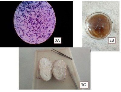
(A) Gram positive short coccobacilli in groups, pairs as well as single bacterial morphology on gram staining result that resemble Corynebacterium pseudotuberculosis (B) On blood agar growth I observed small, white, dry, opaque and concentrically ringed characteristics of bacterial colony that resemble Corynebacterium pseudotuberculosis (C) Appearance of crosssectioned excised mass.
The exploratory puncture was the highly diagnostic technique in the case of cutaneous and subcutaneous abscesses.1,8 Similarly, in the present case, the exploratory puncture was done. An abscessis classified as either an extended abscess without limitations or well-circumscribed, firm, and movable under the skin based on its nature.7,11 This is in line with the present case, where the encountered abscess was circumscribed and hard under the skin. Surgical treatment was dependent on the nature of the abscess. In the present case, treatment of abscess was performed by en bloc excision along with antibacterial therapy and allowed the skin wound to healby the first intention. A similar surgical correction of treatment was followed by Misk et al7 .
CONCLUSION
In conclusion that management of well-circumscribed, firm, and movable superficial skin abscess in cattle through en bloc excision along with antibacterial therapy and allowing the skin wound to heal by the first intention should be effective.
ACKNOWLEDGEMENTS
The authors are thankful to Dr. Shiferaw Moltumo for his help through provision of all necessary materials to perform surgical procedure and Dereje Gudeta for his assistance with the post-operative management of this case. The authors also acknowledge Addis Ababa University College of Veterinary Medicine and agriculture for providing material and necessary facilities to do this case report.
AUTHOR CONTRIBUTIONS
Both of us were contributed to the manuscript preparation and final submission. The main surgical procedure and original manuscript writing, the manuscript structure modification and editing were performed by Zerihun Mulatu Tuji, while Dese Kefyalew Woldegeorgis and Cheru Telila were participated as assistant surgeon and Bashahun Gebremichael, Sagni Abdisa and Jiregna Dugasa were participated as reviewing process. Finally, we all authors read and approved the final manuscript submission.
ETHICS CONSIDERATION
The approval of ethical committee and the institutional review board was taken for conducting this study and followed all the animal ethics and welfare guidelines.
CONFLICTS OF INTEREST
The authors declare that they have no conflicts of interest.


