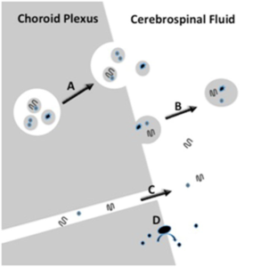
1. Potter R, Patterson BW, Elbert DL, et al. Increased in vivo amyloid-beta42 production, exchange, and loss in presenilin mutation carriers. Science translational medicine. 2013; 5: 189ra177. doi: 10.1126/scitranslmed.3005615
2. Parada C, Gato A, Bueno D. Mammalian embryonic cerebrospinal fluid proteome has greater apolipoprotein and enzyme pattern complexity than the avian proteome. Journal of proteome research. 2005; 4: 2420-2428. doi: 10.1021/pr050213t
3. Lehtinen MK, Walsh CA. Neurogenesis at the brain-cerebrospinal fluid interface. Annual review of cell and developmental biology. 2011; 27: 653-679. doi: 10.1146/annurev-cellbio-092910-154026
4. Weber JA, Baxter DH, Zhang S, et al. The microRNA spectrum in 12 body fluids. Clinical chemistry. 2010; 56: 1733-1741. doi: 10.1373/clinchem.2010.147405
5. Burgos KL, Javaherian A, Bomprezzi R, et al. Identification of extracellular miRNA in human cerebrospinal fluid by nextgeneration sequencing. Rna. 2013; 19: 712-722. doi: 10.1261/ rna.036863.112
6. Lehtinen MK, Zappaterra MW, Chen X, et al. The cerebrospinal fluid provides a proliferative niche for neural progenitor cells. Neuron. 2011; 69: 893-905. doi: 10.1016/j.neuron.2011.01.023
7. Fencl V, Koski G, Pappenheimer JR. Factors in cerebrospinal fluid from goats that affect sleep and activity in rats. The Journal of physiology. 1971; 216: 565-589. doi: 10.1113/jphysiol.1971. sp009541
8. Pappenheimer JR, Fencl V, Karnovsky ML, Koski G. Peptides in cerebrospinal fluid and their relation to sleep and activity. Research publications – Association for Research in Nervous and Mental Disease. 1974; 53: 201-210.
9. Martin FH, Seoane JR, Baile CA. Feeding in satiated sheep elicited by intraventricular injections of CSF from fasted sheep. Life sciences. 1973; 13: 177-184.doi: 10.1016/0024-3205(73)90193-8
10. Pedrazzoli M, D’Almeida V, Martins PJ, et al. Increased hypocretin-1 levels in cerebrospinal fluid after REM sleep deprivation. Brain research. 2004; 995: 1-6. doi: 10.1016/j. brainres.2003.09.032
11. Hollander JA, Pham D, Fowler CD, Kenny PJ. Hypocretin-1 receptors regulate the reinforcing and reward-enhancing effects of cocaine: pharmacological and behavioral genetics evidence. Frontiers in behavioral neuroscience. 2012; 6: 47. doi: 10.3389/ fnbeh.2012.00047
12. Cason AM, Aston-Jones G. Role of orexin/hypocretin in conditioned sucrose-seeking in female rats. Neuropharmacology. 2014; 86: 97-102. doi: 10.1016/j.neuropharm.2014.07.007
13. Nixon JP, Mavanji V, Butterick TA, Billington CJ, Kotz CM, Teske JA. Sleep disorders, obesity, and aging: the role of orexin. Ageing research reviews. 2015; 20: 63-73. doi: 10.1016/j. arr.2014.11.001
14. Fronczek R, Overeem S, Lee SY, et al. Hypocretin (orexin) loss and sleep disturbances in Parkinson’s Disease. Brain : a journal of neurology. 2008; 131: e88. doi: 10.1093/brain/ awm222
15. Fronczek R, van Geest S, Frölich M, et al. Hypocretin (orexin) loss in Alzheimer’s disease. Neurobiology of aging. 2012; 33: 1642-1650. doi: 10.1016/j.neurobiolaging.2011.03.014
16. Johanson CE, Duncan JA, Stopa EG, Baird A. Enhanced prospects for drug delivery and brain targeting by the choroid plexus-CSF route. Pharmaceutical research. 2005; 22: 1011- 1037. doi: 10.1007/s11095-005-6039-0
17. Lun M, Monuki ES, Lehtinen MK. Development and functions of the choroid plexus–cerebrospinal fluid system. Nature reviews. Neuroscience. 2015; 16: 445-457. doi: 10.1038/ nrn3921
18. Brown PD, Davies SL, Speake T, Millar ID. Molecular mechanisms of cerebrospinal fluid production. Neuroscience. 2004; 129: 957-970. doi: 10.1016/j.neuroscience.2004.07.003
19. Larocca D, Burg MA, Jensen-Pergakes K, Ravey EP, Gonzalez AM, Baird A. Evolving phage vectors for cell targeted gene delivery. Current pharmaceutical biotechnology. 2002; 3: 45- 57. doi: 10.2174/1389201023378490
20. Strazielle N, Ghersi-Egea JF. Physiology of blood-brain interfaces in relation to brain disposition of small compounds and macromolecules. Molecular pharmaceutics. 2013; 10, 1473- 1491. doi: 10.1021/mp300518e
21. van der Pol E, Boing AN, Harrison P, Sturk A, Nieuwland R. Classification, functions, and clinical relevance of extracellular vesicles. Pharmacological reviews. 2012; 64: 676-705. doi: 10.1124/pr.112.005983
22. Turturici G, Tinnirello R, Sconzo G, Geraci F. Extracellular membrane vesicles as a mechanism of cell-to-cell communication: advantages and disadvantages. American journal of Physiology Cell physiology. 2014; 306: C621-C633. doi: 10.1152/ ajpcell.00228.2013
23. Kosaka N, Iguchi H, Yoshioka Y, Takeshita F, Matsuki Y, Ochiya T. Secretory mechanisms and intercellular transfer of microRNAs in living cells. J Biol Chem. 2010; 285: 17442- 17452. doi: 10.1074/jbc.M110.107821
24. Cai J, Han Y, Ren H, et al. Extracellular vesicle-mediated transfer of donor genomic DNA to recipient cells is a novel mechanism for genetic influence between cells. Journal of molecular cell biology. 2013; 5: 227-238. doi: 10.1093/jmcb/ mjt011
25. Alvarez-Erviti L, Seow Y, Yin HF, Betts C, Lakhal S, Wood MJA. Delivery of siRNA to the mouse brain by systemic injection of targeted exosomes. Nature biotechnology. 2011; 29: 341- 345. doi: 10.1038/nbt.1807
26. Sawamoto K, Wichterle H, Gonzalez-Perez O, et al. New neurons follow the flow of cerebrospinal fluid in the adult brain. Science. 2006; 311: 629-632. doi: 10.1126/science.1119133
27. Ariza J, Steward C, Rueckert F, et al. Dysregulated iron me- tabolism in the choroid plexus in fragile X-associated tremor/ ataxia syndrome. Brain research. 2015; 1598: 88-96. doi: 10.1016/j.brainres.2014.11.058
28. Grapp M, Wrede A, Schweizer M, et al. Choroid plexus transcytosis and exosome shuttling deliver folate into brain parenchyma. Nature communications. 2013; 4: 2123. doi: 10.1038/ ncomms3123
29. Serot JM, Zmudka J, Jouanny P. A possible role for CSF turnover and choroid plexus in the pathogenesis of late onset Alzheimer’s disease. Journal of Alzheimer’s disease: JAD. 2012; 30: 17-26. doi: 10.3233/JAD-2012-111964
30. Buendia I, Egea J, Parada E, et al. The Melatonin-N,NDibenzyl(N-methyl)amine hybrid ITH91/IQM157 affords neuroprotection in an in vitro alzheimer’s model via hemo-oxygenase-1 induction. ACS chemical neuroscience. 2015; 6(2): 288-296. doi: 10.1021/cn5002073
31. Bateman RJ, Munsell LY, Morris JC, Swarm R, Yarasheski KE, Holtzman DM. Human amyloid-beta synthesis and clearance rates as measured in cerebrospinal fluid in vivo. Nature medicine. 2006; 12: 856-861. doi: 10.1038/nm1438
32. Bloom GS. Amyloid-beta and tau: the trigger and bullet in Alzheimer disease pathogenesis. JAMA neurology. 2014; 71: 505-508. doi: 10.1001/jamaneurol.2013.5847
33. Krzyzanowska A, Carro E. Pathological alteration in the choroid plexus of Alzheimer’s disease: implication for new therapy approaches. Frontiers in pharmacology. 2012; 3: 75. doi: 10.3389/fphar.2012.00075
34. Tricoire H, Malpaux B, Moller M. Cellular lining of the sheep pineal recess studied by light-, transmission-, and scanning electron microscopy: morphologic indications for a direct secretion of melatonin from the pineal gland to the cerebrospinal fluid. The Journal of comparative neurology. 2003; 456: 39-47. doi: 10.1002/cne.10477
35. Anton-Tay F, Wurtman RJ. Regional uptake of 3H-melatonin from blood or cerebrospinal fluid by rat brain. Nature. 1969; 221: 474-475. doi: 10.1038/221474a0
36. Tan DX, Manchester LC, Sanchez-Barcelo E, Mediavilla MD, Reiter RJ. Significance of high levels of endogenous melatonin in Mammalian cerebrospinal fluid and in the central nervous system. Current neuropharmacology. 2010; 8: 162-167. doi: 10.2174/157015910792246182
37. He H, Dong W, Huang F. Anti-amyloidogenic and anti-apoptotic role of melatonin in Alzheimer disease. Current neuropharmacology. 2010; 8: 211-217. doi: 10.2174/157015910792246137
38. Masilamoni JG, Jesudason EP, Dhandayuthapani S, et al. The neuroprotective role of melatonin against amyloid beta peptide injected mice. Free radical research. 2008; 42: 661-673. doi: 10.1080/10715760802277388
39. Matyszak MK, Lawson LJ, Perry VH, Gordon S. Stromal macrophages of the choroid plexus situated at an interface between the brain and peripheral immune system constitutively express major histocompatibility class II antigens. Journal of neuroimmunology. 1992; 40: 173-181. doi: 10.1016/0165- 5728(92)90131-4
40. Nataf S, Strazielle N, Hatterer E, Mouchiroud G, Belin MF, Ghersi-Egea JF. Rat choroid plexuses contain myeloid progenitors capable of differentiation toward macrophage or dendritic cell phenotypes. Glia. 2006; 54: 160-171. doi: 10.1002/ glia.20373
41. Vercellino M, Votta B, Condello C, et al. Involvement of the choroid plexus in multiple sclerosis autoimmune inflammation: a neuropathological study. Journal of neuroimmunology. 2008; 199: 133-141. doi: 10.1016/j.jneuroim.2008.04.035
42. Petito CK. Human immunodeficiency virus type 1 compartmentalization in the central nervous system. Journal of neurovirology. 2004; 10(Suppl 1): 21-24.doi: 10.1080/753312748
43. Meeker RB, Boles JC, Bragg DC, Robertson K, Hall C. Development of neuronal sensitivity to toxins in cerebrospinal fluid from HIV-type 1-infected individuals. AIDS research and human retroviruses. 2004; 20: 1072-1078. doi: 10.1089/ aid.2004.20.1072
44. Belujon P, Grace AA. Hippocampus, amygdala, and stress: interacting systems that affect susceptibility to addiction. Annals of the New York Academy of Sciences. 2011; 1216: 114-121. doi: 10.1111/j.1749-6632.2010.05896.x
45. Fowler CD, Lu Q, Johnson PM, Marks MJ, Kenny PJ. Habenular alpha5 nicotinic receptor subunit signalling controls nicotine intake. Nature. 2011; 471: 597-601. doi: 10.1038/nature09797
46. LavezziAM, Matturri L, Del Corno G, Johanson CE. Vulnerability of fourth ventricle choroid plexus in sudden unexplained fetal and infant death syndromes related to smoking mothers. International journal of developmental neuroscience: the official journal of the International Society for Developmental Neuroscience. 2013; 31: 319-327. doi: 10.1016/j.ijdevneu.2013.04.006
47. Li MD, Kane JK, Matta SG, Blaner WS, Sharp BM. Nicotine enhances the biosynthesis and secretion of transthyretin from the choroid plexus in rats: implications for beta-amyloid formation. The Journal of neuroscience: the official journal of the Society for Neuroscience. 2000; 20: 1318-1323.doi: 10.1523/JNEUROSCI.20-04-01318.2000
48. Schreiber G, Aldred AR, Jaworowski A, Nilsson C, Achen MG, Segal MB. Thyroxine transport from blood to brain via transthyretin synthesis in choroid plexus. Am J Physiol. 1990; 258: R338-R345. doi: 10.1152/ajpregu.1990.258.2.R338


