Cancer Studies and Molecular Medicine

Open journal

ISSN 2377-1518

Elimination of Tumor Suppressor Proteins during Liver Carcinogenesis

Nikolai A. Timchenko* and Kyle Lewis

Nikolai A. Timchenko. PhD

Division of Pediatric Surgery Cincinnati Children’s Hospital Medical Center, 3333 Burnet Avenue Cincinnati, OH, 45229, USA; Tel. 513-636-0129; E-mail: Nikolai.Timchenko@cchmc.org

INTRODUCTION

The development of hepatocellular carcinoma (HCC) has a long history of affecting mainly adults. In the majority of cases, HCC develops in patients which have chronic liver diseases and/or are under chemical treatments. These chronic diseases affect many signaling pathways leading to liver cancer. One of the critical events in the development of HCC is the loss of hepatocytes to properly control proliferation mainly associated with inability of hepatocytes to stop proliferation. This failure to terminate liver proliferation in HCC patients is associated with the reduction or neutralization of a negative control of liver proliferation. In this review, we summarize recent publications which provide new insight into mechanisms of termination of liver proliferation under normal conditions when liver proliferates but does not develop liver cancer and recent reports that show how these mechanisms of termination are eliminated during development of HCC leading to continued proliferation and tumor growth. Mechanisms of normal liver proliferation/termination have been investigated in several models including liver proliferation/termination during postnatal development, liver proliferation/termination after surgical resections (partial hepatectomy) and liver proliferation after acute treatments with carbon tetrachloride (CCl4). These systems provided general principles of termination of liver proliferation under conditions when liver does not develop cancer. Investigations of liver cancer in animal models were mainly focused on the development of liver cancer after treatments with diethylnitrosamine (DEN), while fewer studies have been done with the chronic treatments by CCl4.

PARTIAL HEPATECTOMY AS A MODEL FOR THE STUDY OF MECHANISMS WHICH TERMINATE LIVER PROLIFERATION

One of the key characteristics of liver cancer is uncontrolled liver proliferation. It is well recognized that malignant cells lose the ability to stop proliferation. The understanding of mechanisms which stop liver proliferation is important for development of therapeutic approaches to treat liver cancer. One of the best systems for the studies of mechanisms that terminate liver proliferation is Partial Hepatectomy (PH). The most common model of PH involves resections of 2/3 of the liver which leads to initiation of liver proliferation and restoration of the original size. While mechanisms of initiation of liver proliferation after PH are well investigated and are described in several recent reviews,1,2,3,4,5,6 very little is known about the mechanisms that terminate liver regeneration. Global gene profiling of the liver 3 weeks after PH has identified alterations in cell cycle, apoptosis, TGFβ and angiogenesis signaling.7 PPAR signaling and lipid metabolism have also been implicated in the termination of liver regeneration.8 It has been shown that certain micro RNAs may be involved in the termination of liver regeneration.9,10 In addition, the ablation of integrin-linked kinase leads to enhanced liver proliferation.11 A recent paper by Koral et al. have shown that leukocyte-specific protein (LPS) serves as a tumor suppressor and inhibits proliferation of hepatoma cell lines.12 It has been shown that termination of liver regeneration after PH and after liver injury requires a tight cooperation of chromatin remodeling proteins and a family of C/EBP proteins and that disorganization of this cooperation leads to a failure of the liver to stop regeneration.13 A number of key regulators of liver biology are under control of C/EBP family proteins and are properly regulated during liver development, differentiation and regeneration. These proteins include SIRT1, PGC1α, p53, FXR, TERT, enzymes of glucose metabolism PEPCK, G6Phase, Glut2 and Glut4 as well as enzymes of triglyceride syntheses.14,15,16,17,18 The ability of C/ EBP proteins to activate or repress these genes depends on their association with p300 or with HDAC1. Using specific knockin animal models, Jin et al. found that these known targets are mis-regulated in the liver if the C/EBP-chromatin remodeling complexes are not controlled in a proper way which leads to the lack of termination of liver regeneration.13,19 Among additional candidates for the termination of liver proliferation, Yap (Yes-associated protein) has been implicated in the regulation of tissue growth and size.20 It has been shown that Yap protein is activated in the liver after surgical resections and in hepatocellular carcinoma.21,22 The expression of Yap is under tight control of Hippo signaling which is also changed after PH and in hepatocellular carcinoma.22 Most important, Yimlamai et al. have shown that Hippo-Yap pathway is critical for maintenance of differentiation state of hepatocytes.23 In summary, studies of liver regeneration after PH have identified several candidates which might terminate liver proliferation, but are eliminated by liver cancer. Although these studies are important and useful for understanding of mechanisms of liver cancer, it has become clear that development of liver cancer includes several additional pathways to block termination of proliferation. In this review, we focus on the mechanisms by which liver cancer eliminates liver-specific tumor suppressor proteins.

LIVER SPECIFIC TUMOR SUPPRESSOR GENES

The quiescent status of the liver is supported by many Tumor Suppressor Genes (TSG). It has been shown that the activity of more than 20 different TSGs is lost in HCC due to mutations or due to hyper-methylation of their promoters.24 The TSGs include micro-RNAs which behave as tumor suppressors.25,26,27 Epigenetic control is also involved in support of TSGs as it has been shown by genome-wide methylation analysis.28,29 Further studies provided convincing evidence that many of these TSGs are involved in the protection of liver from development of cancer. Detailed information for these tumor suppressor genes of the liver has been discussed in several recent reviews.24,30 Therefore, we will here briefly discuss some of these TSGs which are related to the focus of our review. One of the important TSGs is Deleted in Liver Cancer (DLC1) tumor suppressor gene. This gene is located on chromosome 8p22 and plays a critical role in multiple liver functions. It has been shown that DLC1 is deleted in 40% human HCC31,32 and that restoration of its expression resulted in inhibition of liver proliferation and reduction of the development of tumors after xenografting HCC cells into nude mice.33 Exomic sequencing of hepatitis C virus (HCV)-associated HCCs has identified novel mutations in AT-Rich Interactive Domain 2 (ARID2) protein which has been further shown to be a liver tumor suppressor protein.34 A family of Suppressors of Cytokine Signaling (SOCS), are inhibitors of cytokine signaling. It has been shown that the liver specific deletion of a member of this family, SOCS3, leads to the increased liver proliferation and formation of hepatocellular carcinoma.35 Among more than 20 known tumor suppressor proteins of the liver, Rb, p53, HNF4α C/EBPα and p16, are investigated in great detail and have been shown to be most critical inhibitors of liver proliferation.

TUMOR SUPPRESSOR PROTEIN P53

P53 is a transcription factor which regulates expression of many genes by direct binding to their promoters.36 Under conditions when liver is challenged by surgical resections or treatments with drugs, expression of p53 is elevated leading to growth arrest, induction of apoptosis, or  senescence.37,38 It has been also shown that p53 regulates ploidy of hepatocytes. Using p53 KO mice, Barton’s group has shown that ploidy levels increased during regeneration of both Wild-Type (WT) and p53(-/-) hepatocytes, but only WT hepatocytes were able to dynamically resolve ploidy levels and return to normal by the end of regeneration. Kurrina et al. identified multiple cell cycle and mitotic regulators (Foxm1, Aurka, Lats2, Plk2, and Plk4) as direct targets of p53 in the liver.37 The expression and activity of p53 is significantly reduced in the majority of cancers including hepatocellular carcinoma.39,40 In about 50% of patients with HCC, the reduction of p53 levels and activity is mediated by mutations within the coding region or within the p53 promoter.40 However, a number of recent studies revealed that the elimination of p53 by ubiquitin proteasome system contributes to the loss of p53 tumor suppressor functions in cancers.41 The main ligase that triggers p53 degradation is MDM2 which targets six key lysine amino acids on p53.42 In addition to MDM2, there are other ligases that target p53 degradation such as CHIP (Cterminus of HSP70 interaction protein).41,43 It is interesting that MDM2 is a transcriptional target for p53 which creates an auto regulation loop that works under conditions of DNA damage. The DNA damage stabilizes p53 protein, but it is degraded by MDM2-proteasome pathway by activation of its own inhibitor at the time when cells recover after stress and do not need p53 anymore.44,45,46The MDM2-dependent degradation of p53 involves other proteins which cooperate with MDM247 or control levels of MDM2. This review is focused on the one of these regulators, Gankyrin, which stabilizes MDM2 and facilitates degradation of p53 during development of liver cancer (see below).

P16/RB/E2F PATHWAY IN LIVER PROLIFERATION AFTER PH AND IN LIVER CANCER

Cell cycle progression in proliferating livers is stimulated by E2F transcription factors which activate several key S-phase specific genes.4 The E2F family consists of eight members, five of which (E1F1-E2F5) interact with Rb, while E2F6- E2F8 do not and work as a repressor of E2F-dependent genes. It has been shown that E2F1 plays an overlapping role in HCC48 and E2F2-E2F7 promote cancer.49 E2F8 transcription factor is a unique member of the family which represses promoters without interactions with Rb. It has been shown that inactivation of both Rb and E2F8 works synergistically to trigger DNA replication.50 In addition, E2F8 is essential for polyploidization in mammalian cells.51 The detailed information for the role of E2F family in cancer has been described in a recent review.49 Similar to other quiescent tissues, the activity of E2F transcription factors is inhibited in quiescent livers by retinoblastoma, (Rb) protein. Among several members of E2F family, E2F2 seems to be a most important regulator of liver proliferation and timely liver regeneration after PH.52 It is important to emphasize that C/EBPα is one of the critical regulators of Rb-E2F complexes and that aged livers have a weak proliferation after PH due to C/ EBPα-mediated enhancement of Rb-E2F repression function.53,54 C/EBPα also regulates E2F complexes with another member of Rb family, p107, which brings about growth arrest in hepatocytes.55 Although C/EBPα -mediated regulation of Rb-E2F complexes is involved in the control of liver proliferation, the most significant pathway of regulation of Rb-E2F complexes is associated with cyclin dependent kinases cdk4 and cdk6. Upon stimulation of liver proliferation by surgical resections, cdk4/ cdk6 kinases are activated by cyclin D1 and phosphorylate Rb leading to the dissociation of Rb-E2F complexes.56 The activities of cdk4/6 are negatively regulated by a member of inhibitors of cdk (INK) proteins, p16. Despite numerous studies of p16 in the liver, very little is known about its role in liver proliferation after PH. Lee et al. showed that p16 undergoes methylation after PH which correlated with liver proliferation.57 Another study of liver proliferation in aged mice revealed that p16 is elevated in livers of old mice and contributes to the weak proliferative response of livers to PH.58 Studies of 130 old human patients who underwent hepatectomy showed that these patients had much higher levels of p16 and that these levels negatively correlated with liver regeneration.59

Examination of mutation/expression of p16 and Rb proteins in human liver cancer and in animal models of carcinogenesis strongly indicated that the loss of functions of these proteins is involved in development of scevere liver cancer. It has been shown that p16 is inactivated at early stages of hepatocarcinogenesis.60 It has been also shown that p16INK4a pathway is altered in rat liver tumors induced by NNK.61 The inactivation of p16 and Rb in human HCC samples has been shown in many publications which are summarized in several reviews.62,63.64 These reviews emphasized that p16, cyclin D1 and Rb pathways are commonly targeted in various cancers. To determine the role of the disruption of these three pathways in HCC, Azichi et al. have analyzed p16, pRB and cyclin D1 in 47 patients with HCCs. The authors have shown that inactivation of p16 was detected in 64% of HCCs; while Rb was inactivated in 28% of HCC samples. Importantly, several patients had inactivation both of these pathways.65 In this study, over expression of cyclin D1 was detected in 11% of examined samples. These observations showed critical role of p16-Rb pathway in protection of liver from development of cancer. In agreement with these observations, Viatour et al. have deleted three members of Rb family (Rb, p107 and p130) and found that these triple knockout mice develop liver cancer with gene expression profile similar to that of human HCC.66 Further studies from this group revealed that Hippo pathway is activated at later stages in these mice.67

C/EBPα: A STRONG INHIBITOR OF LIVER PROLIFERATION AND A TUMOR SUPPRESSOR PROTEIN

C/EBPα belongs to the C/EBP family of proteins, βZIP proteins which contain basic region and luecine zipper region.4,68 These proteins are transcription factors which dimerize with each other and control multiple functions in different tissues.

Numerous studies revealed that C/EBPα is a strong inhibitor of liver proliferation.69,70,71,72,73,74 Despite the fact that C/EBPα is a transcription factor, its activities are regulated on the levels of protein-protein interactions and post-translational modifications. Growth inhibitory activity of C/EBPα is tightly regulated in the liver. One of the critical pathways that control the growth inhibitory activity of C/EBPα is phosphorylation at Ser193. It has been shown that ph-S193 isoform of C/EBPα is a strong growth inhibitory protein, while un-ph-193 isoform has reduced activity to inhibit liver proliferation.75,76,77 Generation of C/EBPα knockin models with substitution of Ser193 to Ala (S193A) and to Asp (S193D) further confirmed the critical role of modifications of S193 in the biological functions of C/EBPα.13,14,15,16,17,18 While liver proliferation after PH is almost completely inhibited in S193D mice, the S193A mice showed an early entry in cell cycle and lack of termination of proliferation after surgeries.13,15 The tumor suppression activity of C/EBPα has been demonstrated in several animal models. Tan et al. have generated C/EBPα knockin mice in which C/EBPα is expressed from the alpha-fetoprotein promoter (which is active in HCC) and have shown that the elevated expression of C/EBPα inhibits liver carcinogenesis.74 Examination of liver cancer in C/EBPα S193D mice under conditions of DEN-mediated carcinogenesis revealed that C/EBPα is a critical tumor suppressor protein because its degradation by Gankyrin causes early development of liver cancer.15 A recent paper by Habib’s group showed that activation of C/EBPα in cirrhotic livers with HCC inhibits liver cancer.78 Regarding levels of C/EBPα in human cancer; C/EBPα was also examined in several reports of human HCC. Examination of levels of C/EBPα in liver tumor sections and non-tumor sections of the same patients has found a significant reduction of C/EBPα mRNA in tumor sections.79 It has been also shown that the reduced expression of C/EBPα in hepatocellular carcinoma is associated with advanced tumor stage and with shortened patient survival.80 In addition to transcriptional down-regulation of C/EBPα and degradation of the protein, liver cancer neutralizes the activity of C/EBPα by de-phosphorylation of C/EBPα at S193.75 Taken together, these studies showed that C/EBPα is a tumor suppression protein and that elimination of growth inhibitory activity of C/EBPα is a critical step in development of liver cancer. C/EBPα- S193D mutant completely inhibits liver proliferation after PH15 and given this strong growth inhibitory activity of S193D mutant in partial hepatectomy studies, one should assume that these mutant mice should be resistant to the development of liver cancer. However, further studies of DEN-mediated liver cancer in the S193D mice revealed that liver cancer developed a mechanism for complete elimination of C/EBPα by Gankyrin.

LIVER-SPECIFIC TUMOR SUPPRESSOR PROTEIN HNF4α

Hepatocyte nuclear factor 4α (HNF4α), regulates several liver functions including proliferation and differentiation of hepatocytes. HNF4α has been a subject of intensive investigations for almost 20 years. These studies demonstrated that HNF4α is a master regulator of liver biology.81 In addition to the key role of HNF4α in adult livers; HNF4α is a critical regulator of pre-natal liver development. The studies by Duncan’s group revealed that HNF4α controls the development of a hepatic epithelium, liver morphogenesis and the sinusoidal organization of the liver during prenatal liver development.82, 83 The HNF4α gene contains two promoters, P1 and P2, each produces 6 and 3 HNF4α isoforms correspondingly by alternative splicing.81 Although the functional relevance of these isoforms is unknown, examination of 450 human colon cancer specimens showed that P1-HNF4α isoforms are lost or localized in the cytoplasm of 80% of examined samples.84 This paper also showed that phosphorylation of HNF4α by Src tyrosine kinase decreases stability of HNF4α and that this mechanism is likely activated in patients with colon cancer.84 These observations suggested that HNF4α is involved in protection of cancer. In agreement with these results, the possible role of HNF4α in development of human HCC has been demonstrated by examination of patients with HCC which showed that the expression of HNF4α correlates with epithelialmesenchymal transition which is involved in metastatic tumor formation.85 A recent paper by Zhang et al. added additional evidence for the role of reduction of HNF4α in development of HCC.86 The role of HNF4α in liver cancer was examined in WT mice and in several genetically modified animal models. The studies in mice have shown a critical role of HNF4α in the liver functions of adult animals. These functions include regulation of expression of genes involved in lipid and bile acid synthesis, gluconeogenesis, blood coagulation, differentiation and proliferation. In this review, we focus on the discussion of HNF4α functions in liver proliferation and cancer. Examination of liver biology in acute HNF4α knockout mice demonstrated up-regulation of genes which are associated with liver proliferation and cell cycle control.87 These studies identified several new direct targets of HNF4α which include Bmp7 and Perp, a regulator of p53-dependent apoptosis. In agreement with these observations, it has been shown that the transient inhibition of HNF4α initiates hepatocellular transformation through microRNA feedback loop circuit.88 It is interesting that once this circuit is activated, it inhibits expression of HNF4α leading to cancer. Tumor suppressor functions of HNF4α have been demonstrated in rat and mouse livers. Ning et al. have found that HNF4α levels are progressively decreased in the livers of DEN-induced rats and that forced expression of HNF4α blocked development of HCC.89 The mechanism of this inhibition of liver cancer involves the block of activation of β-catenin signaling. Consistent with this report, Apte’s group has shown that hepatocyte-specific deletion of HNF4α in adult mice causes increased hepatocyte proliferation and activation of cell cycle genes.90 Examination of liver cancer in these hepatocyte-specific knockout mice after DEN injections showed that the deletion of HNF4α significantly increases the number and size of hepatic tumors.90 While in rat livers HNF4α protected development of liver cancer through inhibition of β-catenin signalling,89 it appears that in mouse livers HNF4α represses tumor through inhibition of both β-catenin and c-myc expression.91,92 In the liver, HNF4α is under control of several pathways alterations of which might reduce levels of HNF4α and cause liver cancer. One of these pathways is Hippo signaling. Using in vivo mouse liver development model, Alder et al. have recently shown that Hippo signaling affects hepatocyte differentiation through HNF4α.93 It has been also shown that mutations in isocitrate dehydrogenease 1 (IDH1) and IDH2 cause intrahepatic cholangiocarcinoma via complete silencing HNF4α and subsequent impaired hepatocyte differentiation.94

GANKYRIN: A POWERFUL ACTIVATOR OF LIVER CANCER

As we mentioned above, quiescent livers express more than 20 tumor suppressor genes. How does liver cancer eliminate activity of these TSGs? Examination of early events in the development of liver cancer in chemical models has identified elevation of Gankyrin.95, 96 Gankyrin (gann-ankyrin repeat protein; gann means cancer in Japanese; also known as p28, p28GANK, PSMD10, and Nas6p) is a non-ATPase subunit of the 26S proteasome and is an oncogene consisting of seven ankyrin repeats that is expressed in several cancer types, particularly HCC in which it was first discovered.95, 97 Recent studies have shown Gankyrin is up-regulated during initiation and progression of HCC and is correlated with capsular invasion, intrahepatic metastasis, and decreased apoptosis.95,98,99 Furthermore, siRNA to Gankyrin has been shown to decrease tumor cell growth in nude mice and higher levels of Gankyrin expression have been correlated with poor prognosis in HCC.100, 101 It has been recently found that the histone deacetylase inhibitor panabinostat (LBH589) inhibits proliferation and metastasis of hepatocellular carcinoma through inhibition of Gankyrin.101 Li et al. have recently identified microRNA-605 as a potent repressor of Gankyrin which also leads to inhibition of liver cancer.102 Many studies have investigated the role of Gankyrin in HCC and several pathways have been elucidated. Jiang et al. have shown that Gankyrin is repressed by FXR in quiescent liver and FXR expression is decreased in HCC. This interaction depends on downstream targets of FXR: C/EBPβ and HDAC1, which form a complex to inhibit Gankyrin expression in quiescent tissue.103 This paper also showed that FXR-mediated prevention of Gankyrin activation in DEN-mediated carcinogenesis inhibits liver cancer.103 Taken together, these papers clearly demonstrated that the inhibition of Gankyrin leads to inhibition of liver cancer.

MECHANISMS OF GANKYRIN-MEDIATED LIVER CANCER

Investigations of mechanisms by which Gankyrin causes development of HCC showed that Gankyrin has two main cancer-promoting activities. The first activity is associated with the neutralization of at least five tumor suppressor proteins and subsequent support of proteins that promote liver cancer. (Figure 1) summarizes signaling pathways which Gankyrin uses to diminish expression/activities of the tumor suppressor proteins and support high levels of cdk4 and Oct4 which promote liver cancer. It has been shown that Gankyrin binds to MDM2/ HDM2 and enhances ubiquitination and degradation of p53.104 During the initial discovery of Gankyrin, it was discovered that it is capable of binding Rb through an LXCXE domain and that this leads to increased phosphorylation of Rb and its subsequent degradation.105 This interaction is involved in conferring anchorage-independent growth in NIH 3T3 fibroblasts. In addition to the interaction with Rb, Gankyrin also binds to D-type kinase, cdk4, and replaces p16INK4a from cdk4 leading to the activation of cdk4.106 The Gankyrin-mediated elimination of p53, Rb and p16 in liver cancer has been confirmed in many other reports.2,15,95,103 Recent studies identified two additional targets of Gankyrin; tumor suppressor proteins C/EBPα and HNF4α. As we noted above, C/EBPα is a strong tumor suppressor protein when it is phosphorylated at Ser193. Gankyrin specifically recognizes ph-Ser193 isoform of C/EBPα and S193D mutant and triggers their degradation through the ubiquitin proteasome system. During development of liver cancer in WT mice treated with DEN, C/EBPα is almost completely converted into ph-S193 isoform and becomes a target for Gankyrin.15 In C/EBPα -S193D mice, Gankyrin eliminates the mutant C/EBPα much earlier leading to fast development of liver cancer.15,103 Several recent publications from Dr. Wang’s group identified HNF4α as additional target of Gankyrin. Using established hepatoma cell lines, this group showed that down-regulation of Gankyrin promotes differentiation of hepatoma cells and that this differentiation is mediated by stabilization of HNF4α. The inverse correlation of Gankyrin and HNF4α was observed in DEN-mediated cancer and in human HCC.107 In addition to degradation of HNF4α, Gankyrin-dependent dedifferentiation of hepatocytes in tumor initiating cells includes stabilization of Oct4 through Gankyrin competitively binding to WWP2, the ubiquitin ligase that normally marks Oct4 for degradation.108

Figure 1: A summary of signaling pathways by which Gankyrin diminishes expression/activities of tumor suppressor proteins and by which it supports high levels/activities of cdk4 and Oct4 promoting liver cancer. Gankyrin directly interacts with C/EBPα, Rb and HNF4α and triggers their degradation. Gankyrin causes degradation of p53 through stabilization of MDM2 ubiquitin ligase. Gankyrin-mediated neutralization of p16 is associated with the disruption of p16-cdk4 complexes and subsequent activation of cdk4 by cyclins D1 and D3. Gankyrin also stabilizes Oct4 by interaction with WWP2 which marks Oct4 degradation.

CSMMOJ-1-104Fig1

The second liver cancer promotion activity of Gankyrin is associated with activation of signaling pathways which initiate liver cancer. It has been shown that Gankyrin promotes liver tumor growth and metastases through activation of Il-6/STAT3 signaling.109 Gankyrin also activates IL-8 during development of liver cancer.110 Two key pathways of liver cancer, β-catenin and c-myc, are also activated by Gankyrin.111 In addition, several reports showed that Gankyrin-mediated liver cancer includes activation of PI3K/Akt pathway and Rho/ROCK/PTEN signalling.112,113 Interestingly, the activation of some of these pathways correlates with expression of stemness factors.114 Although elevation of Gankyrin in HCC is well documented, very little is known about mechanisms by which liver cancer activates Gankyrin. Our work revealed that Gankyrin is expressed in normal livers at very low levels due to FXR-dependent silencing, but it is activated in liver cancer by the reduction of FXR signalling.103 FXR supports high levels of chromatin-remodeling complexes C/EBPα-HDAC1 which bind and partially repress the Gankyrin promoter in quiescent liver. Upon treatments with DEN, FXR is reduced leading to de-repression of the promoter.103 A recent paper suggested an additional mechanism of increase of Gankyrin which is associated with activation of interleukin-1α/ IRAK-1 inflammation signaling and subsequent activation of the Gankyrin promoter by NF-Y-p300 complexes.115 (Figure 2) summarizes current knowledge about activation of Gankyrin in liver cancer and Gankyrin-dependent activities which contribute to development of liver cancer. The activation of Gankyrin in rodent models of carcinogenesis is mediated perhaps by two important events: de-repression of the Gankyrin promoter by reducing FXR signaling and subsequent activation by interleukin1α/IRAK-1signaling. The elevation of Gankyrin causes elimination of 5 tumor suppressor proteins and activation of positive regulators of cancer such as β-catenin and c-myc. These global alterations contribute to the development of liver cancer.

Figure 2: Activation of Gankyrin in liver cancer. Gankyrin is activated by carcinogens using two main pathways: 1) reduction of FXR signaling leading to a release of repression of the Gankyrin promoter; and 2) activation of Interleukin-1α/IRAK-1 pathway and subsequent activation of the Gankyrin promoter by JNK and NF-Y/p300/CBP transcriptional complex. Once activated, Gankyrin displays two main cancer-promoting activities: 1) elimination of tumor suppressor proteins; and 2) activation of tumor-promoting Oct4, c-myc, β-catenin, PI3K-Akt and Rho/ROCK pathways.

CSMMOJ-1-104Fig2

TREATMENTS AND PREVENTION OF LIVER CANCER BY I NHIBITION OF GANKYRIN AND BY RESTORATION OF ACTIVITIES OF TSGs

Current studies of liver cancer using global profiling of gene expression, chromatin remodeling and proteomics revealed multiple alterations in the liver biology which are associated with each other. This situation suggests that it is unlikely to generate a single-gene therapeutic approach to cure liver cancer. However, literature data also show that Gankyrin is one of the critical components of the development of liver cancer because it controls multiple pathways of liver cancer (Figures 1 and 2). This fact raises a unique possibility to correct/prevent liver cancer by targeting of Gankyrin or by activation of FXR/inhibition of interleukin-1α/IRAK signaling. Among those possibilities, the promising approach might be the activation of FXR because it has been shown that long-lived little mice express high levels of FXR and do not develop liver cancer with age and after treatments with DEN.103 It has been shown that high levels of FXR prevent activation of Gankyrin and rescue expression of tumor suppressor genes protecting from development of cancer.103 Moreover, our unpublished results revealed that direct activation of FXR by specific ligand GW4064 rescues tumor suppressor proteins and prevents liver cancer (Lewis and Timchenko, unpublished results).Very promising observations have been recently found in the studies of liver cancer in rat models of cirrhosis and HCC by Habib’s group. Using short activating RNA (saRNA) strategy, the authors activated C/EBPα in rats with severe cirrhosis and HCC and found significant inhibition of liver cancer and dramatic improvement of liver functions.78 Examination of cancer pathways in hepatoma cell lines after activation of C/EBPα by saRNA revealed that correction of C/ EBPα expression increased levels of 18 tumor suppressor gene including HNF4α, p53, Rb, DLC1, ARID2 and SOCS3. saRNA- mediated activation of C/EBPα also down-regulated several canonical pathways of liver cancer such as HFG, β-catenin and c-myc signaling. Several critical drivers of liver proliferation were also down-regulated including cyclin D1 and Stat3.78 Importantly, activation of C/EBPα by saRNA improved liver functions. (Figure 3) summarizes positive effects of activation of C/EBPα in livers with HCC on liver biology and functions.

Figure 3: Rescue of C/EBPα expression in HCC inhibits liver cancer. The diagram summarizes observations published in a recent paper78 and suggests possible mechanisms of C/ EBPα -mediated inhibition of liver cancer (see text).

CSMMOJ-1-104Fig3

These observations show that C/EBPα is a master regulator of many tumor suppressor genes, critical repressor of tumor promoting pathways, and a positive regulator of liver functions. These observations place C/EBPα in a unique position to be a therapeutic target for the treatments of patients with liver functions. How does the correction of one protein correct so many cancer associated dysfunctions in the liver? Although this issue requires further examination of molecular pathways in livers after activation of C/EBPα, literature data and data in our lab suggest some of these pathways such as a possible feedback loop leading to down-regulation of Gankyrin. We have shown that the Gankyrin promoter contains two high affinity C/EBP sites.103 Therefore, it is possible that activated C/EBPα represses the Gankyrin promoter in complexes with HDAC1 leading to the rescue of TGS and to repression of c-myc and β -catenin signaling (Figure 3). In agreement with this hypothesis, some of the up-regulated TSGs, c-myc and β-catenin are targets of Gankyrin see Figure 2. Regardless of the mechanisms, it is clear that C/EBPα is a key tumor suppressor protein in the liver.

CONCLUSION

Development of liver cancer involves multiple alterations of liver biology on several levels of gene expression complicating development of therapeutic approaches to treat cancer. Although these multiple changes are not easy to correct, recent progress in investigations of tumor suppressor proteins and mechanisms of their elimination in cancer provides a possibility to develop approaches which might reduce liver cancer at advanced stages and improve liver functions. It is likely that tumor suppressor proteins communicate with each other through different signaling pathways and rescue/protection of one of them is sufficient for inhibition of liver cancer. In this regard, tumor suppressor protein C/EBPα is a promising candidate, correction of which inhibits liver cancer. We think that, similar to C/EBPα, correction of HNF4α might also have beneficial effects on the liver since HNF4α regulates liver differentiation and many liver functions. It is also interesting that activities of both these proteins are regulated by specific phosphorylation pathways which also might be considered as possible tools for correction of C/ EBPα and HNF4α. However, the most hopeful strategy seems to be activation of their promoters and prevention of their degradation by Gankyrin. Specifically, drug-mediated activation of FXR and subsequent block of Gankyrin elevation could be considered for inhibition of liver cancer in human patients. Some of the known drug-activators of FXR are already in trials for NAFLD and might be quickly incorporated in the trails for patients with HCC.

ACKNOWLEDGEMENT

This work is supported by NIH R01CA159942 and R01 GM551888 grants and by Internal Development Funds (CCHMC).

1. Fausto N, Campbell JS, Riehle KJ. Liver regeneration. J Hepatol. 2012; 57: 692-694. doi: 10.1002/jcp.21172

2. Jones K, Timchenko L, Timchenko NA. The role of CUGBP1 in age-dependent changes of liver functions. Ageing Research Reviews. 2012; 11: 442-449. doi: 10.1016/j.arr.2012.02.007

3. Riehle KJ, Dan YY, Campbell JS, Fausto N. New concepts in liver regeneration. J Gastroenterol Hepato.l 2011; 26 Suppl 1: 203-212. doi: 10.1111/j.1440-1746.2010.06539.x

4. Timchenko NA. Aging and liver regeneration. Trends Endocrinol Metab. 2009; 20: 171-176. doi: 10.1016/j. tem.2009.01.005

5. Michalopoulos G. Principles of liver regeneration and Growth Homeostasis. Comprehensive Physiology. 2013; 3: 485-513. doi: 10.1002/cphy.c120014

6. Michalopoulos G. Advances in liver regeneration. Expert Review of Gastroenterology. & Hepatology. 2014; 26: 1-11. doi: 10.1586/17474124.2014.934358

7. Nygard IE, Mortensen KE, Hedegaard J, et al. The genetic regulation of the terminating phase of liver regeneration. Comp Hepatol. 2012; 11: 3. doi: 10.1186/1476-5926-11-3

8. Rychtrmoc D, Hubalkova L, Viskova A, Libra A, Buncek M, Cervinkova Z. Transcriptome temporal and functional analysis of liver regeneration termination. Physiol Res. 2012; 61 Suppl 2: S77-S92.doi: 10.33549/physiolres.932393

9. Chen H, Sun Y, Dong R, et al. Mir-34a is upregulated during liver regeneration in rats and is associated with the suppression of hepatocyte proliferation. PLoS One. 2011; 6: e20238. doi: 10.1371/journal.pone.0020238

10. Yuan B, Dong R, Shi D, et al. Down-regulation of miR-23b may contribute to activation of the TGF-beta1/Smad3 signalling pathway during the termination stage of liver regeneration. FEBS Lett. 2011; 585: 927-934. doi: 10.1016/j.febslet.2011.02.031

11. Apte U, Gkretsi V, Bowen WC, et al. Enhanced liver regeneration following changes induced by hepatocyte-specific genetic ablation of integrin-linked kinase. Hepatology. 2009; 50: 844- 851. doi: 10.1002/hep.23059

12. Koral K, Paranjpe S, Bowen WC, Mars W, Luo J, Michalopoulos GK. Leukocyte specific protein-1: A novel regulator of hepatocellular proliferation and migration deleted in human HCC. Hepatology. 2014. doi: 10.1002/hep.27444

13. Jin J, Hong IH, Lewis K, et al. Cooperation of C/EBP family proteins and chromatin remodeling proteins is essential for termination of liver regeneration in mice. Hepatology. 2014. doi: 10.1002/hep.27295

14. Jin J, Wang GL, Iakova P, et al. Epigenetic changes play critical role in age-associated dysfunctions of the liver. Aging Cell. 2010; 9: 895-910. doi: 10.1111/j.1474-9726- .2010.00617.x

15. Wang GL, Shi X, Haefliger S, et al. Elimination of C/EBPalpha through the ubiquitin-proteasome system promotes the development of liver cancer in mice. J Clin Invest. 2010; 120: 2549-2562. doi: 10.1172/JCI41933

16. Hong IH, Lewis K, Iakova P, et al. Age-associated Change of C/EBP Family Proteins Causes Severe Liver Injury and Acceleration of Liver Proliferation after CCl4 Treatments. J Biol Chem. 2014; 289: 1106-1118. doi: 10.1074/jbc.M113.526780

17. Jin J, Iakova P, Breaux M, et al. Increased expression of enzymes of triglyceride synthesis is essential for the development of hepatic steatosis. Cell Rep. 2013; 3: 831-843. doi: 10.1016/j. celrep.2013.02.009

18. Jin J, Iakova P, Jiang Y, et al. Transcriptional and translational regulation of C/EBPbeta-HDAC1 protein complexes controls different levels of p53, SIRT1, and PGC1alpha proteins at the early and late stages of liver cancer. J Biol Chem. 2013; 288: 14451-14462. doi: 10.1074/jbc.M113.460840

19. Michalopoulos G. Terminating hepatocyte proliferation during liver regeneration: The roles of two members of the same family (C/EBP α and β) with opposing actions. Hepatology. 2014. doi: 10.1002/hep.27329

20. Zhao B, Wei X, Li W, et al. Inactivation of YAP oncoprotein by the Hippo pathway is involved in cell contact inhibition and tissue growth control. Genes Dev. 2007; 21(21): 2747-2761. doi: 10.1101/gad.1602907

21. Wang C, Zhang L, He Q, et al. Differences in Yes-associated protein and mRNA levels in regenerating liver and hepatocellular carcinoma. Mol Med Rep. 2012; 5(2): 410-414. doi: 10.3892/ mmr.2011.640

22. Grijalva JL, Huizenga M, Mueller K, et al. Dynamic alterations in Hippo signaling pathway and YAP activation during liver regeneration. Am J Physiol Gastrointest Liver Physiol. 2014; 307(2): G196-G204. doi: 10.1152/ajpgi.00077.2014

23. Yimlamai D, Christodoulou C, Galli GG, et al. Hippo pathway activity influences liver cell fate. Cell. 2014; 157(6): 1324- 1338. doi: 10.1016/j.cell.2014.03.060

24. Martin J, Dufour JF. Tumor suppressor and hepatocellular carcinoma. World J Gastroenterol. 2008; 14(11): 1720-1733. doi: 10.3748/wjg.14.1720

25. Callegari E, Gramantieri L, Domenicali M, D’Abundo L, Sabbioni S, Negrini M. MicroRNAs in liver cancer: a model for investigating pathogenesis and novel therapeutic approaches. Cell Death Differ. 2014. doi: 10.1038/cdd.2014.136

26. Yin H, Peng X, Ren P, Cheng B, Li S, Qin C. MicroRNAs as a novel class of diagnostic biomarkers in detection of hepatocellular carcinoma: a meta-analysis. Tumour Biol. 2014. doi: 10.1007/s13277-014-2544-2

27. Khare S, Zhang Q, Ibdah JA. Epigenetics of hepatocellular carcinoma: role of microRNA. World J Gastroenterol. 2013; 19(33): 5439-5445. doi: 10.3748/wjg.v19.i33.5439

28. Revill K, Wang T, Lachenmayer A, et al. Genome-wide methylation analysis and epigenetic unmasking identify tumor suppressor genes in hepatocellular carcinoma. Gastroenterology. 2013; 145(6): 1424-35.e1-25. doi: 10.1053/j.gastro.2013.08.055

29. Xue W, Kitzing T, Roessler S, et al. SWA cluster of cooperating tumor-suppressor gene candidates in chromosomal deletions. Proc Natl Acad Sci U S A. 2012; 109(21): 8212-8217. doi: 10.1073/pnas.1206062109

30. Aguirre E, Renner O, Narlik-Grassow M, Blanco-Aparicio C. Genetic Modeling of PIM Proteins in Cancer: Proviral Tagging and Cooperation with Oncogenes, Tumor Suppressor Genes, and Carcinogens. Front Oncol. 2014; 4: 109. doi: 10.3389/ fonc.2014.00109. eCollection 2014

31. Wolosz D, Walczak A, Wilczynski GM, Szparecki G, Wilczek E, Gornicka B. Deleted in liver cancer 1 expression and localization in hepatocellular carcinoma tissue sections. Oncol Lett. 2014; 8(2): 785-788. doi: 10.3892/ol.2014.2216

32. Zimonjic DB, Popescu NC. Role of DLC1 tumor suppressor gene and MYC oncogene in pathogenesis of human hepatocellular carcinoma: potential prospects for combined targeted therapeutics. Int J Oncol. 2012; 41(2): 393-406. doi: 10.3892/ ijo.2012.1474

33. Zhou X, Thorgeirsson SS, Popescu NC. Restoration of DCL-1 gene expression induces apoptosisand inhibits both cell growth and tumorigenecity in human hepatocarcinima cells. Oncogene. 2014; 23: 1308-1313. doi: 10.1038/sj.onc.1207246

34. Zhao H, Wang J, Han Y, et al. ARID2: a new tumorsuppressor gene in hepatocellular carcinoma. Oncotarget. 2011; 2(11): 886-891.doi: 10.18632/oncotarget.355

35. Baltayiannis G, Baktayiannis N, Tsianov EV. Suppressors of cytokine signaling as tumor suppressors. Silencing of SOC3 facilitates tumor formation and growth in lung and liver. J Boun. 2008; 13: 263-265.

36. Vousden KH, Prives C. Blinded by light: the growing complexity of p53. Cell. 2009; 137: 413-431. doi: 10.1016/j. cell.2009.04.037

37. Kurinna S, Stratton SA, Coban Z, et al. p53 regulates a mitotic transcription program and determines ploidy in normal mouse liver. Hepatology. 2013; 57(5): 2004-2013. doi: 10.1002/ hep.26233

38. Kirstein MM, Vogel A. The pathogenesis of hepatocellular carcinoma. Dig Dis. 2014; 32(5): 545-553. doi: 10.1159/000360499

39. Hernandez-Boussard T, Rodrigez-Tome P, Montesano R, Hainaout P. IARC p53 mutation database: a rational database to compile and analyze p53 mutations in human tumors and cell lines. International Agency fo Research on Cancer. Human Mutat. 1999; 14: 1-8. doi: 10.1002/(SICI)1098-1004- (1999)14:1<_x0031_:_x003a_AID-HUMU1>3.0.CO;2-H

40. Vaughan C, Pearsall I, Yeudall A, Deb SP, Deb S. p53: Its Mutations and Their Impact on Transcription. Subcell Biochem. 2014; 85: 71-90. doi: 10.1007/978-94-017-9211-0_4

41. Pant V, Lozano G. Limiting the power of p53 through the ubiquitin proteasome pathway. Genes Dev. 2014; 28(16): 1739- 1751. doi: 10.1101/gad.247452.114

42. Rodriguez MS, Desterro JM, Lain S, Lane DP, Hay RT. Multiple C-terminal lysine residues target p53 for ubiquitin-proteasome-mediated degradation. Mol Cell Biol. 2000; 20(22): 8458- 8467. doi: 10.1128/MCB.20.22.8458-8467.2000

43. Lukashchuk N, Vousden KH. Ubiquitination and degradation of mutant p53. Mol Cell Biol. 2007; 27: 8284-8295. doi: 10.1128/MCB.00050-07

44. Barak Y, Juven T, Haffner R, Oren M. mdm2 expression is induced by wild type p53 activity. EMBO J. 1993; 12(2): 461- 468. doi: 10.1101/gad.1941710

45. Wu X, Bayle JH, Olson D, LevineAJ. The p53-mdm-2 auto regulatory feedback loop. Gen & Dev. 1993; 7: 1126-1132.doi: 10.1101/gad.7.7a.1126

46. Saucedo LG, Carsten BP, Seavey SE, Albee LD Perry ME. Regulation of transcriptional activity of p53 gene by p53 in response to UV radiation. Cell Growth Differ. 1998; 9: 119-130.

47. Pand V, Lozano G. Dissecting the p53-mdm2 feedback loop in vivo: uncoupling the role of p53 stability and activity. Oncotarget. 2014; 5: 1149-1156.doi: 10.18632/oncotarget.1797

48. Conner EA, Lemmer ER, Omori M, Wirth PJ, Factor VM, Thorgeirsson SS. Dual functions of E2F-1 in a transgenic mouse model of liver carcinogenesis. Oncogene. 2000 19(44): 5054- 5062.doi: 10.1038/sj.onc.1203885

49. Zhan L, Huang C, Meng XM, et al. Promising roles of mammalian E2Fs in hepatocellular carcinoma. Cell Signal. 2014; 26(5): 1075-1081. doi: 10.1016/j.cellsig.2014.01.008

50. Ghazaryan S, Sy C, Hu T, et al. Inactivation of Rb and E2f8 synergizes to trigger stressed DNA replication during erythroid terminal differentiation. Mol Cell Biol. 2014; 15: 2833-2847. doi: 10.1128/MCB.01651-13

51. Pandit SK, Westendorp B, Nantasanti S, et al. E2F8 is essential for polyploidization in mammalian cells. Nat Cell Biol. 2012; 11: 1181-1191. doi: 10.1038/ncb2585

52. Delgado I, Fresnedo O, Iglesias A, et al. A role for transcription factor E2F2 in hepatocyte proliferation and timely liver regeneration. Am J Physiol Gastrointest Liver Physiol. 2011; 301(1): G20-G31. doi: 10.1152/ajpgi.00481.2010

53. Iakova P, Awad SS, and Timchenko NA. Aging reduces proliferative capacities of liver by switching pathways of C/ EBP growth arrest. Cell. 2003; 113: 495-506. doi: http://dx.doi. org/10.1016/S0092-8674(03)00318-0

54. Timchenko NA. Old livers: C/EBP meets new partners. Cell Cycle. 2003; 2: 445-446. doi: 10.4161/cc.2.5.467

55. Timchenko NA, Wilde M, Darlington GJ. C/EBP regulates formation of S-phase specific E2F/p107 complexes in livers of newborn mice. Mol Cell Biol. 1999; 19:2936-2945.doi: 10.1128/MCB.19.4.2936

56. Rickheim DG, Nelsen CJ, Fassett JT, Timchenko NA, Hansen LK, Albrecht JH. Differential regulation of cyclins D1 and D3 in hepatocyte proliferation. Hepatology. 2002; 36(1): 30-38. doi: 10.1053/jhep.2002.33996

57. Lee K, Lee KM, Kim TJ, et al. The nuclear 16-kD protein methylation increases in the early period of liver regeneration in a hepatectomized rat. Exp Mol Med. 2004; 36(6): 563-571. doi: 10.1038/emm.2004.72

58. Wang MJ, Chen F, Li JX, et al. Reversal of hepatocyte senescence after continuous in vivo cell proliferation. Hepatology. 2014; 60(1): 349-361. doi: 10.1002/hep.27094

59. Zhu C, Ikemoto T, Utsunomiya T, et al. Senescence-related genes possibly responsible for poor liver regeneration after hepatectomy in elderly patients. J Gastroenterol Hepatol. 2014; 29(5): 1102-1108. doi: 10.1111/jgh.12468

60. Hui AM, Makuuchi M, Li X, Cell cycle regulators and human hepatocarcinogenesis. Hepatogastroenterology. 1988; 45: 1635-1642

61. Pulling LC, Klinge DM, Belinsky SA. p16INK4a and β-catenin alterations in rat liver tumors induced by NNK. Carcinogenesis. 2001; 22(3): 461-466. doi: 10.1093/carcin/22.3.461

62. Nishida N, Kudo M. Recent advancements in comprehensive genetic analyses for human hepatocellular carcinoma. Oncology. 2013; 84 Suppl 1: 93-97. doi: 10.1159/000345897

63. Nishida N, Goel A. Genetic and epigenetic signatures in human hepatocellular carcinoma: a systematic review. Curr Genomics. 2011; 12(2): 130-137. doi: 10.2174/138920211795564359

64. Dominguez-Malagón H, Gaytan-Graham S. Hepatocellular carcinoma: an update. Ultrastruct Pathol. 2001; 25(6): 497- 516.doi: 10.1080/019131201753343539

65. Azechi H, Nishida N, Fukuda Y, et al. Disruption of the p16/ cyclin D1/retinoblastoma protein pathway in the majority of human hepatocellular carcinomas. Oncology. 2001; 60(4): 346- 354. doi: 10.1159/000058531

66. Viatour P, Ehmer U, Saddic LA, et al. Notch signaling inhibits hepatocellular carcinoma following inactivation of the RB pathway. J Exp Med. 2011; 208(10): 1963-1976. doi: 10.1084/ jem.20110198

67. Ehmer U, Zmoos AF, Auerbach RK, et al. Organ size control is dominant over Rb family inactivation to restrict proliferation in vivo. Cell Rep. 2014; 8(2): 371-381. doi: 10.1016/j. celrep.2014.06.025

68. Johnson PE. Molecular stop signs: regulation of cell-cycle arrest by C/EBP transcription factors. J Cell Science. 2005; 118: 2545-2455.doi: 10.1242/jcs.02459

69. Timchenko NA, Harris TE, Wilde M, et al. CCAAT/enhancer binding protein alpha regulates p21 protein and hepatocyte proliferation in newborn mice. Mol Cell Biol. 1997; 17: 7353- 7361.doi: 10.1128/mcb.17.12.7353

70. Flodby PC, Barlow H., Kalefjord L, Ahrlund-Richer L, Xanthopolous KG. Increased hepatic cell proliferation and lung abnormalities in mice deficient in CCAAT/Enhancer binding protein α. J Biol Chem. 1996; 271: 24753-24760. doi: 10.1074/ jbc.271.40.24753

71. Soriano HE, Kang DC, Finegold M, et al. Lack of C/EBPα gene expression results in increased DNA synthesis and in an increased frequency of immortalization of freshly isolated mouse hepatocytes. Hepatology. 1998; 27: 392-401.doi: 10.1002/hep.510270212

72. Wang H, Goode T, Iakova P, Albrecht J, Timchenko NA. C/ EBPα triggers proteasome-dependent degradation of cdk4 during growth arrest. EMBO J. 2002; 21: 930-941. doi: 10.1093/ emboj/21.5.930

73. Wang H, Iakova P, Wilde M, et al. C/EBPα arrests cell proliferation through direct inhibition of cdk2 and cdk4. Molecular Cell. 2001; 8: 817-828. doi: http://dx.doi.org/10.1016/S1097- 2765(01)00366-5

74. Tan EH, Hooi SC, Laban M, et al. CCAAT/Enhancer Binding Protein Knock-in Mice Exhibit Early Liver Glycogene Storage and Reduced Susceptibility to Hepatocellular Carcinoma. Cancer Res. 2005; 65: 10330-10337. doi: 10.1158/0008-5472. CAN-04-4486

75. Wang G-L, Iakova P, Wilde M, Awad S, Timchenko NA. Liver tumors escape negative control of proliferation via PI3K/ Akt-mediated block of C/EBPα growth inhibitory activity. Gen & Dev. 2004: 18:912-925. doi: 10.1101/gad.1183304

76. Wang G-L, Shi X, Salisbury E, et al. Cyclin D3 maintains growth-inhibitory activity of C/EBPα by stabilizing C/EBPαcdk2 and C/EBPα-Brm complexes. Mol Cell Biol. 2006; 26: 2570-2582. doi: 10.1128/MCB.26.7.2570-2582.2006

77. Wang G-L, Shi X, Salisbury E, Timchenko NA. Regulation of apoptotic and growth inhibitory activities of C/EBPα in different cell lines. Exp Cell Research. 2008; 314: 1626-1639. doi: 10.1016/j.yexcr.2008.01.028

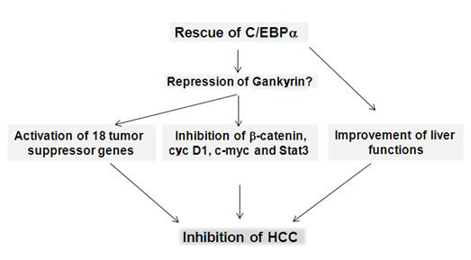
78. Reebye V, Sætrom P, Mintz PJ, et al. Novel RNA oligonucleotide improves liver function and inhibits liver carcinogenesis in vivo. Hepatology. 2014; 59(1): 216-227. doi: 10.1002/ hep.26669

79. Tomizawa M, Watanabe K, Saicho H, Nakagawara A, Tagawa M. Down-regulated expression of the CCAAT/enhancer binding protein alpha and beta in human hepatocellular carcinoma: a possible prognostic marker. Anticancer Res. 2003; 23: 351-354.

80. Tseng HH, Hwang YH, Yeh KT, Chang JG, Chen YL, Yu HS. Reduced expression of C/EBPα protein in hepatocellular carcinoma is associated with advanced tumor stage and shortened patient survival. J Cancer Res Clin Oncol. 2009; 135: 241-247. doi: 10.1007/s00432-008-0448-5

81. Babeu JP, Boudreau F. Hepatocyte nuclear factor 4-alpha involvement in liver and intestinal inflammatory networks. World J Gastroenterol. 2014; 20(1): 22-30. doi: 10.3748/wjg.v20.i1.22

82. Parviz F, Matullo C, Garrison WD, et al. Hepatocyte nuclear factor 4alpha controls the development of a hepatic epithelium and liver morphogenesis. Nat Genet. 2003; 34(3): 292-296. doi: 10.1038/ng1175

83. Lemaigre F, Zaret KS. Liver development update: new embryo models, cell lineage control, and morphogenesis. Curr Opin Genet Dev. 2004; 14(5): 582-950. doi: 10.1016/j. gde.2004.08.004

84. Chellappa K, Jankova L, Schnabl JM, et al. Src tyrosine kinase phosphorylation of nuclear receptor HNF4α correlates with isoform-specific loss of HNF4α in human colon cancer. Proc Natl Acad Sci U S A. 2012; 109(7): 2302-2307. doi: 10.1073/ pnas.1106799109

85. Yao D, Peng S, Dai C. The role of hepatocyte nuclear factor 4alpha in metastatic tumor formation of hepatocellular carcinoma and its close relationship with the mesenchymalepithelial transition markers. BMC Cancer. 2013; 13: 432. doi: 10.1186/1471-2407-13-432

86. Zhang B, Wang J, Wang X, et al. Proteogenomic characterization of human colon and rectal cancer. Nature. 2014; 513(7518): 382-387. doi: 10.1038/nature13438

87. Bonzo JA, Ferry CH, Matsubara T, Kim JH, Gonzalez FJ. Suppression of hepatocyte proliferation by hepatocyte nuclear factor 4α in adult mice. J Biol Chem. 2012; 287(10): 7345-7356. doi: 10.1074/jbc.M111.334599

88. Hatziapostolou M, Polytarchou C, Aggelidou E, et al. An HNF4α-miRNA inflammatory feedback circuit regulates hepatocellular oncogenesis. Cell. 2011; 147(6): 1233-1247. doi: 10.1016/j.cell.2011.10.043

89. Ning BF, Ding J, Yin C, et al. Hepatocyte nuclear factor 4 alpha suppresses the development of hepatocellular carcinoma. Cancer Res. 2010; 70(19): 7640-7651. doi: 10.1158/0008-5472. CAN-10-0824

90. Walesky C, Gunewardena S, Terwilliger EF, et al. Hepatocyte-specific deletion of hepatocyte nuclear factor-4α in adult mice results in increased hepatocyte proliferation. Am J Physiol Gastrointest Liver Physiol. 2013; 304(1): G26-37. doi: 10.1152/ ajpgi.00064.2012

91. Walesky C, Edwards G, Borude P, et al. Hepatocyte nuclear factor 4 alpha deletion promotes diethylnitrosamine-induced hepatocellular carcinoma in rodents. Hepatology. 2013; 57(6): 2480-2490. doi: 10.1002/hep.26251

92. Yang M, Li SN, Anjum KM, et al. A double-negative feedback loop between Wnt-β-catenin signaling and HNF4α regulates epithelial-mesenchymal transition in hepatocellular carcinoma. J Cell Sci. 2013; 126(Pt 24): 5692-5703. doi: 10.1242/ jcs.135053

93. Alder O, Cullum R, Lee S, et al. Hippo Signaling Influences HNF4A and FOXA2 Enhancer Switching during Hepatocyte Differentiation. Cell Rep. 2014 Sep 24. pii: S2211-1247- (14):00722-00730. doi: 10.1016/j.celrep.2014.08.046

94. Saha SK, Parachoniak CA, Ghanta KS, et al. Mutant IDH inhibits HNF-4α to block hepatocyte differentiation and promote biliary cancer. Nature. 2014; 513(7516): 110-114. doi: 10.1038/ nature13441

95. Dawson S. Hepatocellular carcinoma and ubiquitin-proteasome system. Biochem. Biophys Acta. 2008; 1782: 775-784. doi: 10.1016/j.bbadis.2008.08.003

96. Lim IK. Spectrum of molecular changes during hepatocvarcinogenesis induced by DEN and other chemical in Fishes 344 rats. Mech. Ageing Dev. 2003; 124: 679-708.doi: 10.1016/s0047-6374(02)00087-8

97. Krzywda S, Brzozowski AM, Higashitsuj H, et al. The crystal structure of Gankyrin, an oncoprotein found in complexes with cyclin-dependent kinase 4, a 19 S proteasomal ATPase regulator, and the tumor suppressors Rb and p53. J Biol Chem. 2004; 279(2): 1541-1545. doi: 10.1074/jbc.M310265200

98. Fu HY, Wang HY, Tan L, Liu SQ, Cao HA, Wu MC. Overexpression of p28/Gankyrin in human hepatocellular carcinoma and its clinical significance. World J. Gastroenterol. 2002; 8: 638-643.doi: 10.3748/wjg.v8.i4.638

99. Jing H, Zhang G, Meng L, Meng Q, Mo H, Tai Y.Gradually elevated expression of Gankyrin during human hepatocarcinogenesis and its clinicopathological significance. Sci Rep. 2014; 4: 5503. doi: 10.1038/srep05503

100. Li H, Fu X, Chen Y, et al. Use of adenovirus-delivered siRNA to target oncoprotein p28GANK in hepatocellular carcinoma. Gastroenterology. 2005; 128: 2029–2041. doi: http://dx.doi.org/10.1053/j.gastro.2005.03.001

101. Song X, Wang J, Zheng T, et al. LBH589 Inhibits proliferation and metastasis of hepatocellular carcinoma via inhibition of Gankyrin/STAT3/Akt pathway. Mol Cancer. 2013; 12(1): 114. doi: 10.1186/1476-4598-12-114

102. Li J, Tian F, Li D, Chen J, Jiang P, Zheng S, Li X, Wang S. MiR-605 represses PSMD10/Gankyrin and inhibits intrahepatic cholangiocarcinoma cell progression. FEBS Lett. 2014; 588(18): 3491-3500. doi: 10.1016/j.febslet.2014.08.008

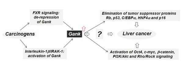
103. Jiang Y, Iakova P, Jin J, et al. Farnesoid X receptor inhibits Gankyrin in mouse livers and prevents development of liver cancer. Hepatology. 2013; 57: 1098-1106. doi: 10.1002/hep.26146

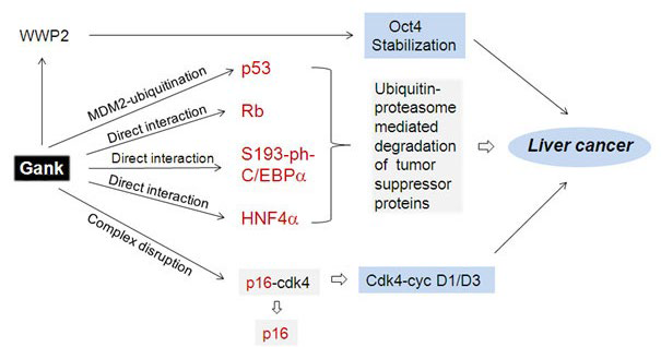
104. Higashitsuji H, Itoh K, Sakurai T, et al. The oncoprotein Gankyrin binds to MDM2/HDM2, enhancing ubiquitylation and degradation of p53. Cancer Cell. 2005; 8: 75-87. doi: http://dx.doi.org/10.1016/j.ccr.2005.06.006

105. Higashitsuji H, Itoh K, Nagao T, et al. Reduced stability of retinoblastoma protein by Gankyrin, an oncogenic ankyrinrepeat protein overexpressed in hepatomas. Nature Medicine. 2000; 6: 96-99.doi: 10.1038/71600

106. Li J, Tsai MD. Novel insights into the INK4-CDK4/6-Rb pathway: counter action of Gankyrin against INK4 proteins regulates the CDK4-mediated phosphorylation of Rb. Biochemistry. 2002; 41: 3977-3983. doi: 10.1021/bi011550s

107. Sun W, Ding J, Wu K, et al. Gankyrin-mediated dedifferentiation facilitates the tumorigenicity of rat hepatocytes and hepatoma cells. Hepatology. 2011; 54(4): 1259-1572. doi: 10.1002/ hep.24530

108. Qian YW, Chen Y, Yang W, et al. p28(GANK) prevents degradation of Oct4 and promotes expansion of tumor-initiating cells in hepatocarcinogenesis. Gastroenterology. 2012; 142: 1547-1558. doi: 10.1053/j.gastro.2012.02.042

109. Zheng T, Hong X, Wang J, et al. Gankyrin promotes tumor growth and metastasis through activation of IL-6/STAT3 signaling in human cholangiocarcinoma. Hepatology. 2014; 59(3): 935-946. doi: 10.1002/hep.26705

110. Bai Z, Tai Y, Li W, et al. Gankyrin activates IL-8 to promote hepatic metastasis of colorectal cancer. Cancer Res. 2013; 73(14): 4548-4558. doi: 10.1158/0008-5472.CAN-12-4586

111. Dong LW, Yang GZ, Pan YF, et al. The oncoprotein p28GANK establishes a positive feedback loop in β-catenin signaling. Cell Res. 2011; 21(8): 1248-1261. doi: 10.1038/ cr.2011.103

112. Fu J, Chen Y, Cao J, et al. p28GANK overexpression accelerates hepatocellular carcinoma invasiveness and metastasis via phosphoinositol 3-kinase/AKT/hypoxia-inducible factor1α pathways. Hepatology. 2011; 53(1): 181-192. doi: 10.1002/ hep.24015

113. Man JH, Liang B, Gu YX, et al. Gankyrin plays an essential role in Ras-induced tumorigenesis through regulation of the RhoA/ROCK pathway in mammalian cells. J Clin Invest. 2010; 120(8): 2829-2841. doi: 10.1172/JCI42542

114. Mine H, Sakurai T, Kashida H, et al. Association of Gankyrin and stemness factor expression in human colorectal cancer. Dig Dis Sci. 2013; 58(8): 2337-2344. doi: 10.1007/ s10620-013-2627-8

115. Su B, Luo T, Zhu J, et al. Interleukin-1β/IRAK-1 inflammatory signaling contributes to persistent Gankyrin activation during hepatocarcinogenesis. Hepatology. 2014. doi: 10.1002/ hep.27551

LATEST ARTICLES

Practical Pointers for Drug Development and Medical Affairs

Gerald L. Klein*, Roger E. Morgan, Shabnam Vaezzadeh, Burak Pakkal and Pavle Vukojevic

doi.

10.17140/CTPOJ-7-125

Prevalence and Risk Factors of Subclinical Mastitis of Goats in Banadir Region, Somalia

Omar M. Salah*, Yasin H. Sh-Hassan, Moktar O. S. Mohamed, Mohamed A. Yusuf and Abas S. A. Jimale

doi.10.17140/VMOJ-9-184

Use of Black Soldier Fly (Hermetia illucens) Prepupae Reared on Organic Waste

Maggot Debridement Therapy: A Natural Solution for Wound Healing

Isayas A. Kebede*, Haben F. Gebremeskel and Gelan D. Dahesa,

doi.10.17140/VMOJ-9-183

Figure 11. Risk Map for the Introduction of Ruminant Diseases at Borders

Ovine Network in Morocco: Epizootics Spread Prevention and Identification of the At-Risk Areas for “Peste des Petits Ruminants” and “Foot and Mouth Disease”

Yassir Lezaar*, Mehdi Boumalik, Youssef Lhor, Moha El-Ayachi, Abelilah Araba and Mohammed Bouslikhane

doi.

10.17140/EPOJ-8-131

The Impact of Family Dynamics on Palliative Care at the End-of-Life

Neil A. Nijhawan*, Rasha Mustafa and Aqeela Sheikh

doi.10.17140/PMHCOJ-10-154

Long-Term Follow-Up After Laparoscopic Radical Prostatectomy for Localized and Locally Advanced Prostate Cancer

Shrenik J. Shah*, Abhishek Jha, Chirag Davara, Rushi Mistry and Kapil Kachhadiya

doi.

10.17140/UAOJ-7-147

LATEST ARTICLES

Pie Chart Showing Overall Proportions of Diagnostic Category of FNAC, JUMC

Retrospective Study

2024 Apr

Abel Tefera*, Lemlem Terefe and Kitesa Biresa
Prevalence (%) of Types of Anthropometric Failure among Previous and Present Studied Tribal Children

Original Research, peer reviewed

2024 Apr

Biswajit Mahapatra and Kaushik Bose*

Opinion

2024 Apr

Gerald L. Klein*, Roger E. Morgan, Shabnam Vaezzadeh, Burak Pakkal and Pavle Vukojevic