Neuro

Open journal

ISSN 2377-1607

CNS Complication of Group A Streptococcal Meningitis in Children: a Comprehensive Case-Based Literature Review

Surya N. Gupta*, Vikash S. Gupta and Nirali Borad

Surya N. Gupta, MD

Associate Professor, Department of Pediatric Neurology, Section of Child Neurology, Women’s and Children’s Hospital, Charleston Area Medical Center 415 Morris Street, Suite 300, Charleston, WV 25301, USA; Tel. 304-388-6441; Fax: 304-388-6445; E-mail: suryangupta@rediffmail.com

INTRODUCTION

Streptococcal infections are common in children. Unlike Group B Streptococci septicemia,1 Group A Streptococcus (S. pyogenes) typically affects children beyond the neonatal period. Occasionally, this causes acute meningitis, glomerulonephritis, or rheumatic heart disease.2 Group A Streptococcus infection is a rare cause of bacterial meningitis. Hydrocephalus is a relatively rare complication of acute bacterial meningitis in children. Nonetheless, it has been reported in 20% of infants and children. Acute Central Nervous System (CNS) complications including hydrocephalus are not unique to Stretococcus pyogenes causing meningitis. They have been reported in children with viral and other bacterial meningitis.3

We report an unusual association of acute hydrocephalus in a 6-year-old girl with Group A Streptococcal meningitis and provide the results of a comprehensive literature review on acute CNS complications and neurological outcome in children 17years and under.

CASE REPORT

A 6-year-old girl presented to the Emergency Department with vomiting and neck pain for 24 hours. She had headache for five days and intermittent fever and sore throat for the past two weeks. She had no seizures and had received no antibiotics. A non-contrast Computerized Tomography (CT) scan of the brain prior to the lumbar puncture was normal. Treatment with intravenous vancomycin and ceftriaxone was initiated.

On admission to the Pediatric Intensive Care Unit, her vital signs were stable and temperature was 37.6 degree C. Her throat was red and congested but the tympanic membranes were normal. Her skin had no lesions. She had decreased alertness and her neck was hyper-extended in a fixed position. Pupils were 4 mm and reactive to light. Fundoscopic examination revealed no papilledema. Deep tendon reflexes were hyperreflexic and on plantar stimulation, her toes were up going.

The Cerebrospinal fluid (CSF) studies revealed an elevated intracranial pressure, a white blood cell count of 7644 mm3, red blood cells 10,000 mm3, protein 440 mg/dl and glucose 20 mg/dl. Serum glucose was 102 mg/dl. CSF smears revealed Gram-positive cocci in pairs and chains. The blood and CSF culture yielded S. pyogenes. Genotyping for the streptococcal isolates was not obtained. The CSF polymerase chain reaction for herpes simplex virus type 1 and 2 DNA for both, was negative.

Hospital Course

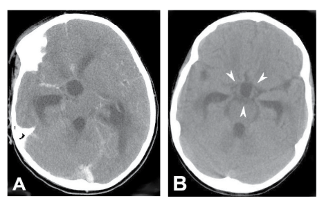
Ten hours after admission, she had an acute increase in heart rate from 80 to 133 beats per minute and blood pressure from 124/69 to 156/47 mm Hg. Her pupils became asymmetric and reacted sluggishly to light. Her Glasgow Coma Scale score declined from 13 to 9. Following endotracheal intubation, mechanical ventilation, and administration of 1 gram/kilogram of mannitol, a contrast-enhanced brain CT scan was performed which revealed communicating hydrocephalus with no herniation or abscess (Figure 1A). An urgent frontal external ventricular drain was placed. A follow up brain CT scan revealed relief of the hydrocephalus (Figure 1B).


Figure 1: An axial Computerized Tomography (CT) of the brain at the fourth ventricle level shows communicating hydrocephalus, figure A was obtained before and figure B was after surgical placement of a ventricular drain which is not seen at this level. A. Contrasted CT scan of the brain 10 hours after initiating parenteral antibiotics shows a global enlargement of the ventricular system. Note the complete effacement of the basal cisterns.
B. A follow up non-contrasted CT scan of the brain 8 hours after surgical placement of a ventricular drain shows a decrease in ventricular size, reappearance of basal cisterns (arrows heads), and also the disappearance of the left parietal hypodensity suggesting no parietal infarction.

NOJ-2-108 Fig 1


The ventriculostomy tube was clamped on day 12 and was removed on day 15. Intravenous ceftriaxone was continued for 21 days. She received physical, occupational, and speech therapies throughout the hospitalization. She was discharged on the 23rd day of admission with no focal motor, cerebellar, or sensory deficits.

DISCUSSION

Increased intracranial pressure caused by communicating hydrocephalus may be the result of long standing hydrocephalus, increased CSF production, decreased CSF absorption, or tonsillar herniation. Streptococcal infection produces many virulence factors and toxins.4

A clinical and epidemiologic study for the last 45 years reported the incidence of GAS meningitis in pediatric population 0.06 cases per 100,000 children per year. The case fatality rate was 43%. Neonatal age and the presence of an associated toxic shock syndrome were identified as risk factors for death. A distant focus of infection was present 36% in the Brazilian case series which were more than half of the patients in the literature. No single virulence determinant could be associated with death.5

Literature Search and the Results

We performed PubMed online search for English literature for Streptococcal pyogenes meningitis in children 0-17 years and under. We used terms “Group A Streptococcal meningitis”, “Streptococcal pyogenes meningitis”, “with and without” and “CNS complications” and “acute and chronic hydrocephalus” with a variable combination. Bibliography of the reports was searched for additional information.

The results of our literature search with CNS complications are shown in Table 1.


Table 1: Lists increasing age-based case Reports of central nervous system (CNS) complicationsin children with Group A Streptococcal meningitis since 1966 [6-38].
Case # Age/ Sex Infection Focus Central Nervous System Complication Year [Reference]
Acute Chronic or Outcome
1 6 d/M Erysipelas Seizures No sequellae 19666
2 8 d/M Unknown Seizures Death 19677
3 13 d/F Cellulitis of both feet Focal Seizures Death 19848
4 14 d/F Cystic gingival lesion Seizures, No sequellae 19839
5 24 d/F Unknown Seizures, Brain Abscesses Death 200010
6 26 d/M Paronychia Seizures, Communicating Hydrocephalus No sequellae 19848
7 28 d/M Unknown Seizures and

Brain Abscess

No sequellae 200811
8 1 m/F Unknown Seizures Death 19677
9 1 m/F Unknown Meningitis and Multiple Brain Abscess NAa 198812
10 1.5 m/M Otitis Media Seizures Severe Motor Deficits 197913
11 NA/NA Unknown NA Hydrocephalus, Porencephalic cyst 198614
12 2 m/M Tonsillitis Seizures and Subdural effusions Moderate Motor Deficit, Cystic Hygroma 199815
13 2 m/NA Ulcerated and Infected hemangioma Infantile spasm and Seizures Encephalomalacia,

Infantile spasms,Profound Developmental Delay

200416
14 2.5 m/M None Seizures and SIADH Seizure Disorder, Optic atrophy, Microcephaly, Psychomotor Retardation 19839
15 2.5 m/F Infected hemangioma Seizures


Psychomotor Retardation 198717
16 2.5 m/M Pharyngitis Focal Seizures Hearing loss 198818
17 3 m/M Infected BCG cscar Generalized Seizures No sequelae 200019
18 3 m/F Chicken pox Hydrocephalus No sequelae 200320
19 7 m/F None Seizures Death 199221
20 11 m/NA Unknown NA Bilateral Subdural Hygroma 197622
21 17 m/M Otitis media Generalized Seizure,

Brain Abscess

No sequelae 200623
22 18 m/M Dermal sinus of nose and Otitis Media Recurrent Meningitis No sequelae 197624
23 2.5 yr/M None Seizures and Increased intracranial pressure Death 200125
24 3 yr/M Otitis media Seizures Partial blindness, Deafness, Mental Retardation 198426
25 3.5 yr/M Pharyngitis and Mastoiditis Cerebral Abscess Left sensorineural hearing Loss 201227
26 4 yr/M Otitis media SIADH No sequelae 19839
27 4.5 yr/NA Cochlear implantation Hearing Loss Language Delay 200528
28 5 yr/NA Right Mastoiditis Right Facial Nerve Palsy No sequelae 200729
29 6 yr/NA Unknown NA Cranial Nerve IV Palsy 197622
30 6 yr/F Pharyngitis Communicating hydrocephalus No sequelae Present
31 8 yr/M Otitis media, Pneumonia Seizures


Neuropsychological deficit 198130
32 8 yr/M Sore throat Seizures No sequelae 19839
33 8 yr/F Unknown Coma No sequelae 19839
34 8 yr/F Unknown (Toxic Shock-Like Syndrome) Death 199431
35 8 yr/M Otitis Media, Pharyngitis Subdural effusion, Hemiparesis, and

Seizures

Headaches and Attention Deficit Disorder 201032
36 10 yr/M Unspecified illness Brain Abscess No sequelae (abscess required surgical drainage ) 200133
37 11 yr/NA Multiple Otitis media Cranial Nerve III

Palsy

Cranial Nerve III Palsy, Subdural Hygroma 200134
38 12 yr/F Pharyngitis,

Otitis media

Coma and

Hemianopia

Persistent

Hemianopia

19839
39 12 yr/F None Brain Abscess No sequelae 198835
40 13 yr/F Sinusitis Seizures Moderate Motor Deficit 199236
41 14 yr/NA Posttraumatic meningitis Cerebrospinal fluid leak Severe Psychomotor Retardation 199037
42 15 yr/M Occipital Fracture Cerebrospinal fluid leak No sequelae 199938
43 15 yr/F Tonsillitis Seizures and Cerebral abscess No sequelae 201032
44 17 yr/M Pharyngitis, sinusitis Cavernous Sinus

Thrombosis

No sequelae 199938
d: days; M: male; F: female; m: month; yr: year ;
aNA, data not available; bSIADH, Syndrome of Inappropriate Anti-Diuretic Hormone; cBCG, Bacillus Calmette–Guérin
The table is based on the results of our search on PubMed for English literature for children age 17years and under with central nervous system complications caused by Streptococcal A meningitis. Our search identified 40 studies with 43 children, excluding the current report (Case # 30). Twenty seven (68%) studies were isolated case reports. Six reports reference number 7, 9, 22, 32, 38 and 8, had 2 cases each. The study number 8 included 6 cases. The children who presented with menigeal manifestations of headache, photophobia, and neck stiffness, but no CNS complications were excluded from this report.


Clinical Demography

With the exception of 7 neonates, the mean age for children with CNS complications was 6.1 years. Of 44 children, 30 (68%) children had a known focus of infection. In fourteen (32%) children the focus of infection was unknown or was not described. Amongst children with known focus, 13 (43%) had otitis media and/or pharyngitis. Tonsillitis, infected hemangioma, cellulitis, or erysipelas, and 1 patient had sore throat. Other minor but significant foci of infection included skull fracture, infected dermal sinus, and mastoiditis.

Central nervous system complication

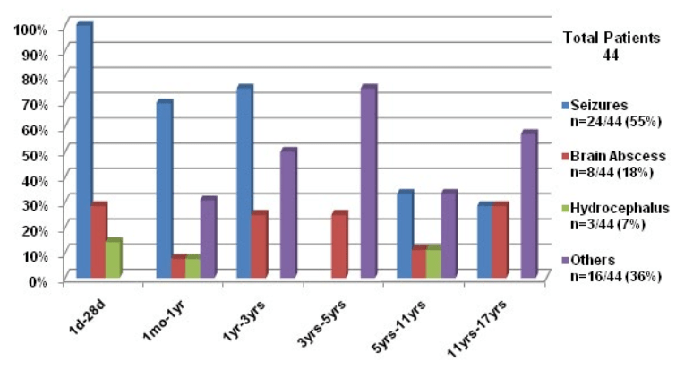
The most common acute CNS complication secondary to Streptococcal meningitis was seizure 24/44 (55%) followed by brain abscess 8/44 (18%). Other acute complications included hydrocephalus, CSF leak, meningitis, cranial nerve palsy and subdural effusion, except one case, the complication was unknown.

Neurological Outcome

Regarding outcome, 18/44 (41%) patients including ours had no sequelae. 10/44 (23%) cases developed a slowing of speech or physical movement with or without a decreased cognitive ability, 3/44 (7%) cases with hearing loss, 2/44 (4.5%) cases with cranial nerve palsy, 2/44 (4.5%) cases developed partial or complete blindness, only 1/44 (2.3%) developed chronic hydrocephalus. In one case the outcome was unknown (See Bar Graph).

In addition, 7/44 (16%) children died from complications of Group A Streptococcus meningitis, secondary to cardio respiratory insufficiency, Waterhouse Friderichsen syndrome, and Toxic shock-like syndrome. It is unclear why previously healthy children with Group A streptococcus meningitis developed significant CNS complications?

In contrast to our case, two previous cases of acute hydrocephalus were reported during early infancy. Acute onset hydrocephalus in our case was likely due to the combined effect of untreated prolonged pharyngitis, and initial delay in diagnosis and treatment. We suggest that a high CSF protein together with pleocytosis impeded absorption of CSF by arachnoid villi at the superior sagittal sinus. Such functional blockage causing chronic hydrocephalus has been reported in the presence of proteinaceous and cellular debris in CSF39 and also after intracranial hemorrhage.40

Alternatively, the term “communicating hydrocephalus” which could be acute or chronic is defined to mean that the CSF within the ventricles communicating with subarachnoid space CSF. But the CT scan panel A shows CSF within the ventricles and an obliterated subarachnoid space. Arguably, there is no evidence that the ventricles and subarachnoid space are in communication with each other (a broader definition of obstructive hydrocephalus), and therefore, this is not “communicating hydrocephalus”. Additionally, hydrocephalus is not caused by increased CSF production or by tonsillar herniation. Instead, in our case, the non-communicating hydrocephalus is caused by ventriculitis resulting in obstruction to the outflow of CSF through the fourth ventricle.

CONCLUSION

Our case illustrates that Group A Streptococcal meningitis induced acute hydrocephalus is rare. A timely instituted surgical intervention may prevent other CNS complication. The review of published cases reveals that central nervous system complications including hydrocephalus are rare. Since prompt antibiotic therapy can minimize risk for acute CNS complications, prompt identification and emergent medical and neurosurgical interventions should be undertaken for an optimum outcome in children with Group A Streptococcal meningitis.


NOJ-2-108 graph


Bar Graph: The bar graph shows age-based CNS complications of Group A Streptococcal meningitis in children age 0-17 years

Others complications reported were meningitis, subdural effusions, infantile spasms, Syndrome of Inappropriate Antidiuretic Hormone secretion (SIADH), increased intracranial pressure, hearing loss, third and forth cranial nerve palsies, coma, and cavernous sinus thrombosis. Seizure, the most common CNS complication, occurred between births to 12 months. Hydrocephalus and cerebral abscess occurred between births to 10 years. Cerebrospinal fluid leak exclusively occurred between 10 to17 years of age. There were no reports of intracranial hemorrhage secondary to Group A w.

ACKNOWLEDGEMENT

Author thanks Jeffery Maisels, MD for his review of this manuscript.

CONFLICTS OF INTEREST

The authors have no financial considerations to disclose or competing interests in relation to this article.

1. Bedford Russell AR, Breathnach A, Sender P. Confirmed group B streptococcus infection: tip of the iceberg. Arch Dis Child Fetal Neonatal. 2001; 84: F140. doi: 10.1136/fn.84.2.F140b

2. Patterson MJ. Streptococcus. In: Baron S, ed. Medical Microbiology. 4th ed. Galveston (TX): University of Texas Medical Branch at Galveston; 1996. 13.

3. Srinivasan L. Ramanan R. Hydrocephalus secondary to group B streptococcal meningitis. Journal of Maternal-Fetal & Neonatal Medicine. 2004; 16(1): 67-68. doi: 10.1080/14767050412331283148

4. Robbins SL, Cotran RS. Pathological Basis of Disease. 7th ed. Copyright©2005, Saunders, An Imprint of Elsevier. 373.

5. Torres RS, Fedalto LE, Torres RF, Steer AC, Smeesters PR. Group Astreptococcus meningitis in Children. Pediatr Infect Dis J. 2013; 32(2): 110-114. doi: 10.1097/INF.0b013e31826fd4af

6. Dillon HC Jr. Group A type 12 streptococcal infection in a newborn nursery. Successfully treated neonatal meningitis. Am J Dis Child. 1966; 112(3): 177-184. doi: 10.1001/archpedi.1966.02090120045002

7. Ramanathan K, Grossman A. Neonatal Streptococcal meningitis. Ill Med J. 1967; 132: 705-707.

8. Coulter JB, Buchannon CR, Vellodi A, Hart CA, Sills JA. Group-A streptococcal infection in the newborn. Lancet. 1984; 2(8398): 355-356.

9. Murphy DJ Jr. Group A streptococcal meningitis. Pediatrics. 1983; 71(1): 1-5.

10. Verboon-Maciolek MA, Krediet TG, Ertbruggen IV, Gerards LG, Fleer A. Severe neonatal group A streptococcal disease. Eur J Pediatr. 2000; 159(6): 450-452. doi: 10.1007/s004310051305

11. Lardhi A. Neonatal group A streptococcal meningitis: a case report and review of the literature. Cases J. 2008; 1(1): 108. doi: 10.1186/1757-1626-1-108

12. Wong VK, Wright HT. Group A beta-hemolytic streptococci as a cause of bacteremia in children. Am J Dis Child. 1988; 142: 831-833. doi: 10.1001/archpedi.1988.02150080037016

13. McMahon BJ, Barrett DH, Bender TR, Brenneman GR, Hurwitz RM, Cannon W. Group A streptococcal meningitis in an infant: report of a case associated with a community outbreak due to M-type 4 organisms. Alaska Med. 1979; 21(4): 57-59.

14. Congeni BL, Bradely J, Hammerschlag MR. Safety and efficacy of once daily ceftriaxone for the treatment of bacterial meningitis. Paediatr Infect Dis J. 1986; 5(3): 293-297. doi: 10.1097/00006454-198605000-00004

15. Moses AE, Beeri M, Engelhard D. Group A streptococcal meningitis: reports of two cases. J Infect. 1998; 36(1): 116-118. doi: 10.1016/S0163-4453(98)93558-7

16. Rezvani M, Jerome Y. Yager AE, Hartfield DS. Group A streptococcal meningitis as a complication of an infected capillary haemangioma. Eur J Pediatr. 2004; 163(1): 19-21. doi: 10.1007/s00431-003-1334-4

17. Yagupsky P, Giladi Y. Group A beta-hemolytic streptococcal bacteremia in children. Pediatr Infect Dis J. 1987; 6(11): 1036-1039.

18. Harden A, Lennin D. Serious supportive group A streptococcal infections in previously healthy children. Pediatr Infect Dis J. 1988; 7(10): 714-718.

19. Melzer M, Joashi U, Murdoch I, Gransden W. Group a streptococcal meningitis with an infected BCG scar. JR Soc Med. 2000; 93(12): 641. doi: 10.1177%2F014107680009301211

20. Brandt C, Kitz R, Lutticken R, Brade V. Streptococcus pyogenes Meningitis Complicating Varicella in a 3-month-old Child. Scand J Infect Dis. 2003; 34: 876-878. doi: 10.1080/00365540310016844

21. Gertner M, Rodriguez L, Barnett SH, Shah K. Group A beta-hemolytic Streptococcus and Waterhouse-Friderichsen syndrome. Pediatr Infect Dis J. 1992; 11(7): 595-596.

22. Burech DL, Koranyi KI, Haynes RE. Serious group A streptococcal diseases in children. J Paediatr. 1976; 88(6): 972-974. doi: 10.1016/S0022-3476(76)81052-9

23. Dehority W, Uchiyama S, Khosravi A, Nizet V. Brain abscess caused by Streptococcus pyogenes in a previously healthy child. J Clin Microbiol. 2006; 44(12): 4613-4615. doi: 10.1128/JCM.01724-06 h

24. Shadravan I, Fishbein J, Hebert LJ. Streptococcal meningitis with an unusual port of entry. Am J Dis Child. 1976; 130(2): 214-215. doi: 10.1001/archpedi.1976.02120030104021

25. Shetty AK, Frankel LR, Maldonado Y, Flaco DA, Lewis DB. Group A streptococcal meningitis: report of case and review of literature since 1976. Paediatr Emerg Care. 2001; 17(6): 430-434. doi: 10.1097/00006565-200112000-00007

26. Rotimi VO, Grange AO, Lawal SF, Eke PI. Childhood meningitis caused by group A streptococci belonging to a new serotype: a case report. East Afr Med J. 1984; 61(8): 648-651.

27. Paul SP, Jerwood S. Group A streptococcal septicemia, meningitis and cerebral abscess: case report and literature review. Turk J Pediatr. 2012; 54(2): 180-183.

28. Pettersen G, Ovetchkine P, Tapiero B. Group A streptococcal meningitis in a pediatric patient following cochlear implantation: report of the first case and review of the literature. J Clin Microbial. 2005; 43: 5816-5818. doi: 10.1128/JCM.43.11.5816-5818.2005

29. Arnoni M, Berezin E, Safadi M, Almeida F, Lopes C. Streptococcus pyogenes Meningitis in Children: Report of Two Cases and Literature Review. The Brazilian Journal of Infectious Diseases. 2007; 11(3): 375-377. doi: http://dx.doi.org/10.1590/S1413-86702007000300015

30. Brown CC, Pickering LK, Baker CJ. Group A streptococcal meningitis without predisposing factors. South Med J. 1981; 74(8): 1029-1030. doi: 10.1097/00007611-198108000-00049

31. Jevon GP, Dunne WM Jr, Hawkins HK, Armstrong DL, Musser JM. Fatal group A streptococcal meningitis and toxic shock-like syndrome: case report. Clin Infect Dis. 1994; 18(1): 91-93. doi: 10.1093/clinids/18.1.91

32. Bruun T, Kittang BR, Mylvaganam H, Lund-Johansen M, Skrede S. Clinical, microbiological and molecular characteristics of six cases of group A streptococcal meningitis in western Norway. Scand J Infect Dis. 2010; 42(9): 665-671. doi: 10.3109/00365548.2010.483477

33. Khan MA, Viagappan GM, Andrews J. Group A streptococcal brain abscess. Scand J Infect Dis. 2001; 33(2): 159.

34. Steppberger K, Adams I, Deutscher J, Müller H, Kiess W. Meningitis in a girl with recurrent otitis media caused by Streptococcus pyogenes–otitis media has to be treated appropriately. Infection. 2001; 29(5): 286-288. doi: 10.1007/s15010-001-1065-2

35. Jagdis F. Group A streptococcal meningitis and brain abscess. Paediatr Infect Dis J. 1988; 7(12): 885-886.

36. Levy EN, Griffith JA, Carvajal HF. Localized meningoencephalitis and group A streptococcal bacteremia. Clin Pediatr (Phila). 1992; 31(7): 438-441. doi: 10.1177/000992289203100712

37. Wilson NW, Copeland B, Bastian JF. Posttraumatic meningitis in adolescents and children. Pediatr Neurosurg. 1990-1991; 16(1): 17-20. doi: 10.1159/000120497

38. Sommer R, Rohner P, Garbino J, Auckenthaler R, Malinverni R, Lew D, Kaiser L. Group A beta-hemolytic streptococcus meningitis: clinical and microbiological features of nine cases. Clin Infect Dis. 1999; 29(4): 929-931. doi: 10.1086/520461

39. Mandell, Bennett, Dollin. Principles and Practice of Infectious Diseases. 5th ed. Copy right 2005 Churchill Livingston. 287-288.

40. Ronald EH, Hill A. Intraventricular Hemorrhage and Post Hemorrhagic Hydrocephalus. Current and potential Future Intervention. Clin perinatal. 1997; 24(3): 589-605.

LATEST ARTICLES

Quality Assurance of General Purpose, Keratin Based and Dye lock Hair Shampoos

Saima Siddique*, Zahida Parveen, Zeeshan Ali and Sidra Mehmood

doi.

Blood Sample from the Patient

Hypertriglyceridemia-Induced Pancreatitis: A Case Report and Literature Review

Maarten Bulterys, Melvin Willems* and Agnes Meersman

doi.

From Neck Pain to a Life-Threatening Condition: A Case Report

Floris Vandewoude* and Sören Verstraete

doi.

LATEST ARTICLES

Original Research

2024 Aug

Saima Siddique*, Zahida Parveen, Zeeshan Ali and Sidra Mehmood

Cross Sectional Study, peer reviewed

2024 Jul

Amanuel P. Beta, Dereje Abera, Legese Belayneh and Isayas A. Kebede

Case Report, peer reviewed

2024 Jul

Syeda Rukh*, Sathyanarayana Machani and Milind Awale