Pathology and Laboratory Medicine

Open journal

ISSN 2996-5942

Circulating Tumor Cells: Beyond Isolation and Detection

Michelle J. McNamara*

Received: October 15th, 2019; Accepted: November 6th, 2019; Published: November 8th, 2019

INTRODUCTION

Cancer is the second leading cause of death in the United States,1 and most cancer-related deaths are a result of metastatic disease.2,3 Thousands of circulating tumor cells (CTCs) are shed by tumors daily and are the means by which these cancers metastasize.4 There has been emerging evidence that the presence of CTCs in the peripheral circulation correlates with decreased progression-free and overall survival in many cancers. Although the 5-year-survival rate for most types of metastatic cancer is fairly dismal,5 the complexity of the metastatic process makes it difficult to completely understand their prognostic significance. Additionally, their clinical relevance is not limited to their metastatic potential and their prognostic impact, and their assessment can possibly be expanded to other applications with development of improved isolation and detection technologies.

CLINICAL RELEVANCE OF CTCS

CTCs have been shown to provide prognostic information in patients with metastatic cancer. The presence of CTCs in early breast cancer is predictive of decreased progression-free and overall survival.6-11 This data has proven to be sufficiently compelling that the American Joint Committee on Cancer (AJCC) has incorporated the presence of CTCs into the staging for breast cancer.12,13 CTC evaluation is a valuable tool for early screening, prognostic assess ment, monitoring of treatment efficacy, and monitoring of disease progression or relapse.14-17 Furthermore, studies being conducted on other tumor types, including lung, prostate, and colorectal cancer, have yielded similar results, however, research is still ongoing to completely characterize the presence of CTCs and its relationship to disease progression.18-22

Although biopsy is considered the “gold standard” for cancer diagnosis and for characterization of tumors,23 there are many advantages to using CTCs for evaluation. Often patients are unable to undergo biopsy procedures due to poor clinical status, or the tumor may be in an inaccessible location. It is also common for tumors to demonstrate significant intra-and/or intertumoral heterogeneity and frequently, primary and metastatic tumors are phenotypically and genetically discordant.24,25,26 Therefore, CTCs are likely more representative of the overall tumor status and disease progression than a biopsy, particularly since tumors frequently continue to undergo genetic evolution as they progress.24,25,26 The evidence also suggests that CTC enumeration alone is insufficient, and that enumeration combined with downstream analysis would be ideal to provide comprehensive prognostic and therapeutic information.11,27

Most studies to date have focused primarily on the prognostic impact of CTCs via molecular characterization or enumeration using a very limited number of tumor markers to determine cell lineage or biomarker status. However, there are many other potential clinical applications for the isolation and detection of disease beyond mere enumeration.

CTCs can be assessed for specific biomarkers to determine if a patient is a candidate for specific types of treatment, and they can be used to monitor for efficacy of treatment, either by decreasing CTC counts or by evaluation of genetic evolution by itself or as a surrogate for resistance to therapy.28 CTCs have also been found to have preferential sites of metastasis depending on the primary tumor type, and thus may lend itself to the prediction of metastatic locations.29 Since CTCs have been found in the peripheral circulation of patients without clinically detectable disease,30,31 it has been postulated that they can be used to detect early metastases.32 Finally, since platelets mediate the survival of CTCs in the circulation, studies are being conducted to exploit this and use genetically-modified platelets that express tumor necrosis factor-related apoptosis-inducing ligand (TRAIL) to induce apoptosis of these tumor cells.33

ISOLATION AND DETECTION TECHNIQUES

Molecular Characterization

Reverse transcriptase-polymerase chain reaction (RT-PCR) is the most commonly used technique for molecular characterization and is considered highly sensitive assay, although this may vary depending on tumoral heterogeneity, contamination by genetic material from leukocytes or other cells, illegitimate transcription of cancer-associated markers in non-malignant cells, the presence of polymerase chain reaction (PCR) inhibitors, or down regulation of target genes after therapy. Also, although some tumors may have tumor-specific abnormalities, most have no tissue-specific markers. Another limitation to PCR is that it requires cell lysis which prevents CTC enumeration or other analyses.

Physical Properties

Size and deformability:

Although many CTCs are typically much larger than most cells in the peripheral blood, there is significant variability in their morphologic appearance. The size of leukocytes ranges from 8-11 µm in diameter, and some CTCs can be of similar size which can make it difficult to separate them based on size alone. CTCs have decreased deformability, or a decreased ability to change their shape in relation to blood cells, however, they are more deformable than benign epithelial cells.34,35 Therefore, it is postulated that increased deformability correlates with increased metastatic potential.36 ISET®, Isolation-by-Size-of-Epithelial-Tumour (Rarecells, France), is a filtration device that separates CTCs in the peripheral blood based on size, which are then analyzed by standard cytomorphological microscopy.37 The advantages to this technique are that it is not dependent on Epithelial cell adhesion molecule (EpCAM) expression or expression of other epithelial markers, however, it can have CTC loss, and it can under-represent small CTCs.

Density:

Density gradient centrifugation is based on the fact that the various constituents of whole blood have different compositions and therefore, different densities and will separate into different layers when subjected to centrifugation (Figure 1). Essentially, there are three layers, the bottom layer where heavier particles are, such as red blood cells and neutrophils, the top layer consisting of plasma and platelets, and the middle layer, which is the buffy coat containing the mononuclear cells and the CTCs.38 Although this is often referred to with the generic term “ficoll,” this terminology was derived from a common product called Ficoll-Paque media (GE Healthcare, Chicago, IL, USA). The advantage to this is that it is inexpensive, and it does not rely on EpCAM or other epithelial markers. The disadvantages are that there may be loss of CTCs, the specimen must be processed shortly after collection, there is contamination with other blood elements, and CTCs may separate into the plasma layer.

Figure 1. Density Gradient Centrifugation

PLMOJ-1-108Fig1

CTCs can be separated into the plasma layer instead of the mononuclear cell layer

Another centrifugation system, OncoQuick® (PA, USA) uses a porous barrier to prevent the separation medium from mixing with the specimen prior to centrifugation. During density gradient centrifugation, the CTCs and the mononuclear cells pass through the barrier, with the higher density red blood cells and granulocytes remaining below the barrier. While the recovery rate is similar to that of ficoll, the OncoQuick® system results in less contamination by blood elements and thus a higher concentration of evaluable CTCs on the slide.39

Electrical properties:

Changes in the cellular content, particularly proteins, nucleic acids, and peptides, alters the dielectric properties of CTCs from that of leukocytes or benign epithelial cells. This is exploited by the DEPArray™ (Silicon Biosystems, Italy), which is an automated microfluidic system that includes an automated instrument, a disposable microfluidic cartridge, and proprietary analysis software. It has the advantage of being automated, not being dependent on expression of EpCAM or other epithelial markers, and the cells are viable and available for downstream analysis.40 However, there is some CTC loss, and the system is expensive.

Immunoaffinity-based methods:

This will not be an exhaustive discussion of enrichment methods, but a few are listed in Table 1. Immunoaffinity-based methods of CTC detection are based on marker expression and use labelled antibodies to isolate or sort the CTCs. These methods use positive or negative enrichment in which the tumor cells are enriched, or the non-tumor cells are depleted from the specimen, respectively. Often these methods result in non-viable cells that cannot be used for downstream analysis, although some microfluidic technologies have overcome this limitation. Most of the immunoaffinity-based methods use a combination of positive and negative enrichment, but EasySep™ (Chennai, India) is an immunomagnetic CTC separation kit based on depletion of leukocytes by CD45 (Figure 2). The advantage to this method is that it is easy to use and offers batch separation, however, there may be CTC loss and contamination with other blood elements.

Table 1. Examples of CTC Enrichment Methods

Method

Principle Advantages Pitfalls

References

ISET® Filtration Inexpensive, fast, easy, captures aggregates, not dependent on surface markers May under-represent smaller CTCs, possible CTC loss, need to pretreat specimens, clogged membranes

14,37,77-79

DEPArray Dielectrophoresis Automated, cells are available for downstream analysis Possible CTC loss, fluorescent imaging without morphological assessment.

14,78,80,81

Ficoll Density gradient centrifugation Inexpensive, fast, easy, not dependent on surface markers CTCs can be in the plasma layer, low purity

4,14,82,83

OncoQuick® Density gradient centrifugation and size Relatively inexpensive, fast, easy, not dependent on surface markers CTCs can be in the plasma layer, low purity  

4,14,82,83

CELLSEARCH® Immunoaffinity, positive and negative enrichment FDA-approved, automated Relies on EpCAM, does not detect EpCAMnegative cells, fluorescent imaging without morphological assessment, expensive

4,8,30,43,78,80,84

EasySep Immunoaffinity, negative enrichment Easy to use, batch separation Possible CTC loss, low purity

85-87

Figure 2. Immunoaffinity-Based Method

PLMOJ-1-108Fig2

Immunoaffinity-based method for CTC isolation and detection
using immunomagnetic beads.

The most frequently used marker for CTC isolation is EpCAM which is not expressed on all CTCs due to a phenomenon called epithelial-mesenchymal transition (EMT), which is part of the complex metastatic cascade that confers the ability for tumor cells to metastasize to distant sites (Figure 3). Although the process by which CTCs are formed and the mechanisms involved in the development of metastases are not the focus of this review, a brief overview provides a basic understanding of this limitation. It involves phenotypic alterations that are significant when considering the development of methodologies to isolate and detect CTCs. In order for CTCs to develop the capacity to metastasize, they require the ability to alter their phenotype from epithelial to mesenchymal through EMT.4,14-17 This involves the selective loss of epithelial adhesion molecules, such as E-cadherin and integrins, that allow the tumor cells to detach from the adjacent cells, digest the extacellular matrix, change their shape and deformability, and migrate to and enter the blood circulation, a process called intravasation. Although thousands of CTCs can be shed by tumors daily,41 most do not survive in the circulation due to hemodynamic forces, physical damage from interaction from other blood elements, destruction by immune cells, or apoptosis.4,14-17 Of the rare cells that do survive, some may become dormant for prolonged periods of time and may not lead to metastasis, or a metastasis may arise years after the CTCs entered the circulation.4,14-17 Once in the circulation, CTCs are often coated by activated platelets which promote the survivability of these cells by preventing NK cell destruction.42 At some point, the CTCs can extravasate into a target organ or tissue, undergo mesenchymal-epithelial transition (MET) whereby they regain their epithelial characteristics and their ability to proliferate, creating a metastatic tumor.4,14-17 This concept of EMT and MET partially contributes to the phenotypic heterogeneity seen in tumors and in CTCs in particular.

Figure 3. Epithelial-Mesenchymal Transition and Mesenchymal-Epithelial Transition in the Metastatic Process

PLMOJ-1-108Fig3

Epithelial cells gain mesenchymal characteristics enabling them to invade, intravate into the circulation, where they may undergo apoptosis or remain dormant for varying periods of time. They are capable of extravasating into a target tissue, regaining epithelial characteristics and establishing a metastatic lesion.

The CELLSEARCH® system (Veridex, Warren, NJ, USA) is the only Food and Drug Administration (FDA)-approved detection system for the enumeration of CTCs in breast cancer patients. It is an immunoaffinity-based system which uses immunomagnetic enrichment to detect cells labeled with antibodies to EpCAM, CD45, and DAPI conjugated to fluorochromes analyzed with a cytometry-based automated instrument. The cells are visualized using a fluorescent imaging system that images the DAPIstained nuclei for adjunctive nuclear evaluation to confirm that the DAPI-positive, EpCAM-positive, CD45-negative cells have nuclei with the size and shape consistent with carcinoma cells. The sensitivity of this method for detecting EpCAM-positive CTCs is ≥85%.9,30,43,44 A CTC count of ≥5 per 7.5 mL of blood is associated with significantly shorter progression-free and overall survival.6-11,44 A meta-analysis evaluating the literature published from 1990 to 2012 confirmed this and also showed that it was not influenced by the detection method or the time point at which the CTCs were assessed.45 Further, a study to analyze pooled individual patient data showed that the prognostic value is superior to that of serum markers (CEA and CA15.3), and that it is unrelated the tumor histologic type, the number of metastases, or the type of treatment.46

A prospective multicenter study comparing the outcomes of patients with metastatic breast, colorectal, and prostate cancer confirmed that the presence of CTCs was a strong predictor of poor overall survival.47 In a separate study involving a small number of samples, CELLSEARCH® detected varying percentages of CTCs in the peripheral blood of patients with a variety of metastatic tumor types, including 64% in colon cancer, 33% in gastric cancer, 66% in rectal cancer, 60% in ovarian cancer, and 20% in prostate cancer.48 This illustrates one major limitation of EpCAMbased assays in that not all epithelial CTCs express EpCAM or other epithelial markers. The success of the CELLSEARCH® system is variable depending on the type of cancer and the stage of disease. Some other studies, particularly those for other tumor types, have used other tumor markers with some success.49-54 However, the challenge of detecting CTCs is in large part due to their phenotypic heterogeneity.55 Therefore, a combination of isolation techniques involving selection based on physical properties along with positive and negative enrichment, with immunological-based methods would yield the most sensitive and specific results.

CYTOMORPHOLOGY

Most reports in the literature regarding tumoral heterogeneity refer to clonal genetic alterations expressed in different parts of a tumor. However, there are many different ways in which tumor heterogeneity can manifest, and these phenotypic changes can be detected or measured in a number of different ways. Tumoral heterogeneity is simply different phenotypic features seen within the same tumor, whether it involves different histological patterns, altered protein expression, or genetic mutations, that can be routinely evaluated using a combination of morphological evaluation in conjunction with ancillary studies, including immunohistochemical staining, immunofluorescence, flow cytometry, in situ hybridization, or a variety of molecular tests. Some of the biomarkers that are targeted are evaluated for diagnostic purposes, and some have prognostic or therapeutic relevance. To date, most investigations regarding CTCs have focused on their prognostic significance, however, CTCs are also present in patients with cancers of unknown primary (CUP), both in body fluids, as well as, in the peripheral circulation. In such cases, performing molecular tests to determine the clonal genetic status in the absence of a definitive diagnosis and identification of the primary tumor is insufficient.

Methodologies such as the CELLSEARCH® system or other methodologies that only evaluate the immunophenotypical or genetic characteristics of CTCs are limited by their inability to provide a true morphological evaluation of the cells, which demonstrate characteristic features that confirm malignancy.56-58 Morphological evaluation can overcome the limitations related to the histological characteristics, such as size, shape, and nuclear and cytoplasmic features, which can be characteristic of malignant cells. This is important because of the morphological heterogeneity demonstrated by CTCs, some of which can be the size of leukocytes, and especially because benign epithelial cells have been found in the peripheral circulation of healthy subjects with inflammatory conditions or benign neoplasms.30,59,60 It is unknown whether or not these cells represent malignant cells in the early stages of cancer development.32

Although morphological evaluation may not be completely necessary in patients with a known primary tumor, this does not exclude the possibility of a new or a concurrent malignancy, and it would be essential in cases of CUP or initial or early diagnoses. In fact, malignant cells detected in effusion cytology is often the first indication of malignancy and require pathological workup for a diagnosis whichmost frequently includes immunohistochemistry and possibly flow cytometry, in situ hybridization, or other molecular testing.61,62

Cytology of pleural effusions and ascitic fluid is able to diagnose malignancy in approximately 70% of cases.63-66 Often the morphological features of CTCs resemble that of the primary tumor type.57,67 This is of particular importance considering that less than 30% of lung cancer patients are able to undergo resection and that many cases are diagnosed by cytology alone.68-70 Although diagnostic molecular profiling can be performed on tumor cells, the specificity can be as low as 75% due to genetic heterogeneity or genetic evolution.71 Therefore, despite the trend towards smaller specimens and the necessity for molecular testing for prognostic biomarkers, the diagnostic algorithm remains the same with histological evaluation with limited immunohistochemical stains followed by molecular testing being standard.72 Historically, cell block specimens have been preferred over cytological smears because the original diagnostic slides can be archived, and ancillary tests can be added on after the initial diagnosis.73 However, recent studies have shown that cytology smears have sufficient, and sometimes higher, cellularity than cell block specimens and yield results concordant with biopsies and resection specimens.68,73,74,75

In 2013, the College of American Pathologists (CAP), the International Association for the Study of Lung Cancer, and the Association for Molecular Pathology (AMP) issued updated guidelines regarding molecular testing in lung cancer that were endorsed by the American Society of Clinical Oncology (ASCO). Direct smears were recommended as the specimen of choice for molecular testing provided that they had adequate cellularity and preservation.76 This makes it possible to use non-cell block specimens, such as direct smears, cytospin preparations, touch preparations, and liquid based cytology for molecular testing in lung cancer. The rationale is because cell blocks are limited by variable cellularity, the inability to perform on-site adequacy assessments, and decreased nucleic acid quality due to formalin fixation.76 This provides the opportunity to expand testing to more specimen types that may be more readily available in certain cancer types.

CONCLUSION

With the evolving understanding of the mechanisms involved in the development of metastases, CTCs have become increasingly relevant, not only regarding their prognostic significance in breast, lung, prostate, colorectal, and other cancers, but their potential to be applied to other clinical applications. The development of methodologies for isolation and detection is the first step in a pathway that incorporates the identification and histological, immunohistochemical, and molecular characterization to enable clinicians to obtain the comprehensive diagnostic, prognostic and therapeutic information necessary to provide appropriate personalized care to cancer patients.

1. Siegel RL, Miller KD, Jemal A. Cancer statistics, 2019. CA Cancer J Clin. 2019; 69(1): 7-34. doi: 10.3322/caac.21551

2. American Cancer Society. Cancer Facts & Figures 2019. Website. https://www.cancer.org/research/cancer-facts-statistics/allcancer-facts-figures/cancer-facts-figures-2019.html. Accessed October 14, 2019.

3. Taketo MM. Reflections on the spread of metastasis to cancer prevention. Cancer Prev Res (Phila). 2011; 4(3): 324-328. doi: 10.1158/1940-6207.CAPR-11-0046

4. Kowalik A, Kowalewska M, Góźdź S. Current approaches for avoiding the limitations of circulating tumor cells detection methods—implications for diagnosis and treatment of patients with solid tumors. Transl Res. 2017; 185: 58-84.e15. doi: 10.1016/j. trsl.2017.04.002

5. National Cancer Institute. SEER Cancer Statistics Review, 1975- 2015. Website. https://seer.cancer.gov/archive/csr/1975_2015/ index.html#contents. Accessed October 14, 2019.

6. Budd GT, Cristofanilli M, Ellis MJ, et al. Circulating tumor cells versus imaging–predicting overall survival in metastatic breast cancer. Clin Cancer Res. 2006; 12(21): 6403-6409. doi: 10.1158/1078- 0432.CCR-05-1769

7. Cristofanilli M, Budd GT, Ellis MJ, et al. Circulating tumor cells, disease progression, and survival in metastatic breast cancer. N Eng J Med. 2004; 351(8): 781-791. doi: 10.1056/NEJMoa040766

8. Cristofanilli M, Hayes DF, Budd GT, et al. Circulating tumor cells: A novel prognostic factor for newly diagnosed metastatic breast cancer. J Clin Oncol. 2005; 23(7): 1420-1430. doi: 10.1200/ JCO.2005.08.140

9. Hayes DF, Cristofanilli M, Budd GT, et al. Circulating tumor cells at each follow-up time point during therapy of metastatic breast cancer patients predict progression-free and overall survival. Clin Cancer Res. 2006; 12(14): 4218-4224. doi: 10.1158/1078-0432. CCR-05-2821

10. Rack B, Schindlbeck C, Jückstock J, et al. Circulating tumor cells predict survival in early average-to-high risk breast cancer patients. J Natl Cancer Inst. 2014; 106(5): pii: dju066. doi: 10.1093/ jnci/dju066

11. Serrano MJ, Rovira PS, Martínez-Zubiaurre I, Rodriguez MD, Fernández M, Lorente JA. Dynamics of circulating tumor cells in early breast cancer under neoadjuvant therapy. Exp Ther Med. 2012; 4(1): 43-48. doi: 10.3892/etm.2012.540

12. American College of Surgeons (ACS). AJCC Cancer Staging Manual. 8th ed: NY, USA: Springer International Publishing; 2017.

13. Giuliano AE, Connolly JL, Edge SB, et al.Ca Breast Cancer— Major changes in the American Joint Committee on Cancer eighth edition cancer staging manual. CA Cancer J Clin. 2017; 67(4): 290- 303. doi: 10.3322/caac.21393

14. Thiele JA, Bethel K, Králíčková M, Kuhn P. Circulating tumor cells: Fluid surrogates of solid tumors. Annu Rev Pathol. 2017; 12(1): 419-447. doi: 10.1146/annurev-pathol-052016-100256

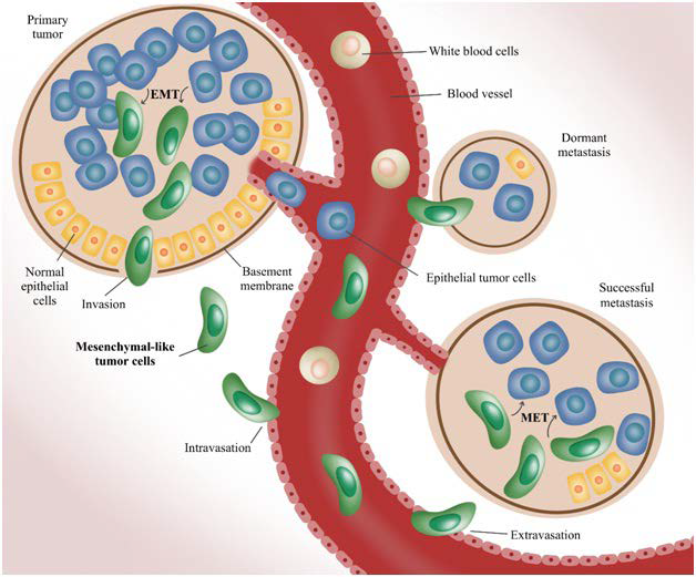
15. Wan L, Pantel K, Kang Y. Tumor metastasis: Moving new biological insights into the clinic. Nat Med. 2013;19:1450-1460. doi: 10.1038/nm.3391

16. Jacob K, Sollier C, Jabado N. Circulating tumor cells: Detection, molecular profiling and future prospects. Expert Rev Proteomics. 2007; 4(6): 741-756. doi: 10.1586/14789450.4.6.741

17. Valastyan S, Weinberg RA. Tumor metastasis: Molecular insights and evolving paradigms. Cell. 2011; 147(2): 275-292. doi: 10.1016/j.cell.2011.09.024

18. Cohen SJ, Punt CJA, Iannotti N, et al. Relationship of circulating tumor cells to tumor response, progression-free survival, and overall survival in patients with metastatic colorectal cancer. J Clin Oncol. 2008; 26(19): 3213-3221. doi: 10.1200/JCO.2007.15.8923

19. Danila DC, Heller G, Gignac GA, et al. Circulating tumor cell number and prognosis in progressive castration-resistant prostate cancer. Clin Cancer Res. 2007; 13(23): 7053-7058. doi: 10.1158/1078- 0432.CCR-07-1506

20. Maheswaran S, Sequist LV, Nagrath S, et al. Detection of mutations in EGFR in circulating lung-cancer cells. N Engl J Med. 2008; 359(4): 366-377. doi: 10.1056/NEJMoa0800668

21. Moreno JG, O’Hara SM, Gross S, et al. Changes in circulating carcinoma cells in patients with metastatic prostate cancer correlate with disease status. Urology. 2001; 58(3): 386-392. doi: 10.1016/s0090-4295(01)01191-8

22. Uen Y-H, Lu C-Y, Tsai H-L, et al. Persistent presence of postoperative circulating tumor cells is a poor prognostic factor for patients with stage I-III colorectal cancer after curative resection. Ann Surg Oncol. 2008; 15(8): 2120-2128. doi: 10.1245/s10434-008- 9961-7

23. King JD, Casavant BP, Lang JM. Rapid translation of circulating tumor cell biomarkers into clinical practice: Technology development, clinical needs and regulatory requirements. Lab Chip. 2014; 14(1): 24-31. doi: 10.1039/c3lc50741f

24. de Bruin EC, McGranahan N, Mitter R, et al. Spatial and temporal diversity in genomic instability processes defines lung cancer evolution. Science. 2014; 346(6206): 251-256. doi: 10.1126/science.1253462

25. Gerlinger M, Rowan AJ, Horswell S, et al. Intratumor heterogeneity and branched evolution revealed by multiregion sequencing. N Engl J Med. 2012; 366(10): 883-892. doi: 10.1056/NEJMoa1113205

26. Navin N, Kendall J, Troge J, et al. Tumour evolution inferred by single-cell sequencing. Nature. 2011; 472(7341): 90-94. doi: 10.1038/nature09807

27. Pantel K, Speicher MR. The biology of circulating tumor cells. Oncogene. 2016; 35(10): 1216-1224. doi: 10.1038/onc.2015.192

28. Tsao SC-H, Wang J, Wang Y, Behren A, Cebon J, Trau M. Characterising the phenotypic evolution of circulating tumour cells during treatment. Nat Commun. 2018; 9(1): 1482. doi: 10.1038/s41467- 018-03725-8

29. Paterlini-Bréchot P. Organ-specific markers in circulating tumor cell screening: An early indicator of metastasis-capable malignancy. Future Oncol. 2011; 7(7): 849-871. doi: 10.2217/fon.11.32

30. Allard WJ, Matera J, Miller MC, et al. Tumor cells circulate in the peripheral blood of all major carcinomas but not in healthy subjects or patients with nonmalignant diseases. Clin Cancer Res. 2004; 10(20): 6897-6904. doi: 10.1158/1078-0432.CCR-04-0378

31. Politaki E, Agelaki S, Apostolaki S, et al. A comparison of Three methods for the detection of circulating tumor cells in patients with early and metastatic breast cancer. Cell Physiol Biochem. 2017; 44(2): 594-606. doi: 10.1159/000485115

32. Klein CA. Parallel progression of primary tumours and metastases. Nat Rev Cancer. 2009; 9(4): 302-312. doi: 10.1038/nrc2627

33. Li J, Sharkey CC, Wun B, Liesveld JL, King MR. Genetic engineering of platelets to neutralize circulating tumor cells. J Control Release. 2016; 228: 38-47. doi: 10.1016/j.jconrel.2016.02.036

34. Cross SE, Jin Y-S, Rao J, Gimzewski JK. Nanomechanical analysis of cells from cancer patients. Nat Nanotechnol. 2007; 2(12): 780-783. doi: 10.1038/nnano.2007.388

35. Shaw Bagnall J, Byun S, Begum S, et al. Deformability of tumor cells versus blood cells. Sci. Rep. 2015; 5: 18542. doi: 10.1038/ srep18542

36. Guck J, Schinkinger S, Lincoln B, et al. Optical deformability as an inherent cell marker for testing malignant transformation and metastatic competence. Biophys J. 2005; 88(5): 3689-3698. doi: 10.1529/biophysj.104.045476

37. Farace F, Massard C, Vimond N, et al. A direct comparison of CellSearch and ISET for circulating tumour-cell detection in patients with metastatic carcinomas. Br J Cancer. 2011; 105: 847-853. doi: 10.1038/bjc.2011.294

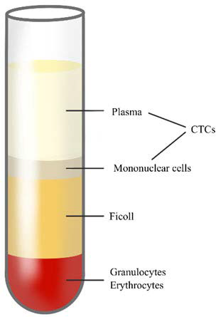
38. GE Healthcare. Isolation of mononuclear cells: Methodology and applications. Website. https://www.sigmaaldrich.com/content/dam/sigma-aldrich/docs/Sigma-Aldrich/General_Information/1/ge-isolation-of-mononuclear-cells.pdf. Accessed October 14, 2019.

39. Gertler RR, Fuehrer K, Dahm M, Nekarda H, Siewert JR. Detection of circulating tumor cells in blood using an optimized density gradient centrifugation. In: Allgayer HMM, Schildberg FW, eds. Staging of Cancer. Recent Results in Cancer Research. Berlin, Heidelberg: Springer; 2003. doi: 10.1007/978-3-642-59349-9_13

40. Di Trapani M, Manaresi N, Medoro G. DEPArray™ system: An automatic image-based sorter for isolation of pure circulating tumor cells. Cytometry A. 2018; 93(12): 1260-1266. doi: 10.1002/cyto.a.23687

41. Nagrath S, Sequist LV, Maheswaran S, et al. Isolation of rare circulating tumour cells in cancer patients by microchip technology. Nature. 2007; 450(7173): 1235-1239. doi: 10.1038/nature06385

42. Placke T, Örgel M, Schaller M, et al. Platelet-derived MHC class I confers a pseudonormal phenotype to cancer cells that subverts the antitumor reactivity of natural killer immune cells. Cancer Res. 2012; 72(2): 440-448. doi: 10.1158/0008-5472.CAN-11-1872

43. Andree KC, van Dalum G, Terstappen LW. Challenges in circulating tumor cell detection by the CellSearch system. Mol Oncol. 2016; 10(3): 395-407. doi: 10.1016/j.molonc.2015.12.002

44. Riethdorf S, Fritsche H, Müller V, et al. Detection of circulating tumor cells in peripheral blood of patients with metastatic breast cancer: A validation study of the CellSearch system. Clin Cancer Res. 2007; 13(3): 920-928. doi: 10.1158/1078-0432.CCR-06-1695

45. Zhang L, Riethdorf S, Wu G, et al. Meta-analysis of the prognostic value of circulating tumor cells in breast cancer. Clin Cancer Res. 2012; 18: 5701-5710. doi: 10.1158/1078-0432.CCR-12-1587

46. Bidard F-C, Peeters DJ, Fehm T, et al. Clinical validity of circulating tumour cells in patients with metastatic breast cancer: A pooled analysis of individual patient data. Lancet Oncol. 2014; 15(4): 406-414. doi: 10.1016/S1470-2045(14)70069-5

47. Miller MC, Doyle GV, Terstappen LW. Significance of circulating tumor cells detected by the CellSearch system in patients with metastatic breast colorectal and prostate cancer. J Oncol. 2010; 2010: 617421-617421. doi: 10.1155/2010/617421

48. Balic M, Dandachi N, Hofmann G, et al. Comparison of two methods for enumerating circulating tumor cells in carcinoma patients. Cytometry B Clin Cytom. 2005; 68B(1): 25-30. doi: 10.1002/cyto.b.20065

49. Allen JE, El-Deiry WS. Circulating tumor cells and colorectal cancer. Curr Colorectal Cancer Rep. 2010; 6(4): 212-220. doi: 10.1007/s11888-010-0069-7

50. Fehm T, Müller V, Aktas B, et al. HER2 status of circulating tumor cells in patients with metastatic breast cancer: a prospective, multicenter trial. Breast Cancer Res Treat. 2010; 124(2): 403-412. doi: 10.1007/s10549-010-1163-x

51. Maly V, Maly O, Kolostova K, Bobek V. Circulating tumor cells in diagnosis and treatment of lung cancer. In Vivo. 2019; 33(4): 1027-1037. doi: 10.21873/invivo.11571

52. Matthew EM, Zhou L, Yang Z, et al. A multiplexed markerbased algorithm for diagnosis of carcinoma of unknown primary using circulating tumor cells. Oncotarget. 2016; 7(4): 3662-3676. doi: 10.18632/oncotarget.6657

53. Wong SC, Chan CM, Ma BB, et al. Clinical significance of cytokeratin 20-positive circulating tumor cells detected by a refined immunomagnetic enrichment assay in colorectal cancer patients. Clin Cancer Res. 2009; 15(3): 1005-1012. doi: 10.1158/1078-0432. CCR-08-1515

54. Wong SCC, Ng SSM, Cheung MT, et al. Clinical significance of CDX2-positive circulating tumour cells in colorectal cancer patients. Br J Cancer. 2011; 104(6): 1000-1006. doi: 10.1038/ bjc.2011.32

55. Alix-Panabières C, Pantel K. Clinical applications of circulating tumor cells and circulating tumor DNA as liquid biopsy. Cancer Discov. 2016; 6(5): 479-491. doi: 10.1158/2159-8290.CD-15-1483

56. Marrinucci D, Bethel K, Bruce RH, et al. Case study of the morphologic variation of circulating tumor cells. Hum Pathol. 2007; 38(3): 514-519. doi: 10.1016/j.humpath.2006.08.027

57. Marrinucci D, Bethel K, Luttgen M, Bruce RH, Nieva J, Kuhn P. Circulating tumor cells from well-differentiated lung adenocarcinoma retain cytomorphologic features of primary tumor type. Arch Pathol Lab Med. 2009; 133(9): 1468-1471. doi: 10.1043/1543- 2165-133.9.1468

58. Demay RM. The Art & Science of Cytopathology. 2nd ed. Chicago, USA: ASCP Press; 2011.

59. Hardingham JE, Hewett PJ, Sage RE, et al. Molecular detection of blood-borne epithelial cells in colorectal cancer patients and in patients with benign bowel disease. Int J Cancer. 2000; 89(1): 8-13.

60. Pantel K, Denève E, Nocca D, et al. Circulating epithelial cells in patients with benign colon diseases. Clin Chem. 2012; 58(5): 936- 940. doi: 10.1373/clinchem.2011.175570

61. Motherby H, Nadjari B, Friegel P, Kohaus J, Ramp U, Böcking A. Diagnostic accuracy of effusion cytology. Diagn Cytopathol. 1999; 20(6): 350-357. doi: 10.1002/(sici)1097- 0339(199906)20:6<_x0033_50:_x003a_aid-dc5>3.0.co;2-7

62. Cakir E, Demirag F, Aydin M, Unsal E. Cytopathologic differential diagnosis of malignant mesothelioma, adenocarcinoma and reactive mesothelial cells: A logistic regression analysis. Diagnos Cytopathol. 2009; 37(1): 4-10. doi: 10.1002/dc.20938

63. Light RW. Pleural effusion. N Eng J Med. 2002; 346(25): 1971- 1977. doi: 10.1056/NEJMcp010731

64. Light RW, Erozan YS, Ball WC. Cells in pleural fluid: Their value in differential diagnosis. JAMA Intern Med. 1973; 132: 854-860.

65. Prakash UBS, Reiman HM. Comparison of needle biopsy with cytologic analysis for the evaluation of pleural effusion: Analysis of 414 cases. Mayo Clin Proc. 1985; 60(3): 158-164. doi: 10.1016/ s0025-6196(12)60212-2

66. Pai R, Shenoy K, Minal J, Suresh P, Chakraborti S, Lobo F. Use of the term atypical cells in the reporting of ascitic fluid cytology: A caveat. Cytojournal. 2019; 16(1): 13. doi: 10.4103/cytojournal.cytojournal_37_18

67. Roh MH. Triage of cytologic direct smears for ancillary studies: A case-based illustration and review. Arch Pathol Lab Med. 2013; 137(9): 1185-1190. doi: 10.5858/arpa.2013-0235-CR

68. Kim L, Tsao MS. Tumour tissue sampling for lung cancer management in the era of personalised therapy: What is good enough for molecular testing? Eur Respir J. 2014; 44(4): 1011-1022. doi: 10.1183/09031936.00197013

69. Malapelle U, Bellevicine C, Zeppa P, Palombini L, Troncone G. Cytology-based gene mutation tests to predict response to antiepidermal growth factor receptor therapy: A review. Diagn Cytopathol. 2011; 39(9): 703-710. doi: 10.1002/dc.21512

70. Nicholson AG, Gonzalez D, Shah P, et al. Refining the diagnosis and EGFR status of non-small cell lung carcinoma in biopsy and cytologic material, using a panel of mucin staining, TTF-1, cytokeratin 5/6, and P63, and EGFR mutation analysis. J Thorac Oncol. 2010; 5(4): 436-441. doi: 10.1097/JTO.0b013e3181c6ed9b

71. Bochtler T, Krämer A. Does cancer of unknown primary (CUP) truly exist as a distinct cancer entity? Front Oncol. 2019; 9: 402. doi: 10.3389/fonc.2019.00402

72. Travis WD, Rekhtman N, Riley GJ, et al. Pathologic diagnosis of advanced lung cancer based on small biopsies and cytology: A paradigm shift. J Thorac Oncol. 2010; 5(4): 411-414. doi: 10.1097/JTO.0b013e3181d57f6e

73. da Cunha Santos G, Saieg MA, Geddie W, Leighl N. EGFR gene status in cytological samples of nonsmall cell lung carcinoma. Cancer Cytopathol. 2011; 119(2): 80-91. doi: 10.1002/cncy.20150

74. Sun P-L, Jin Y, Kim H, Lee C-T, Jheon S, Chung J-H. High concordance of EGFR mutation status between histologic and corresponding cytologic specimens of lung adenocarcinomas. Cancer Cytopathol. 2013; 121(6): 311-319. doi: 10.1002/cncy.21260

75. Knoepp SM, Roh MH. Ancillary techniques on direct-smear aspirate slides. Cancer Cytopathol. 2013; 121(3): 120-128. doi: 10.1002/cncy.21214

76. Roy-Chowdhuri S. From cytopathology in focus: Updated NSCLC guideline moves molecular cytopathology forward. CAP Today. Website. https://www.captodayonline.com/cytopathology-infocus-updated-nsclc-guideline-moves-molecular-cytopathology-forward/. Accessed October 14, 2019.

77. Chinen LTD, de Carvalho FM, Rocha BMM, et al. Cytokeratinbased CTC counting unrelated to clinical follow up. J Thorac Dis. 2013; 5(5): 593-599. doi: 10.3978/j.issn.2072-1439.2013.09.18

78. Ferreira MM, Ramani VC, Jeffrey SS. Circulating tumor cell technologies. Mol Oncol. 2016; 10(3): 374-394. doi: 10.1016/j.molonc.2016.01.007

79. Zheng S, Lin HK, Lu B, et al. 3D microfilter device for viable circulating tumor cell (CTC) enrichment from blood. Biomed Microdevices. 2011; 13(1): 203-213. doi: 10.1007/s10544-010-9485-3

80. Lin E, Cao T, Nagrath S, King MR. Circulating tumor cells: Diagnostic and therapeutic applications. Ann Rev Biomed Eng. 2018; 20(1): 329-352. doi: 10.1146/annurev-bioeng-062117-120947

81. Peeters DJE, De Laere B, Van den Eynden GG, et al. Semiautomated isolation and molecular characterisation of single or highly purified tumour cells from CellSearch enriched blood samples using dielectrophoretic cell sorting. Br J Cancer. 2013; 108(6): 1358- 1367. doi: 10.1038/bjc.2013.92

82. Baker MK, Mikhitarian K, Osta W, et al. Molecular detection of breast cancer cells in the peripheral blood of advanced-stage breast cancer patients using multimarker real-time reverse transcriptionpolymerase chain reaction and a novel porous barrier density gradient centrifugation technology. Clin Cancer Res. 2003; 9(13): 4865- 4871.

83. Gabriel MT, Calleja LR, Chalopin A, Ory B, Heymann D. Circulating tumor cells: A review of non–EpCAM-Based approaches for cell enrichment and isolation. Clin Chem. 2016; 62(4) :571-581. doi: 10.1373/clinchem.2015.249706

84. Bankó P, Lee SY, Nagygyörgy V, et al. Technologies for circulating tumor cell separation from whole blood. J Hematol Oncol. 2019; 12(1): 48. doi: 10.1186/s13045-019-0735-4

85. Harouaka RA, Nisic M, Zheng SY. Circulating tumor cell enrichment based on physical properties. J Lab Autom. 2013; 18(6): 455-468. doi: 10.1177/2211068213494391

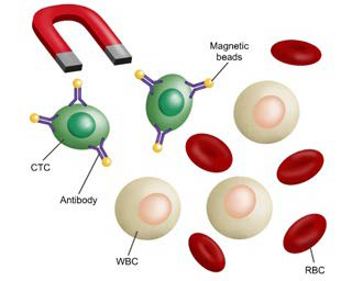
86. Lustberg M, Jatana KR, Zborowski M, Chalmers JJ. Emerging technologies for CTC detection based on depletion of normal cells. Recent Results Cancer Res. 2012; 195: 97-110. doi: 10.1007/978- 3-642-28160-0_9

87. Yin J, Wang Y, Yin H, et al. Circulating tumor cells enriched by the depletion of leukocytes with bi-antibodies in non-small cell lung cancer: Potential clinical application. PLoS One. 2015; 10(8): e0137076-e0137076. doi: 10.1371/journal.pone.0137076

LATEST ARTICLES

Use of Black Soldier Fly (Hermetia illucens) Prepupae Reared on Organic Waste

review

2024 Mar

Isayas A. Kebede*, Haben F. Gebremeskel and Gelan D. Dahesa,
Figure 11. Risk Map for the Introduction of Ruminant Diseases at Borders

Original Research, peer reviewed

2024 Mar

Yassir Lezaar*, Mehdi Boumalik, Youssef Lhor, Moha El-Ayachi, Abelilah Araba and Mohammed Bouslikhane

Commentary, End-of-Life, Family Dynamics, palliative Care, peer reviewed

2024 Mar

Neil A. Nijhawan*, Rasha Mustafa and Aqeela Sheikh

Retrospective Study

2024 Mar

Shrenik J. Shah*, Abhishek Jha, Chirag Davara, Rushi Mistry and Kapil Kachhadiya

Attention, Disaster, Mindfulness, Original Research, peer reviewed

2024 Mar

Christina Hagen*, Lars Lien, Edvard Hauff and Trond Heir

review

2024 Mar

Isayas A. Kebede* and Gelan D. Dahesa

review

2024 Feb

Bekiyad S. Daro*; Yared A. Deneke, Jiregna G. Negasa and Ishetu N. Dibaba