Gastro

Open journal

ISSN 2377-8369

Biphasic Roles of a Small G-Protein, RAC1 in Pancreatic Β-Cell

Mayur S. Parmar, Altaf Ahmed Meer and Ismail Syed*

Ismail Syed, PhD

Division of Endocrinology, Beth Israel Deaconess Medical Center, Harvard Medical School, E/CLS-724, 3 Blackfan Circle, MA 02215, Boston, USA; Tel. (617)735-3327; E-mail: isyed@bidmc.harvard.edu

INTRODUCTION

Diabetes is a metabolic disorder with multiple etiologies characterized by chronic hyperglycemia. This results from dysregulated insulin secretion and/or from the resistance to insulin action in peripheral tissues. In the settings of the metabolic disorder, disturbances in carbohydrate, fat and protein metabolism results in a diverse set of complications associated with pancreas, liver, kidney, heart and other vital organs. As per the National Diabetes Statistics Report, 2014, in 2012, 29.1 million Americans (2.9% of the population) have diabetes, which include 1.25 million type 1 diabetic children and adults. Further, 86 million people of age 20 and above are pre-diabetic, and are at increased risk for developing type 2 diabetes. Over decades of research, a greater understanding of the pancreatic β-cells in physiological insulin release has been made to therapeutically target and treat the metabolic disorder. Insulin secretion from islet β-cells is majorly regulated by glucose and other insulin secretagogues. This is mediated through fluctuations in the intracellular calcium, and interplay of soluble secondary messengers like reactive oxygen species (ROS), cyclic nucleotides and hydrolytic products generated from the phospholipases A2, C and D.113 In addition, adenine nucleotides [e.g., ATP] and guanine nucleotides [e.g., GTP14,15,16,17; regulate physiological insulin secretion. Even though many studies have shown the underlying mechanism[s] involved in stimulus-secretion coupling of glucose stimulated insulin secretion (GSIS), the precise molecular and cellular mechanism still remains unknown. However, role of guanine nucleotide-binding protein (G-protein) has been highly researched for their role in insulin release. The signal-transduction system (Adenylylcyclases, ion channels, and phospholipases) involved in insulin release are linked to the receptors for hormones or stimulatory agents via G-proteins.

CLASSIFICATION OF G-PROTEINS IN β-CELL

Till date three major classes of G-proteins have been identified in pancreatic β-cells.18,19,20,21,22 The first class of G-proteins, heterotrimeric G-proteins assists in coupling membrane-associated receptors to their intracellular effectors adenylyl cyclases, ion channels, and phosphodiesterases.23,24,25 The second class of G-proteins, small monomeric G-proteins [17-30 kDa] play a vital role in protein organization and trafficking of secretory vesicle.26 These small G-proteins undergo posttranslational modifications [isoprenylation and methylation] at their C-terminal residues (CAAX motif)26,27,28,29,30 for their active confirmation. The third class of G-proteins, the elongation factors and Tau proteins are implicated in protein synthesis. 

SMALL G-PROTEINS

Based on the substantial evidences on the regulation of pancreatic islet β-cell function, small G-proteins are categorized into three major groups. Rho, Rac1, Cdc42 and ADP-ribosylation factor-6 [Arf6] fall under the first category of small G-proteins and these play an important role in cytoskeletal remodeling and vesicular fusion.3148 The second category of small G-proteins comprises of Rap1 and RabGTPases (Rab3A and Rab27).49 These Rab GTPases assists in priming and docking of insulin-laden secretory granules on the plasma membrane. Unlike first category of small G-proteins, requisite for posttranslational modifications and mechanism[s] involved in the activation of Rab GTPases under the physiological insulin secretagogues remains elusive. However, Rap1 is activated transiently by glucose50 and undergoes carboxymethylation.18,51 The third group of small G-proteins consists of Rab2, Rhes and Rem2 which are under-studied,52,53,54,55 whereas, RalA appears to draw direct regulatory effects in exocytosis.56 Do you have data about small G-proteins expression in pancreas? 

ACTIVATION AND DEACTIVATION CYCLE OF SMALL G-PROTEINS

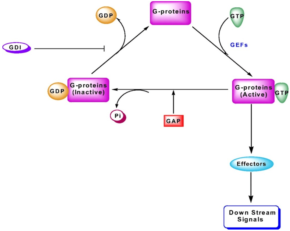
Like heterotrimeric G-proteins, small G-proteins also shuttle between their inactive (GDP-bound) and active (GTP-bound) conformations, and are tightly regulated by various G-protein regulatory factors/proteins. Till date, three regulatory factors have been identified for small G-proteins, viz., Guanine exchange nucleotide factors [GEFs], GDP-dissociation inhibitors [GDIs] and GTPase-activating proteins [GAPs]. GEFs facilitate the translation of the inactive GDP-bound to their active GTP-bound forms, while, the GDIs avert the dissociation of GDP from the G-proteins, thereby keeping them in the inactive conformation (Figure 1). However, GAPs, convert the active GTP-bound to their inactive GDP-bound form by inactivating the intrinsic GTPase activity of the candidate G-proteins. The efficiency of the G-protein activation cascade depends on the relative amounts of active to inactive GTPase. The activity of GTPase can be altered either by accelerating GDP dissociation by GEFs or by inhibiting GDP dissociation by GDIs, or by accelerating GTP hydrolysis by GAPs. Any imbalance in either of the regulatory factors alters the hydrolytic cycle and physiological functions in pancreatic β-cells.49,57,58 

Figure 1: Activation and deactivation cascade of small G-proteins49

Activation and deactivation cascade of small G-proteins


SMALL G-PROTEIN-RAC

Rac was first identified and implicated in cellular function with two cDNAs encoding proteins, Rac1 and Rac2.59 So far three isoforms of Rac proteins, Rac1, Rac2, and Rac3 have been identified in mammals. Both Rac1 and Rac2 share over 90% homology. Rac1 and Rac3 are extensively expressed in diverse tissues, whereas as, Rac2 is restricted to hematopoietic cells. Rac1 and other small G-proteins, Cdc42 and Arf6 have been recognized as key regulatory molecules in vesicle trafficking and organelle dynamics coupled with proliferation and survival of a cell.19,49 In addition, Rac1 has also been shown to play a vital role in various diseased states including cancer and neurological disorders,60,61,62 liver fibrosis63 and diabetes.57,58 Furthermore, Rac1 protein has shown to be associated with GLUT4 translocation in the muscle of diabetic patients.64,65 Furthermore, Rac1 has shown to play an important role in wound healing, bacterial clearance and cell adhesion/migration by regulating actin dynamics in the gut.66 Rac1 together with cdc42 induces intestinal wound closure, mediated by N-formyl peptide receptor (FPR) stimulation leading to enhanced intestinal epithelial cell restitution.67 In addition, many pathogenic bacteria secrete factors that trigger the posttranslational modification and activation of Rho proteins like Rac1 and cdc42 leading to gut epithelial cell death via apoptosis.68 Citalán-Madrid and group have clearly depicted the roles of small G proteins as important signaling molecules in the regulation of epithelial junctions.69 Herein this review, we describe both the positive and negative roles for Rac1 small G-protein in islet β-cell pathophysiology. 

A. Positive Role of Rac1 in Insulin Secretion: Like other small G-proteins, Rac also shuttles between inactive GDP and active GTP conformations to facilitate cellular function. These proteins undergo ADP ribosylation by C3 component of botulinum toxin prior to their association with membrane. However, potential roles for Rac1 in glucose stimulated insulin secretion (GSIS) was first demonstrated by using Clostridium difficile toxins A and B, which irreversibly monoglucosylate and inactivate specific G-proteins (Cdc42 and Rac1).32 Like Cdc42, Rac1 also undergoes posttranslational carboxymethylation and membrane translocation in the presence of stimulatory glucose concentrations.32 Expression of an inactive mutant of Rac1 (N17Rac1) in INS-1 cells resulted in significant morphological changes leading to inhibition of GSIS. These findings also confirmed the involvement of small G-protein Rac1 in cytoskeletal remodeling and reorganization.41 As stated above, Rac1 also requires prenylation for its function. Experiments involving pharmacological and molecular biological inhibition of Rac1 prenylation indicated marked reduction in GSIS in a variety of insulin-secreting β-cells. For an instance, GGTI-2147, a specific inhibitor for geranylgeranylation, one of the post translational modifications, significantly augmented accumulation of Rac1 in cytosol and inhibited GSIS in insulin-producing β-Cell line INS 832/13. Over expression of the regulatory α-subunit of protein prenyltransferase also attenuated glucose-induced insulin secretion in clonal pancreatic β-cells.31 In addition, a recent study has shown that siRNA-mediated knock down of small G-protein Rac1 attenuated GSIS significantly having no effect on the basal insulin secretion, suggesting a positive modulatory roles for Rac1 in insulin secretion.70 The importance of these small G-proteins in insulin secretion has been extensively studied in vitro; however, studies concentrating on in vivo Rac1 knock out models are limited. As Rac1 small G-protein is involved in many physiological processes, knocking out Rac1 might have deleterious effects. In this context, epithelial-specific Rac1-Knockout mice showed epithelial hyperplasia and a reduced basal cell layer.71 Recent study has shown that, Rac1 specific knockout in pancreatic β-cells has no difference in either β-cell mass or pancreatic islet density explaining the possible compensatory mechanisms by other Rho-GTPases.72 However, glucose stimulated insulin secretion was attenuated in these mice lacking Rac1 in β-cells both in vivo and in isolated islets. Furthermore, Rac1-null mice [βRac1−/−] exhibited impaired glucose tolerance and hypoinsulinemia, suggesting key regulatory roles for Rac1 in normal insulin function.43 Taken together, these evidences suggest a positive role for Rac1 protein in islet function. 

RAC1-NOX SIGNALING IN INSULIN SECRETION

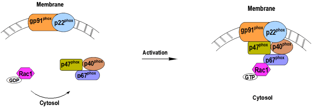
Recent evidence suggests that NADPH oxidase derived tonic increase in reactive oxygen species (ROS) is required for glucose stimulated insulin secretion.50,73,74,75,76 NADPH oxidase (Nox) represent a group of superoxide-generating enzymes which transport electrons through membranes and catalyze the cytosolic NADPH-dependent reduction of molecular oxygen to O2 •-.77 Till date, seven Nox family members have been identified i.e., Nox1, Nox2, Nox3, Nox4, Nox5, DUOX1 and DUOX2.78 The Phagocytic Nox is a multicomponent enzyme complex, composed of membrane components [catalytic glycosylated gp91phox and the regulatory non-glycosylated p22phox], cytosolic proteins [p47phox, p67phox, p40phox] and a small GTPase, Rac ½.78 Activation of Nox requires translocation of cytosolic components to the membrane and association with gp91phox/p22phox complex (Figure 2).79 Furthermore, Nox1 is the first homologue of gp91phox to be described and requires small GTPase Rac for activation.80,81,82,83 In contrast to Nox1, 2 and 3, Nox4 is a constitutively active enzyme and is activated without the necessity for GTPase Rac or the cytosolic components.84 


Figure 2: Activation of NADPH oxidase holoenzyme.

Activation of NADPH oxidase holoenzyme.


In this setting, the functional activation of Rac1 has shown to be critical in holoenzyme assembly and activation of Nox.78,85,86,87,88 In support of this, Gorzalczany and associates have shown the activation of Nox and subsequent generation of ROS by targeting Rac1 to the membrane fraction.89 They also demonstrated that prenylated Rac1 but not the unprenylated form binds to the phagocyte membrane more efficiently to facilitate the superoxide generation. Along these lines, Pi and Collins have overviewed the existing evidence in supporting “secondary messenger” roles of ROS in physiological insulin secretion.50 In addition, studies have also emphasized roles for Nox in physiological insulin secretion. For example, Diphenyleneiodonium [DPI], a selective inhibitor of Nox, inhibited glucose-induced Nox activity and GSIS.75 These observations were further confirmed by Morgan and associates suggesting that DPI or p47phox antisense-induced inhibition of Nox attenuated GSIS under static or perifusion conditions.90 Graciano and co-workers demonstrated regulatory roles for Nox in palmitate-induced superoxide generation and insulin secretion in rat islets.50 Furthermore, recent findings suggests that prenylation and activation of Rac1 are critical for glucose- and mitochondrial fuel-induced Nox-dependent ROS generation in clonal pancreatic β-cells and rodent islets.91 In summary, a tonic increase in intracellular ROS is necessary for normal physiological insulin secretion and Rac1 initiates subsequent signaling steps including Nox activation and insulin release.92 

B. RAC1-Nox Signaling and Metabolic Dysfunction: In addition to the above described beneficial roles for Rac1 in Nox-mediated ROS signaling in islet function, recent evidence also confirmed negative roles for ROS in islet β-cell dysfunction.91 Excessive ROS generation is considered central to the development of diabetes and its associated complications. Under normal physiological conditions, generation of free radicals is relatively low; however increased levels of circulating glucose promote intracellular accumulation of superoxides leading to metabolic dysfunction. Although, mitochondria remain the primary source for free radicals, emerging evidence implicates Nox as one of the major sources of extra-mitochondrial ROS. Immunological localization and functional regulation of Nox have been described in clonal β-cells, rat and human islets.50,75,90,92 Studies by Shen and associates in cardiac myocytes have also suggested regulatory roles for Rac1 in the activation of Nox and associated generation of ROS in animal models of diabetes.93 In addition, significant increase in Nox-mediated oxidative stress and subsequent metabolic dysfunction has been clearly reviewed in a recent article by Kowluru.70 However, very little is known with regard to regulatory roles of Rac1 in the holoenzyme assembly and activation of Nox in islet β-cells following chronic exposure to glucose, saturated fatty acids or cytokines.

In this context, recent findings demonstrated that prenylation of Rac1 is necessary for glucose-induced Nox activation and ROS generation in isolated β-cells.91 In addition, studies have also implicated Nox in metabolic dysfunction of the islet β-cell under conditions of glucolipotoxicity and exposure to cytokines.57,94 Generation of ROS under these conditions appears to be largely due to the activation of Nox, since inhibition of Nox [e.g., DPI, apocynin or siRNA-p47phox] or Rac1 activation [e.g., GGTI-2147, NSC23766] markedly attenuated deleterious effects on pancreatic β-cells. In addition, the activation status of Rac1 was shown to be under precise control of Tiam1, a known guanine nucleotide exchange factor for Rac1, but not Cdc42 and Rho G-proteins in isolated β-cells.95 In further support of this, a marked reduction in high glucose-, high palmitate- cytokine-induced Rac1 and Nox activation and ROS generation in isolated β-cells was observed following treatment with NSC23766, a selective inhibitor of Tiam1/Rac1 signaling axis.57,94 Using selective inhibitors of protein prenylation, Subasinghe, et al. demonstrated a critical requirement of prenylation of Rac1 for Nox-mediated β-cell dysfunction.94

Taken together, these in vitro findings clearly implicate participatory roles of Nox in exerting effects at the mitochondrial level including loss in membrane potential, cytochrome C release and activation of caspase-3 culminating in islet β-cell dysfunction.94,96 In addition, recent studies from Sidarala and colleagues present the evidence that the Rac1-Nox2 signaling is vital in high glucose induced activation of stress activated kinases and loss in GSIS causing islet β-cell dysfunction.97 Despite these in vitro evidences, potential roles for Nox in islet dysfunction in animal models of type 2 diabetes are minimal. However, a recent study systematically examined the functional status of Nox in islets from Zucker Diabetic Fatty [ZDF] rat, which develops obesity, hyperinsulinemia, hyperglycemia and a decline in β-cell function.58 These in vitro observations supported by findings in islets derived from the diabetic rodents [the ZDF rat] and diabetic human islets, form basis for the development of small molecule inhibitors for Rac1 and Nox activation in halting the metabolic defects, thereby retaining normal β-cell mass. In addition, a recent study from Zhou and colleagues also confirmed that the treatment with selective inhibitor NSC23766 attenuated Rac1 expression and oxidative stress in the pancreas in ob/ob mice.98 These findings provide insights into potential therapeutic targets and interventional modalities to prevent the metabolic defects. 

POTENTIAL THERAPEUTIC TARGETS AND INTERVENTIONAL MODALITIES

Based on the above discussion and published evidences, it is clear that Nox-derived reactive oxygen species have both positive and negative roles in the islet β-cell function. Targeting Nox holoenzyme complex could be beneficial in subsiding the excessive generation of ROS during oxidative stress milieu. In this context, a recent study proposed that gp91phox, p47phox and p67phox might serve as potential drug targets due to their selective association in the Nox holoenzyme complex.99 On the contrary, peptide inhibitors blocking Rac1/2 activation and p47phox translocation might not be a good approach, since they are integral members of other NADPH oxidasecomplexes too. However, Mizrahi, et al. developed p47phox-p67phox-Rac1 chimera as a quintessential single molecule activator of Nox100 to study the effects of Nox activation regulatory roles for Rac1. These observations are in agreement with the findings, where researchers have demonstrated a decrease in glucose-mediated Nox-induced ROS generation in the presence of prenylation inhibitors. Developing inhibitors for such quintessential single molecule activators might provide a novel therapeutics to minimize excessive generation of ROS Nox-mediated pancreatic β-cell dysfunction.

Furthermore, an alternate approach to minimize the excessive generation of ROS is to enrich the antioxidant capacity of the islet β-cells. As reviewed by Acharya and Ghaskadbi,101 pancreatic islet β-cells hold a poor antioxidant defense mechanism. And counterbalancing oxidative environment by antioxidant treatment or overexpressing antioxidant enzymes might prove to be successful in regulating islet β-cell function. Indeed, such modalities have been shown to work efficiently both in vivo and in vitro. Along these lines, treatment with antioxidant, α-lipoic acid has been demonstrated to improve insulin sensitivity in type 2 diabetic subjects.102 Moreover, researchers have also shown that vitamin E treatment improves pancreatic physiology under diabetic state.103 Asayama, et al. found that rats deficient in vitamin E, selenium, or both had decreased insulin secretory reserves, suggesting that vitamin E status can directly affect pancreatic islet function. In a mouse model of type 2 diabetes, treatment with vitamin E combined with vitamin C and n-acetyl cysteine resulted in large number of pancreatic islets than controls.104 Furthermore, a recent study in humans has shown that, taurine affectively restored β-cell function and improved insulin sensitivity.105 Together these studies further highlight antioxidant therapy as one of the feasible options in attenuating excessive generation of ROS and subsequent reduction in oxidative stress environment in the islet β-cells.

In addition to the above mentioned strategies for attenuating oxidative stress, inhibitors blocking Tiam1/Rac1/Nox signaling axis,57,94,106 polyphenolic extracts supplementation,107 stress activated kinase inhibitors,108,109,110,111 and angiotensin receptor antagonists112 have proven efficaciously to reduce oxidative stress and improve islet β-cell function. 

CONCLUSION

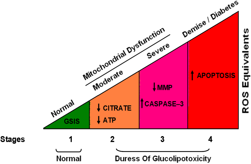
Glucose stimulate insulin secretion (GSIS) involves a series of metabolic events involving interaction between a variety of signaling pathways to facilitate the transport of insulin-laden granules to the plasma membrane for fusion and subsequent insulin release. Compelling evidence supports involvement of small G-proteins like Rac1 and Cdc42 in the cytoskeletal reorganization, which is necessary for GSIS to occur. Recent findings further validate that Tiam1 represents one of the GEFs for Rac1 and that Tiam1/Rac1 signaling axis is requisite for GSIS. Nox appears to be an effector protein for Tiam1/Rac1 signaling and that its activation leads to a tonic increase in the generation of ROS under the stimulatory conditions of glucose and fatty acids leading to insulin release. In addition to this, Tiam1/Rac1 signaling axis appears to play a vital role in Nox-mediated ROS generation under the duress of excessive glucose, palmitate, ceramide and cytokines culminating in oxidative stress and metabolic dysfunction of islet β-cells. Together, these findings suggest positive and negative modulatory roles for Tiam1-Rac1-Nox signaling pathway in islet function. The Figure 3 depicted below is indicative of potential effects of ROS on islet β-cells at different stages. Low levels of generated ROS have a positive effect on glucose stimulated insulin secretion, and as the levels of the ROS increases it causes detrimental effects and β-cell dysfunction.113 Therefore, it may be challenging to draw a line as to how much of ROS generation is beneficial for the normal function of islets as opposed to how much is bad to elicit damaging effects on the pancreatic β-cell. It is likely that there may be a “window of opportunity” or “point of return” for the islet β-cell to recover from the noxious effects of excessive ROS due to accelerated Tiam1-Rac1-Nox signaling pathway in the diabetic states.


Figure 3: Hypothetical model for ROS generation in identifying the effects on pancreatic islet β-cells.

Hypothetical model for ROS generation in identifying the effects on pancreatic islet
β-cells.


CONFLICTS OF INTEREST

The authors declare that they dont have any conflicts of interest or any acknowledgements for this submission.

FINANCIAL INTERESTS

None.

1. Malaisse WJ. Hormonal and environmental modification of islet activity. In: Steiner DF, Frankel N, eds. Handbook of physiology. New York: American Physiological Society. 1972; 237-260.

2. Prentki M, Matschinsky FM. Calcium, cAMP, and phospholipid-derived messengers in coupling mechanisms of insulin secretion. Physiol Rev. 1987; 67: 1185-1248. doi: 10.1152/physrev.1987.67.4.1185

3. MacDonald MJ. Elusive proximal signals of β-cells for insulin secretion. Diabetes. 1990; 39: 1461-1466. doi: 10.2337/diab.39.12.1461

4. Laychock SG. Glucose metabolism, second messengers and insulin secretion. Life Sci. 1990; 47: 2307-2316. doi: 10.1016/0024-3205(90)90269-w

5. Newgard CB, McGarry JD. Metabolic coupling factors in pancreatic beta-cell signal transduction. Annu Rev Biochem. 1995; 64: 689-719. doi: 10.1146/annurev.bi.64.070195.003353

6. Deeney JT, Prentki M, Corkey BE. Metabolic control of β-cell function. Semin Cell Dev Biol. 2000; 11: 267-275. doi: 10.1006/scdb.2000.0175

7. Newgard CB, Lu D, Jensen MV, et al. Stimulus/secretion coupling factors in glucose-stimulated insulin secretion: insights gained from a multidisciplinary approach. Diabetes. 2002; 51(3): S389-S393. doi: 10.2337/diabetes.51.2007.S389

8. Berggren PO, Leibiger IB. Novel aspects on signal transduction in the pancreatic β cell. Nutr Metab Cardiovasc Dis. 2006; 16(1): S7-S10. doi: 10.1016/j.numecd.2005.11.005

9. Metz SA. Membrane phospholipid turnover as an intermediary step in insulin secretion.Putative roles of phospholipases in cell signaling. Am J Med. 1988; 85: 9-21. doi: 10.1016/0002-9343(88)90393-2

10. Metz SA. The pancreatic islet as a Rubik’s cube. Is phospholipid hydrolysis a piece of the puzzle? Diabetes. 1991; 40: 1565-1573. doi: 10.2337/diab.40.12.1565

11. Lawrence M, Shao C, Duan L, McGlynn K, Cobb MH. The protein kinases ERK1/2 and their roles in pancreatic β cells. Acta Physiol (Oxf). 2008; 192: 11-17. doi: 10.1111/j.1748-1716.2007.01785.x

12. Lang J. Molecular mechanisms and regulation of insulin exocytosis as a paradigm of endocrine secretion. Eur J Biochem. 1999; 259: 3-17. doi: 10.1046/j.1432-1327.1999.00043.x

13. Poitout V. Phospholipid hydrolysis and insulin secretion: a step toward solving the Rubik’s cube. Am J Physiol Endocrinol Metab. 2008; 294: E214-E216. doi: 10.1152/ajpendo.00638.2007

14. Metz SA, Rabaglia ME, Pintar TJ. Selective inhibitors of GTP synthesis impede exocytotic insulin release from intact rat islets. J Biol Chem. 1992; 267: 12517-12527.

15. Metz SA, Meredith M, Rabaglia ME, Kowluru A. Small elevations of glucose concentration redirect and amplify the synthesis of guanosine 5-triphosphate in rat islets. J Clin Invest. 1993; 92: 872-882. doi: 10.1172/JCI116662

16. Komatsu M, Noda M, Sharp GW. Nutrient augmentation of Ca2-dependent and Ca2+-independent pathways in stimulus-coupling to insulin secretion can be distinguished by their guanosine triphosphate requirements: studies on rat pancreatic islets. Endocrinology. 1998; 139: 1172-1183. doi: 10.1210/endo.139.3.5859

17. Straub SG, James RF, Dunne MJ, Sharp GW. Glucose augmentation of mastoparan stimulated insulin secretion in rat and human pancreatic islets. Diabetes. 1998; 47: 1053-1057. doi: 10.2337/diabetes.47.7.1053

18. Kowluru A, Seavey SE, Li G, et al. Glucose- and GTP-dependent stimulation of the carboxylmethylation of Cdc42 in rodent and human pancreatic islets and pure β cells: evidence for an essential role for GTP-binding proteins in nutrient-induced insulin secretion. J Clin Invest. 1996; 98: 540-555. doi: 10.1172/JCI118822

19. Kowluru A. Regulatory roles for small G-proteins in the pancreatic β cell: lessons from models of impaired insulin secretion. Am J Physiol Endocrinol Metab. 2003; 285: E669-E684. doi: 10.1152/ajpendo.00196.2003

20. Wang Z, Thurmond DC. Mechanisms of biphasic insulin-granule exocytosis- roles of the cytoskeleton, small GTPases and SNARE proteins. J Cell Sci. 2009; 122: 893-903. doi: 10.1242/​jcs.034355

21. Robertson RP, Seaquist ER, Walseth TF. G proteins and modulation of insulin secretion. Diabetes. 1991; 40: 1-6. doi: 10.2337/diab.40.1.1

22. Seaquist ER, Walseth TF, Redmon JB, Robertson RP. G-protein regulation of insulin secretion. J Lab Clin Med. 1994; 123: 338-345. doi: 10.2337/diab.40.1.1

23. Gilman AG. G proteins: transducers of receptor-generated signals. Annu Rev Biochem.1987; 56: 615-649. doi: 10.1146/annurev.bi.56.070187.003151

24. Birnbaumer L. Receptor-to-effector signaling through G proteins: roles for beta gamma dimers as well as α subunits. Cell. 1992; 71: 1069-1072. doi: 10.1016/S0092-8674(05)80056-X

25. Clapham DE, Neer EJ. New roles for G-protein βγ-dimers in transmembrane signaling. Nature. 1993; 365: 403-406. doi: 10.1038/365403a0

26. Takai Y, Sasaki T, Matozaki T. Small GTP-binding proteins. Physiol Rev. 2001; 81: 153-208. doi: 10.1152/physrev.2001.81.1.153

27. Casey PJ, Seabra MC. Protein prenyltransferases. J Biol Chem. 1996; 271: 5289-5292. doi: 10.1074/jbc.271.10.5289

28. Maurer-Stroh S, Washietl S, Eisenhaber F. Protein prenyltransferases. Genome Biol. 2003; 4: 212.1-212.9. doi: 10.1186/gb-2003-4-4-212

29. Seabra MC, Reiss Y, Casey PJ, Brown MS, Goldstein JL. Protein farnesyltransferase and geranylgeranyltransferase share a common α subunit. Cell. 1991; 65: 429-434. doi: 10.1016/0092-8674(91)90460-G

30. Fu HW, Casey PJ. Enzymology and biology of CaaX protein prenylation. Rec ProgHorm Res. 1999; 54: 315-342.

31. Veluthakal R, Kaur H, Goalstone M, Kowluru A. Dominant negative-subunit of farnesyl-and geranylgeranyltransferase inhibits glucose-stimulated, but not KCl stimulated, insulin secretion in INS 832/13 cells. Diabetes. 2007; 56: 204-210. doi: 10.2337/db06-0668

32. Kowluru A, Li G, Rabaglia ME, et al. Evidence for differential roles of the Rho subfamily of GTP-binding proteins in glucose- and calcium-induced insulin secretion from pancreatic β cells. Biochem Pharmacol. 1997; 54: 1097-1108. doi: 10.1016/S0006-2952(97)00314-6

33. Regazzi R, Kikuchi A, Takai Y, Wollheim CB. The small GTP-binding proteins in the cytosol of insulin-secreting cells are complexed to GDP dissociation inhibitor proteins. J Biol Chem. 1992; 267: 17512-17519. doi: 10.1016/S0021-9258(19)37072-3

34. Kowluru A, Rabaglia ME, Muse KE, Metz SA. Subcellular localization and kinetic characterization of guanine nucleotide binding proteins in normal rat and human pancreatic islets and transformed β cells. Biochim Biophys Acta. 1994; 1222: 348-359. doi: 10.1016/0167-4889(94)90040-x

35. Daniel S, Noda M, Cerione RA, Sharp GW. A link between Cdc42 and syntaxin is involved in mastoparan stimulated insulin release. Biochemistry. 2002; 41: 9663-9671. doi: 10.1021/bi025604p

36. Kowluru A, Chen HQ, Tannous M. Novel roles for the Rho subfamily of GTP-binding proteins in succinate induced insulin secretion from β TC3 cells: further evidence in support of succinate mechanism of insulin release. Endocr Res. 2003; 29: 363-376. doi: 10.1081/erc-120025043

37. Nevins AK, Thurmond DC. Glucose regulates the cortical actin network through modulation of Cdc42 cycling to stimulate insulin secretion. Am J Physiol Cell Physiol. 2003; 285: C698-C710. doi: 10.1152/ajpcell.00093.2003

38. Nevins AK, Thurmond DC. A direct interaction between Cdc42 and vesicle-associated membrane protein 2 regulates SNARE-dependent insulin exocytosis. J Biol Chem. 2005; 280: 1944-1952. doi: 10.1074/jbc.m409528200

39. Nevins AK, Thurmond DC. Caveolin-1 functions as a novel Cdc42 guanine nucleotide dissociation inhibitor in pancreatic β-cells. J Biol Chem. 2006; 281: 18961-18972. doi: 10.1074/jbc.M603604200

40. Wang Z, Oh E, Thurmond DC. Glucose-stimulated Cdc42 signaling is essential for the second phase of insulin secretion. J Biol Chem. 2007; 282: 9536-9546. doi: 10.1074/jbc.M610553200

41. Li J, Luo R, Kowluru A, Li G. Novel regulation by Rac1 of glucose-and forskolin-induced insulin secretion in INS-1 β cell. Am J Physiol Endocrinol Metab. 2004; 286: E818-E827. doi: 10.1152/ajpendo.00307.2003

42. McDonald P, Veluthakal R, Kaur H, Kowluru A. Biologically active lipids promote trafficking and membrane association of Rac1 in insulin-secreting INS832/13 cells. Am J Physiol Cell Physiol. 2007; 292: C1216-C1220. doi: 10.1152/ajpcell.00467.2006

43. Asahara A, Kido Y, Shigeyama Y, et al. Rac1 regulates glucose induced insulin secretion through modulation of cytoskeletal organization in β cells. Diabetes. 2008; 57(1): A55.

44. Greiner TU, Kesavan G, Ståhlberg A, Semb H. Rac1 regulates pancreatic islet morpholgenesis. BMC Dev Biol. 2009; 9: 2. doi: 10.1186/1471-213X-9-2

45. Kowluru A, Amin R. Inhibitors of post-translational modifications of G-proteins as probes to study the pancreatic beta cell function: potential therapeutic implications. Curr Drug Targets Immune Endocr Metabol Disord. 2002; 2: 129-139. doi: 10.2174/1568008023340668

46. Lawrence JT, Birnbaum MJ. ADP-ribosylation factor 6 regulates insulin secretion through plasma membrane phosphatidylinositol 4,5-bisphosphate. Proc Natl Acad Sci. 2003; 100: 13320-13325. doi: 10.1073/pnas.2232129100

47. Grodnitzky JA, Syed N, Kimber MJ, Day TA, Donaldson JG, Hsu WH. Somatostatin receptors signal through EFA6A-ARF6 to activate phospholipase D in clonal β-cells. J Biol Chem. 2007; 282: 13410-13418. doi: 10.1074/jbc.M701940200

48. Hammar E, Tomas A, Bosco D, Halban PA. Role of the Rho-ROCK (Rho-associated kinase) signaling pathway in the regulation of pancreatic-cell function. Endocrinology. 2009; 150: 2072-2079. doi: 10.1210/en.2008-1135

49. Kowluru A. Small G proteins in islet beta-cell function. Endocr Rev. 2010; 31: 52-78. 10.1210/er.2009-0022

50. Graciano MF, Santos LR, Curi R, Carpinelli AR. NAD(P)H oxidase participates in the palmitate-induced superoxide production and insulin secretion by rat pancreatic islets. J Cell Physiol. 2011; 226(4): 1110-1117. doi: 10.1002/jcp.22432

51. Buffa L, Fuchs E, Pietropaolo M, Barr F, Solimena M. ICA69 is a novel Rab2 effector regulating ER-Golgi trafficking in insulinoma cells. Eur J Cell Biol. 2008; 87: 197-209. doi: 10.1016/j.ejcb.2007.11.003

52. Chan SL, Monks LK, Gao H, Deaville P, Morgan NG. Identification of the monomeric G-protein, Rhes, as an efaroxan-regulated protein in the pancreatic β-cell. Br J Pharmacol. 2002; 136: 31-36. doi: 10.1038/sj.bjp.0704680

53. Sharoyko VV, Zaitseva II, Varsanyi M, et al. Monomeric G-protein, Rhes, is not an imidazolineregulated protein in pancreatic β-cells. Biochem Biophys Res Commun. 2005; 338: 1455-1459. doi: 10.1016/j.bbrc.2005.10.145

54. Taylor JP, Jackson DA, Morgan NG, Chan SL. Rhes expression in pancreatic β-cells is regulated by efaroxan in a calcium-dependent process. Biochem Biophys Res Commun. 2006; 349: 809-815. doi: 10.1016/j.bbrc.2006.08.102

55. Lopez JA, Kwan EP, Xie L, He Y, James DE, Gaisano HY. The RalAGTPase is a central regulator of insulin exocytosis from pancreatic islet cells. J Biol Chem. 2008; 283: 17939-17945. doi: 10.1074/jbc.M800321200

56. Syed I, Jayaram B, Subasinghe W, Kowluru A. Tiam1/Rac1 signaling pathway mediates palmitate-induced, ceramide-sensitive generation of superoxides and lipid peroxides and the loss of mitochondrial membrane potential in pancreatic beta-cells. Biochem Pharmacol. 2010; 80: 874-883. doi: 10.1016/j.bcp.2010.05.006

57. Syed I, Kyathanahalli CN, Jayaram B, et al. Increased Phagocyte-Like NADPH Oxidase and ROS Generation in Type 2 Diabetic ZDF Rat and Human Islets. Diabetes. 2011; 60: 2843-2852. doi: 10.2337/db11-0809

58. Didsbury J, Weber RF, Bokoch GM, Evans T, Snyderman R. Rac, a novel ras-related family of proteins that are botulinum toxin substrates. J Biol Chem. 1989; 264(28): 16378-16382.

59. Wang Z, Pedersen E, Basse A, et al. Rac1 is crucial for Ras-dependent skin tumor formation by controlling Pak1-Mek-Erk hyperactivation and hyperproliferation in vivo. Oncogene. 2010; 29: 3362-3373. doi: 10.1038/onc.2010.95

60. Sosa MS, Lopez-Haber C, Yang C, et al. Identification of the Rac-GEF P-Rex1 as an essential mediator of ErbB signaling in breast cancer. Mol Cell. 2010; 40: 877-892. doi: 10.1016/j.molcel.2010.11.029

61. Hayashi-Takagi A, Takaki M, Graziane N, et al. Disruptedin-Schizophrenia 1 (DISC1) regulates spines of the glutamate synapse via Rac1. Nat Neurosci. 2010; 13: 327-332. doi: 10.1038/nn.2487

62. Bopp A, Wartlick F, Henninger C, Kaina B, Fritz G. Rac1 modulates acute and subacutegenotoxin-induced hepatic stress responses, fibrosis and liver aging. Cell Death and Disease. 2013; 4: e558. doi: 10.1038/cddis.2013.57

63. JeBailey L, Wanono O, Niu W, Roessler J, Rudich A, Klip A. Ceramide-and oxidant-induced insulin resistance involve loss of insulin-dependent Rac-activation and actin remodeling in muscle cells. Diabetes. 2007; 56: 394-403. doi: 10.2337/db06-0823

64. Ueda S, Kitazawa S, Ishida K. Crucial role of the small GTPase Rac1 in insulin-stimulated translocation of glucose transporter 4 to the mouse skeletal muscle sarcolemma. FASEB J. 2010; 24: 2254-2261. doi: 10.1096/fj.09-137380

65. Iizuka M, Konno S. Wound healing of intestinal epithelialcells. World J Gastroenterol. 2011; 17: 2161-2171. doi: 10.3748/wjg.v17.i17.2161

66. Babbin BA, Jesaitis AJ, Ivanov AI, et al. Formylpeptide receptor-1 activation enhances intestinal epithelialcell restitution through phosphatidylinositol3-kinase-dependent activation of Rac1 and Cdc42. J Immunol. 2007; 179: 8112-8121. doi: 10.4049/​jimmunol.179.12.8112

67. Lemichez E, Aktories K. Hijacking of Rho GTPasesduring bacterial infection. Exp Cell Res. 2013; 319: 2329-2336. doi: 10.1016/j.yexcr.2013.04.021

68. Citalán-Madrid AF, García-Ponce A, Vargas-Robles H, Betanzos A, Schnoor M. Small GTPases of the Ras superfamily regulateintestinal epithelial homeostasis and barrierfunction via common and unique mechanisms. Tissue Barriers. 2013; 1: e26938: 1-28. doi: 10.4161/tisb.26938

69. Kowluru A. Friendly, and not so friendly, roles of Rac1 in islet β-cell function: lessons learnt from pharmacological and molecular biological approaches. Biochem Pharmacol. 2011; 81(8): 965-975. doi: 10.1016/j.bcp.2011.01.013

70. Benitah SA, Frye M, Glogauer M, Watt FM. Stem cell depletion through epidermal deletion of Rac1. Science. 2005; 309: 933-935. doi: 10.1126/science.1113579

71. Asahara S, Shibutani Y, Teruyama K, et al. Ras-related C3 botulinum toxin substrate 1 (RAC1) regulates glucose-stimulated insulin secretion via modulation of F-actin. Diabetologia. 2013; 56: 1088-1097. doi: 10.1007/s00125-013-2849-5

72. Pi J, Collins S. Reactive oxygen species and uncoupling protein 2 in pancreatic β-cell function. Diabetes Obes Metab. 2010; 12(2): 141-148. doi: 10.1111/j.1463-1326.2010.01269.x

73. Graciano MF, Santos LR, Curi R, Carpinelli AR. NAD(P)H oxidase participates in the palmitate-induced superoxide production and insulin secretion by rat pancreatic islets. J Cell Physiol. 2011; 226(4): 1110-1117. doi: 10.1002/jcp.22432

74. Newsholme P, Morgan D, Rebelato E, et al. Insights into the critical role of NADPH oxidase(s) in the normal and dysregulated pancreatic beta cell. Diabetologia. 2009; 52: 2489-2498. doi: 10.1007/s00125-009-1536-z

75. Oliveira HR, Verlengia R, Carvalho CR, Britto LR, Curi R, Carpinelli AR. Pancreatic beta cells express phagocyte-like NADPH oxidase. Diabetes. 2003; 52: 1457-1463. doi: 10.2337/diabetes.52.6.1457

76. Pi J, Bai Y, Zhang Q, et al. Reactive oxygen species as a signal in glucose-stimulated insulin secretion. Diabetes. 2007; 56: 1783-1791. doi: 10.2337/db06-1601

77. Krause KH. Tissue distribution and putative physiological function of NOX family NADPH oxidases. Jpn J Infect Dis. 2004; 57(5): S28-S29.

78. Geiszt M. NADPH oxidases: new kids on the block. Cardiovasc Res. 2006; 71: 289-299. doi: 10.1016/j.cardiores.2006.05.004

79. Bedard K, Krause KH. The NOX family of ROS generating NADPH oxidases: physiology and pathophysiology. Physiol Rev. 2007; 87: 245-313. doi: 10.1152/physrev.00044.2005

80. Banfi B, Maturana A, Jaconi S, et al. A mammalian H+ channel generated through alternative splicing of the NADPH oxidase homolog NOH-1. Science. 2000; 287(5450): 138-142. doi: 10.1126/science.287.5450.138

81. Sumimoto H, Miyano K, Takeya R. Molecular composition and regulation of the Nox family NAD(P)H oxidases. Biochem Biophys Res Commun. 2005; 338: 677-686. doi: 10.1016/j.bbrc.2005.08.210

82. Cheng G, Diebold BA, Hughes Y, Lambeth JD. Nox1-dependent reactive oxygen generation is regulated by Rac1. J Biol Chem. 2006; 281: 17718-17726. doi: 10.1074/jbc.M51275120

83. Martyn KD, Frederick LM, von Loehneysen K, Dinauer MC, Knaus UG. Functional analysis of Nox4 reveals unique characteristics compared to other NADPH oxidases. Cell Signal. 2006; 18: 69-82. doi: 10.1016/j.cellsig.2005.03.023

84. Hordijk PL. Regulation of NADPH oxidases: the role of Rac proteins. Circ Res. 2006; 98: 453-462. doi: 10.1161/01.res.0000204727.46710.5e

85. Abo A, Pick E, Hall N, Totty N, Teahan CG, Segal AW. Activation of the NADPH oxidase involves the small-GTP binding protein p21rac1. Nature. 1991; 353: 668-670. doi: 10.1038/353668a0

86. Knaus UG, Heyworth PG, Evans T, Curnutte JT, Bokoch GM. Regulation of phagocyte oxygen radical production by the GTP-binding protein Rac 2. Science. 1991; 254: 1512-1515. doi: 10.1126/science.1660188

87. Babior BM. NADPH oxidase: an update. Blood. 1999; 93(5): 1464-1476.

88. Borregaard N, Tauber AI. Subcellular localization of the human neutrophil NADPH oxidase b-cytochrome and associated flavoprotein. J Biol Chem. 1984; 259(1): 47-52.

89. Gorzalczany Y, Sigal N, Itan M, Lotan O, Pick E. Targeting of Rac1 to the phagocyte membrane is sufficient for the induction of NADPH oxidase assembly. J Biol Chem. 2000; 275(1): 40073-40081. doi: 10.1074/jbc.M006013200

90. Morgan D, Rebelato E, Abdulkader F, et al. Association of NAD(P)H oxidase with glucose-induced insulin secretion by pancreatic beta-cells. Endocrinology. 2009; 150(5): 2197-2201. doi: 10.1210/en.2008-1149

91. Syed I, Kyathanahalli CN, Kowluru A. Phagocyte-like NADPH oxidase generates ROS in INS 832/13 cells and rat islets: role of protein prenylation. Am J Physiol Regul Integr Comp Physiol. 2011; 300(3): R756-R762. doi: 10.1152/ajpregu.00786.2010

92. Uchizono Y, Takeya R, Iwase M, et al. Expression of isoforms of NADPH oxidase components in rat pancreatic islets. Life Sci. 2006; 80(2): 133-139. doi: 10.1016/j.lfs.2006.08.031

93. Shen E, Li Y, Li Y, et al. Rac1 is required for cardiomyocyte apoptosis during hyperglycemia. Diabetes. 2009; 58(10): 2386-2395. doi: 10.2337/db08-0617

94. Subasinghe W, Syed I, Kowluru A. Phagocyte-like NADPH oxidase promotes cytokine-induced mitochondrial dysfunction in pancreatic β-cells: evidence for regulation by Rac1. Am J Physiol Regul Integr Comp Physiol. 2011; 300: R12-R20. doi: 10.1152/ajpregu.00421.2010

95. Veluthakal R, Madathilparambil SV, McDonald P, Olson LK, Kowluru A. Regulatory roles for Tiam1, a guanine nucleotide exchange factor for Rac1, in glucose-stimulated insulin secretion in pancreatic beta-cells. Biochem Pharmacol. 2009; 77: 101-113. doi: 10.1016/j.bcp.2008.09.021

96. Kowluru RA, Kowluru A, Veluthakal R, et al. TIAM1-RAC1 signalling axis-mediated activation of NADPH oxidase-2 initiates mitochondrial damage in the development of diabetic retinopathy. Diabetologia. 2014; 57(5): 1047-1056. doi: 10.1007/s00125-014-3194-z

97. Sidarala V, Veluthakal R, Syeda K, Vlaar C, Newsholme P, Kowluru A. Phagocyte-like NADPH oxidase (Nox2) promotes activation of p38MAPK in pancreatic β-cells under glucotoxic conditions: evidence for a requisite role of Ras-related C3 botulinum toxin substrate 1 (Rac1). Biochem Pharmacol. 2015. doi: 10.1016/j.bcp.2015.04.001

98. Zhou S, Yu D, Ning S, et al. Augmented Rac1 Expression and Activity are Associated with Oxidative Stress and Decline of β Cell Function in Obesity. Cell Physiol Biochem. 2015; 35: 2135-2148. doi: 10.1159/000374019

99. El-Benna J, Dang PM, Périanin A. Peptide-based inhibitors of the phagocyte NADPH oxidase. Biochem Pharmacol. 2010; 80(6): 778-785. doi: 10.1016/j.bcp.2010.05.020

100. Mizrahi A, Berdichevsky Y, Casey PJ, Pick E. The quintessential NADPH oxidase activator: a prenylated p47PHOX-p67PHOX-Rac1 chimera – membrane association and functional capacity. J Biol Chem. 2010; 285(33): 25485-25499. doi: 10.1074/jbc.M110.113779

101. Acharya JD, Ghaskadbi SS. Islets and their antioxidant defense. Islets. 2010; 2: 225-235. doi: 10.4161/isl.2.4.12219

102. Jacob S, Ruus P, Hermann R, et al. Oral administration of RAC-alpha-lipoic acid modulates insulin sensitivity in patients with type-2 diabetes mellitus: a placebo-controlled pilot trial. Free Radic. Biol. Med. 1999; 27: 309-314. doi: 10.1016/S0891-5849(99)00089-1

103. Sena CM, Nunes E, Gomes A, et al. Supplementation of coenzyme Q10 and alpha-tocopherol lowers glycated hemoglobin level and lipid peroxidation in pancreas of diabetic rats. Nutr. Res. 2008; 28: 113-121. doi: 10.1016/j.nutres.2007.12.005

104. Asayama K, Kooy NW, Burr IM. Effect of vitamin E deficiency and selenium deficiency on insulin secretory reserve and free radical scavenging systems in islets: decrease of islet manganosuperoxide dismutase. J. Lab. Clin. Med. 1986; 107: 459-464.

105. Xiao C, Giacca A, Lewis GF. Oral taurine but not N-acetylcysteine ameliorates NEFA-induced impairment in insulin sensitivity and β-cell function in obese and overweight, non-diabetic men. Diabetologia. 2008; 51: 139-146. doi: 10.1007/s00125-007-0859-x

106. Veluthakal R, Palanivel R, Zhao Y, McDonald P, Gruber S, Kowluru A. Ceramide induces mitochondrial abnormalities in insulin-secreting INS-1 cells: potential mechanisms underlying ceramide-mediated metabolic dysfunction of the beta cell. Apoptosis. 2005; 10: 841-850. doi: 10.1007/s10495-005-0431-4

107. Decorde K, Teissedre PL, Sutra T, Ventura E, Cristo JP, Rouanet JM. Chardonnay grape seed procyaidin extract supplementation prevents high-fat diet-induced obesity in hamsters by improving adipokine imbalance and oxidative stress markers. Mol Nutr Food Res. 2009; 53: 659-666. doi: 10.1002/mnfr.200800165

108. Kaneto H, Nakatani Y, Kawamori D, et al. Role of oxidative stress, endoplasmic reticulum stress, and c-Jun N-terminal kinase in pancreatic β-cell dysfunction and insulin resistance. Int J Biochem Cell Biol. 2006; 38: 782-793. doi: 10.1016/j.biocel.2006.01.004

109. de la Rosa LC, Vrenken TE, Hannivoort RA, et al. Carbon monoxide blocks oxidative stress-induced hepatocyte apoptosis via inhibition of the p54 JNK isoform. Free Radic Biol Med. 2007; 44: 1323-1333. doi: 10.1016/j.freeradbiomed.2007.12.011

110. Mosen H, Salehi A, Alm P, et al. Defective glucose-stimulated insulin release in the diabetic Goto-Kakizaki rat coincides with reduced activity of the islet carbon monoxide signaling pathway. Endocrinology. 2005; 146: 1553-1558. doi: 10.1210/en.2004-0851

111. Mosen H, Salehi A, Henningsson R, Lundquist I. Nitric oxide inhibits, and carbon monoxide activates, islet acid α-glucoside hydrolase activites in parallel with glucose-stimulated insulin secretion. J Endocrinol. 2006; 190: 681-693. doi: 10.1677/joe.1.06890

112. Nakayama M, Inoguchi T, Sonta T, et al. Increased expression of NAD(P)H oxidase in islets of animal models of Type 2 diabetes and its improvement by an AT1 receptor antagonist. Biochem Biophys Res Commun. 2005; 332(4): 927-933. doi: 10.1016/j.bbrc.2005.05.065

113. Ismail S. Mechanisms of regulation of islet function by NADPH oxidase. Diss. Wayne State University, 2011.

LATEST ARTICLES

Quality Assurance of General Purpose, Keratin Based and Dye lock Hair Shampoos

Saima Siddique*, Zahida Parveen, Zeeshan Ali and Sidra Mehmood

doi.

Blood Sample from the Patient

Hypertriglyceridemia-Induced Pancreatitis: A Case Report and Literature Review

Maarten Bulterys, Melvin Willems* and Agnes Meersman

doi.

From Neck Pain to a Life-Threatening Condition: A Case Report

Floris Vandewoude* and Sören Verstraete

doi.

LATEST ARTICLES

Original Research

2024 Aug

Saima Siddique*, Zahida Parveen, Zeeshan Ali and Sidra Mehmood

Cross Sectional Study, peer reviewed

2024 Jul

Amanuel P. Beta, Dereje Abera, Legese Belayneh and Isayas A. Kebede

Case Report, peer reviewed

2024 Jul

Syeda Rukh*, Sathyanarayana Machani and Milind Awale