INTRODUCTION
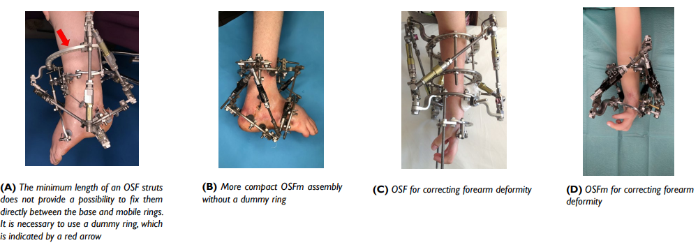
The orthopedic hexapod Ortho-SUV Frame (OSF), like other well-known orthopedic hexapods,1,2,3,4 has such disadvantages as large bulkiness and weight of the hardware versus the Ilizarov apparatus. There is also an objective limitation on working with “short segments” of the limbs, i.e., when the base and mobile rings are located at a close distance less of than 10-12 cm. The use of Z-shaped plates and “dummy” rings do not provide to solve this problem completely.5,6,7,8 A “minimized” version of the OSF, minimized Ortho-SUV Frame (OSFm), was developed for this purpose.3 OSFm differs from its “big brother”, OSF, by a significant reduction in size by 70 cm3 and weight by 314 grams (Figure 1). This study aimed to determine the capabilities of OSFm for angulation and translation as the most significant for deformity correction.
Figure 1. OSF and OSFm
METHODS
The bench test was performed using plastic models of the tibia osteotomized at the middle third of the shaft. One-ring 140 mm diameter modules were used to fix each of the bone fragments. The bone fragments were located in the center of the rings. The initial distance between the base and mobile rings was 200 mm.
The reduction capabilities of OSFm with a standard struts size were studied in the first series of experiments. OSF with a short struts size was used in the second comparison series. Three groups were studied depending on the method of fixing the struts to the rings in each series of bench tests: directly to the ring, using six straight plates, and using six Z-shaped plates. Reduction features were evaluated by the maximum displacement of the mobile bone fragment relative to the base one (Figure 2):
Figure 2. Movements of the Mobile Bone Fragment
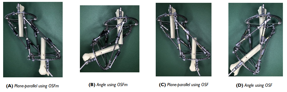
- Plane-parallel in the frontal plane (inward and outward) and in the sagittal plane (anterior and posterior);
- At an angle in the frontal plane (varization and valgization) and in the sagittal plane (antecurvation and recurvation).
The displacement was stopped when any of the struts reached its minimum or maximum possible length, which was a limitation for the subsequent movement of the mobile bone fragment.
Plane-parallel displacements were measured in mm, and angular displacements — in degrees. To obtain statistically significant data, the experiment was repeated five times with each of the six collected models. The obtained quantitative data were statistically processed in the Statistica version 10.1 program. The Mann-Whitney test, median Chi-square (χ2) test, and analysis of variance (ANOVA) test were used to compare biases. Differences between groups were considered statistically significant at p<0.05.
RESULTS
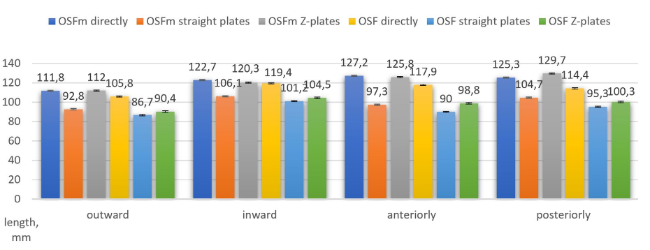
OSFm with fixation of the struts directly to the rings exceeds OSF by 6 mm in terms of the capabilities of plane-parallel movement outward and by 3.3 mm inward. OSFm outperforms OSF outward by 6.1 mm and inward by 5.2 mm using straight plates, and outward by 21.6 mm and inward by 15.8 mm using Z-plates. OSFm with fixation of the struts directly to the rings exceeds OSF during plane-parallel movement anteriorly by 9.8 mm and posteriorly by 11.3 mm. OSFm outperforms OSF anteriorly by 7.3 mm and posteriorly by 8.5 mm using straight plates, and anteriorly by 27.5 mm and posteriorly by 29.4 mm using Z-shaped plates (Figure 3).
Figure 3. Diagrams of the Maximum Possible Values of Plane-Parallel Movement of the Mobile Bone Fragment
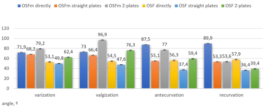
OSFm with fixation of the struts directly to the rings exceeds OSF by 18.8º in terms of the possibilities of creating (and correcting) varus deformity and with valgization by 19.5º. OSFm outperforms OSF with varization by 19.2º and with valgization by 18.1º using straight plates. OSFm is better at 17.4º with varization and 20.1º with valgization using Z-plates. OSFm with fixation of struts directly to the rings exceeds OSF with antecurvation by 17.3º and with recurvation by 17.4º. OSFm outperforms OSF with antecurvation by 18.4º and with recurvation by 17.9º using straight plates. OSFm provides better result with antecurvation by 18.7º, with recurvation OSF by 16.9º using Z-shaped plates (Figure 4).
Figure 4. Diagrams of the Maximum Possible Values of the Angular Movement of the Mobile Bone Fragment
DISCUSSION
Two minimalized modifications of orthopedic hexapods are known: Small Bone System and OSFm. The first one was created based on the Orthex-frame and differs in the reduced size of the elements from its analogue Orthex-frame, Large Bone System.9 Orthex-frame demonstrated clinical efficacy in correcting deformities in children and outperformed the Taylor Spatial Frame (TSF).10 There have been no targeted comparative studies of the reduction capabilities of the minimized version of this hexapod.
The results of the study of the reduction capabilities of OSF and its minimized version, OSFm, demonstrated significant differences for all methods of fixing struts to rings. OSFm outperforms OSF in all types of plane-parallel displacements because the maximum possible OSFm struts length is 12 mm longer than the maximum possible OSF struts length. Likewise, OSFm outperforms OSF in all types of angular misalignment because the minimum possible length of the OSFm struts is less than the minimum possible length of the OSF struts by 12 mm.
In translation OSFm exceeds OSF by 2.8-9.5% fixed the struts directly to the rings. OSFm outperforms OSF by 4.8-9.7% using straight plates, and by 27.3-29.3% using Z-plates. In angulation OSFm with struts fixed directly to the rings exceeds OSF by 33.9-55.4%. Accordingly, OSFm outperforms OSF by 36.9-47.3% using straight plates, and by 29.6-36% using Z-plates.
This study was not intended to determine the maximum possible translation and angulation that could be provided by OSFm and OSF. Each indicator from the study can be increased by changing the distance between the base and mobile rings, the angle of their inclination, changing the struts attachment positions, a combination of straight and Z-shaped plates, as well as using additional “dummy” rings.4,11,12
CONCLUSION
The data obtained indicate that OSFm, in comparison with OSF, has better translation and angulation capabilities. Further research will be related to the study provided by OSFm distraction, rotation possibilities, the bone fragments fixation rigidity, as well as the development of optimal OSFm assemblies for long bones, large joints, and foot deformity correction.
AUTHORS CONTRIBUTIONS
(1) Conception and design: L.N. Solomin; (2) Administrative support: L.N. Solomin; (3) Provision of study materials or patients: D.V. Gavrilov, M.A. Baushev, V.A. Vilensky; (4) Collection and assembly of data: D.V. Gavrilov; (5) Data analysis and interpretation: D.V. Gavrilov; L.N. Solomin (6) Manuscript writing: D.V. Gavrilov; L.N. Solomin; (7) Final approval of manuscript: All authors.
CONFLICTS OF INTEREST
The authors declare that they have no conflicts of interest.


