Osteology and Rheumatology

Open journal

ISSN 2996-5918

A Rare Cause of Shoulder Pain: Ganglion Cyst of the Acromioclavicular Joint

Hakan Sarman*, Metin Celik and Mehmet Murat Bala

Received: August 11th, 2016 Accepted: September 2nd, 2016 Published: September 6th, 2016

INTRODUCTION

A ganglion cyst (GC) may have an acute or chronic onset and is generally related to repeated microtrauma of the wrist dorsum.1,2,3,4 The differential diagnosis of shoulder pain and functional disorders includes shoulder compression syndromes (impingement), glenohumeral instability, rotator cuff diseases, tendonitis, adhesive capsulitis, trauma, cervical radiculopathies, neoplasms, degenerative diseases, acromioclavicular (AC) joint separation, arthritic variations, crystal arthropathies (including Milwaukee’s shoulder) and atypical emplacement of a GC.1,2,5,6,7,8 This paper reports a patient with shoulder pain due to a GC in the AC joint who presented to our clinic along with the post-operative evaluation.

CASE REPORT

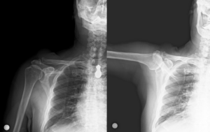
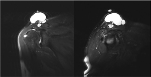
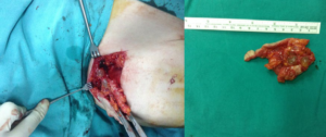
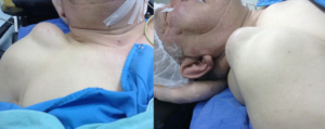
A 65-year-old man presented to our clinic complaining of a slow-growing swelling and right shoulder pain that began one year previously. Physical examination showed a 10×8×5 cm soft, slightly painful mass above the right AC joint, slightly limiting shoulder range of motion (ROM) (Figure 1). A direct x-ray showed arthritic changes in the right AC joint (Figure 2). Magnetic resonance imaging (MRI) showed a 10×8×5 cm mass related to the right AC joint, that was tentatively diagnosed as a GC, and partial rupture of the supraspinatus muscle (Figure 3). The patient’s complaint persisted despite three months of physical therapy and surgery was planned. Under general anesthesia in the chaise longue position, the mass on the right AC joint was entered with a fish-mouth incision (Figure 4). The mass was observed to be connected to the AC joint and an excision biopsy was performed. Histopathology showed that the mass was consistent with GC. There was no recurrence at the 1.5-year follow-up. His pain resolved and ROM was normal despite partial rupture of the supraspinatus.

Figure 1: Clinical demonstration of patient.

ORHOJ-1-107Fig1


Figure 2
: Radiography imaging of patient.

ORHOJ-1-107Fig2


Figure 3
: Magnetic resonance imaging of patient.

ORHOJ-1-107Fig3


Figure 4
: Operation macroscopic imaging.

ORHOJ-1-107Fig4

DISCUSSION

Ganglion cysts occur most commonly at the hand and wrist and are only very rarely observed in the shoulder region and bone.2,3,6,7 The incidence is greatest in women 30-50 years of age. The etiology of GC is not fully known, but joint pathology and microtrauma are thought to contribute.8,9 Our patient was a laborer who used his arm very frequently and often experienced minor trauma. He presented to our clinic complaining of shoulder pain and swelling.

The most widely accepted theory for the pathogenesis of a GC in the AC joint is fluid spreading around the joint in the rotator cuff in conjunction with labrum tears.2,6,10,11,12 Schroder et al13 and Youmet al14 reported that treating the labrum pathology led to regression of the cyst without surgical intervention. Tung et al15 investigated the causes of shoulder pain and identified a paralabral GC visible on MRI imaging in 2.3% of their cases. In our case, an excision biopsy was performed, the shoulder pain resolved, and the shoulder ROM improved.

After total excision, a GC may recur in the same location, especially in cases where the joint capsule in the pedicle isnot sutured with appropriate tension.3,16,17,18 In our case, the GC was excised using an open approach and the mass was observed to be connected to the AC joint. At the 1.5-year follow-up, the patient’s pain was improved and the shoulder ROM had increased. There was no recurrence of the AC joint cyst.

CONCLUSION

Although GCs are rarely observed in the shoulder region, they may cause pain and restrict ROM. Surgical excision may improve symptoms and function with very low rate of recurrence.

CONFLICTS OF INTEREST

The authors declare that they have no conflicts of interest.

CONSENT

The patient has provided written permission for the publication of this case details.

1. Montet X, Zamorani-Bianchi MP, Mehdizade A, Martinoli C, Bianchi S. Intramuscular ganglion arising from the acromioclavicular joint. Clin Imaging. 2004; 28(2): 109-112. doi: 10.1016/s0899-7071(03)00104-9

2. Skedros JG, Knight AN. Massive acromioclavicular ganglionic cyst treated with excision and allograft patch of acromioclavicular region. J Shoulder Elbow Surg. 2012; 21(3): 1-5. doi: 10.1016/j.jse.2011.07.033

3. Sarman H, Işık C, Şahin AA, et al. El ve El bilek tümörlü hastalarda eksizyonel biyopsi sonuçlarinin değerlendirilmesi [In Turkish]. Abant Med J. 2014; 3(1): 55-61. doi: 10.5505/abantmedj.2014.30602

4. Orman G, Yesiladali G, Olcay E, Duymus M. Comparison of ultrasonography and magnetic resonance imaging for diagnosis of soft tissue masses of the hand and wrist. Eur J Gen Med. 2015; 12(1): 38-43. doi: 10.15197/sabad.1.12.07

5. Ferrick MR, Marzo JM. Ganglion cyst of the shoulder associated with a glenoid labral tear and symptomatic glenohumeral instability. A case report. Am J Sports Med. 1997; 25(5): 717-719. doi: 10.1177/036354659702500523

6. Good LM, DiCarlo JB, High WA. An unusual cutaneous manifestation of a ganglion cyst. J Am Acad Dermatol. 2011; 64(6): 1206-1208. doi: 10.1016/j.jaad.2009.09.034

7. Parperis K, Carrera G, Baynes K, et al. The prevalence of chondrocalcinosis (CC) of the acromioclavicular (AC) joint on chest radiographs and correlation with calcium pyrophosphate dihydrate (CPPD) crystal deposition disease. Clin Rheumatol. 2013; 32(9): 1383-1386. doi: 10.1007/s10067-013-2255-x

8. Genta MS, Gabay C. Images in clinical medicine. Milwaukee shoulder. N Engl J Med. 2006; 354(2): e2. doi: 10.1056/NEJMicm050094

9. Matev B, Georgiev GP, Stokov L. A rare case of intraosseous ganglion of the triquetrum. J Clin Exp Invest. 2012; 3(1): 111-112. doi: 10.5799/ahinjs.01.2012.01.0124

10. Haber LH, Waanders NA, Thompson GH, Petersilge C, Ballock RT. Sternoclavicular joint ganglion cysts in young children. J Pediatr Orthop. 2002; 22(4): 544-547. Website. http://journals.lww.com/pedorthopaedics/Abstract/2002/07000/Sternoclavicular_Joint_Ganglion_Cysts_In_Young.24.aspx. Accessed
August 10, 2016

11. Deutsch A, Altchek DW, Veltri DM, Potter HG, Warren RF. Traumatic tears of the subscapularis tendon. Clinical diagnosis, magnetic resonance imaging findings, and operative treatment. Am J Sports Med. 1997; 25(1): 13-22. doi: 10.1177/036354659702500104

12. Kessler MA, Stoffel K, Oswald A, Stutz G, Gaechter A. The SLAP lesion as a reason for glenolabral cysts: A report of five cases and review of the literature. Arch Orthop Trauma Surg. 2007; 127(4): 287-292. doi: 10.1007/s00402-006-0154-1

13. Tirman PF, Feller JF, Janzen DL, Peterfy CG, Bergman AG. Association of glenoid labral cysts with labral tears and glenohumeral instability: Radiologic findings and clinical significance. Radiology. 1994; 190(3): 653-658. doi: 10.1148/radiology.190.3.8115605

14. Westerheide KJ, Dopirak RM, Karzel RP, Snyder SJ. Suprascapular nerve palsy secondary to spinoglenoid cysts: Results of arthroscopic treatment. Arthroscopy. 2006; 22(7): 721-727. doi: 10.1016/j.arthro.2006.03.019

15. Schroder CP, Skare O, Stiris M, Gjengedal E, Uppheim G, Brox JI. Treatment of labral tears with associated spinoglenoid cysts without cyst decompression. J Bone Joint Surg Am. 2008; 90(3): 523-530. doi: 10.2106/jbjs.f.01534

16. Youm T, Matthews PV, El Attrache NS. Treatment of patients with spinoglenoid cysts associated with superior labral tears without cyst aspiration, debridement, or excision. Arthroscopy. 2006; 22(5): 548-552. doi: 10.1016/j.arthro.2005.12.060

17. Tung GA, Entzian D, Stern JB, Green A. MR imaging and MR arthrography of paraglenoid labral cysts. AJR Am J Roentgenol. 2000; 174(6): 1707-1715. doi: 10.2214/ajr.174.6.1741707

18. Satku K, Ganesh B. Ganglia in children. J Pediatr Orthop. 1985; 5(1): 13-15. doi: 10.1097/01241398-198501000-00003

LATEST ARTICLES

Use of Black Soldier Fly (Hermetia illucens) Prepupae Reared on Organic Waste

review

2024 Mar

Isayas A. Kebede*, Haben F. Gebremeskel and Gelan D. Dahesa,
Figure 11. Risk Map for the Introduction of Ruminant Diseases at Borders

Original Research, peer reviewed

2024 Mar

Yassir Lezaar*, Mehdi Boumalik, Youssef Lhor, Moha El-Ayachi, Abelilah Araba and Mohammed Bouslikhane

Commentary, End-of-Life, Family Dynamics, palliative Care, peer reviewed

2024 Mar

Neil A. Nijhawan*, Rasha Mustafa and Aqeela Sheikh

Retrospective Study

2024 Mar

Shrenik J. Shah*, Abhishek Jha, Chirag Davara, Rushi Mistry and Kapil Kachhadiya

Attention, Disaster, Mindfulness, Original Research, peer reviewed

2024 Mar

Christina Hagen*, Lars Lien, Edvard Hauff and Trond Heir

review

2024 Mar

Isayas A. Kebede* and Gelan D. Dahesa

review

2024 Feb

Bekiyad S. Daro*; Yared A. Deneke, Jiregna G. Negasa and Ishetu N. Dibaba