INTRODUCTION
Dystocia is a condition in which parturition is delayed or difficult. It’s the dam’s inability to give birth to her young on her own.1Dystocia is caused by congenital or acquired abnormalities of the pelvis, cervix, vagina, or vulva. Pelvic abnormalities, vulvar or vaginal stenosis, neoplasms of the vagina and vulva, vaginal cystocele, inadequate cervical dilatation, uterine torsion, and ventral displacement of the uterus can all cause constriction/obstruction of the birth canal.2 Small pelvis, exostoses or pelvic deformities, pelvic fracture, osteomalacia, and hypoplasia of the vagina and vulva are some of common pelvic abnormalities that can cause dystocia.2,3,4 Hereditary reasons of pelvic defects, such as tiny size and insufficient pelvic ligament development, or small breeds with outsized fetuses, are other causes of dystocia due to a narrow or defective pelvis. The presence of a small bony pelvis was identified as a symptom of sacral luxation or displacement.3,5 added to the genetic cause of a small-size pelvis.5 The rate of occurrence of dystocia is closely related to the maturity of the dam. It is widely accepted that heifers calving at roughly two years of age have difficulty delivering the calf, even with good feeding and treatment. Often, intervention is required to save the life of the calf and/or cow. When a heifer can give birth to a calf without assistance, however, it is common to notice that labor lasts longer than in older cows.6 Early heifer breeding, breeding of underdeveloped heifers, and breeding of heifers and calves with pelvic fractures can result in a smaller mother’s pelvis, resulting in dystocia at parturition.3 The dimensions of bony pelvis are too small to allow passage of the fetus.7 Feto-maternal dystocia is caused by circumstances such as feto-pelvic disproportion, in which the foetus is either too large to travel through the birth canal or the dam’s pelvis is too narrow to allow easy foetal ejection after parturition.8In such dystocia, parturition may not proceed over first stage, as the chances of calf being struck in the pelvic inlet are more. Surgical intervention is the only way to opt for delivering the calf, as forced traction may endanger life of both dam and calf.4,9 There are various reasons for performing a caesarean section (C-Section), including maternal and fetal reasons. Immature heifers, pelvic abnormalities, failure of cervical dilation, uncorrectable uterine torsion, uterine tear, and prepartum paralysis are among the maternal indicators. When compared to multiparous cows, the risk factors in cattle are raised when the heifer is less than two years old.10 A case of dystocia due to narrow pelvis associated with immaturity in heifer and its successful surgical management is presented in this report.
CASE HISTORY AND FIELD EXAMINATION
On Friday, September 17th, 2021 at midday, the farmer was called to Bedeno Veterinary Clinic and complained that his two-year-old (2-years-old) heifer, with full term of pregnancy, had been unable to deliver a fetus since yesterday mid-night. The owner said that he and other locals attempted unsuccessfully to birth the fetus. The vets traveled by a motorcycle and arrived at the scene. The vets continued to examine the cow and upon field examination, the rectal temperature was 38.80 °C, and tachycardia (130 beats per minute) and dyspnea (16 breaths per minute) were present. There was a rupture of water bag, swelling of vulva, absence of straining, abdominal distension. The heifer was recumbent and unable to stand. After the external genitalia was washed with water and soap solution, then cleaned with savlon (Cetrimide 3%+chlorohexidine gluconate 0.3%), solution (by disolving 250 ml of Savlon with 1000 ml of warm water) and sugar solution (500 grams of sugar disolved with 250 ml water) was made and added to the vulva to remove water content from swollen vulva to ease manual manipulation. Here, sugar is not a standard treament for dystocia but the vets were used in reduction of water from swollen vulva. Accordingly, the water content was drained and the swollen vulvar size was reduced. After the vulvar edema was reduced and lubricated with 100 ml of paraffin oil, per-vaginal delivery was attempted but failed because the maternal bony pelvis was very narrow. Locals damaged the calf ’s lower mussle while attempting to remove the fetus before the veterinarian team arrived. Even fetotomical birth was impossible because the recognized or palpable or reachable part of the fetus was only the fetal mussle. Only C-Section was left as a final resort to save the heifer. As there was no operation room built in the field, the place (floor) where the cow had been recumbent (just nearby the cow) was covered by polyvenyl plastic material (4 m×5 m) about the area of 20 m2 and with the help of many assistances, the heifer was moved on the plastic. Additinally, tent was made over the recumbent cow from the same type of material with the same area coverage. So, by doing this the surgical procedure was conducted under aseptic nature.
SURGICAL MANAGEMENT
The surgical site was prepared aseptically on the left flank region after the cow was secured by tying legs front and back and placed on its right lateral recumbency. Then, using an inverted L-block approach, 30 ml of 2% lidocaine hydrochloride (2% Lignocaine 20 mg/ml, Scott-Edil pharmaceutical Cot. Ltd, India) was injected subcutaneously to generate a wall of analgesia encircling the operative field. Here, the vets do not want to give full dose of lignocaine beacause of the increased heart rate and decreased respiration rate. This may cause unwanted negative effect on compromised animal. A surgical blade was used to make a 45 cm long vertical incision on the skin. To reach the uterus, the dissection continued through subcutaneous tissues, all abdominal muscle layers, and the peritoneum. Subsequent to incision of uterus, the dead fetus was disconnected from the intact umbilical cord and taken from the womb (Figure 1A). The fetal membrane with the intact umbilical cord were left behind in the uterus. the The uterus was dusted with procaine penicillin G powder (Fortified Procaine Pencilline 400,000 I.U., CSPC Zhangnuo Pharmceutical (Shijiazhuang Co., Ltd, Hebei Province, China) and sutured in two layers using absorbable catgut size 2 (6 m) (Shandong Sinorgmed International Co., Ltd, China) in an inversion (Lambert) pattern. The peritoneum was sutured with absorbable catgut sutures in a basic continuous suture pattern (2-0). Interlock pattern with chromic catgut size (3-0) was used to seal the abdominal wall (Assault suture, Switzerland). Finally, using nylon size (2-0) the skin was closed using a simple interrupted suture procedure (Figure 1B). Antibiotics injection (Penstrip 400, a combination of Procaine penicillin G 200,000 IU and dihydrostreptomycin sulphate 200 mg at a dose of 1 ml/20 kgs (15 ml/ 300 kgs body weight, IM), Interchemie Werken, Holland product, Western Australia, Australia) for 5 consecutive days, oxytetracycline aerosol wound spray (oxy voicid 3.58%, provet, Ankara, Turkey) was sprayed on surgical wound of operated heifer once a day until complete healing, along with multivitamin (Introvit, dose; 10-15 ml for cattle, Interchemie Werken, Holland product, Western Australia, Australia) injection were all used as post-operative managements. After 5-hours of operation, the fetal membrane was expelled through birth canal spontaneously. The skin sutures were removed on the 20th day after the operation, the wound had healed and was quite active (Figure 1C). The heifer recovered completely without any post-operative difficulties and now looks like the one in the picture (Figure 1D). The owner was advised to closely monitor the cow and giving good nutrition and bedding (cleaning of cow pen and making comfortable) to facilitate wound healing. The day of operation was too cold. So, the owner was also advised to provide charcoal heat/wood fire in the cow pen so as to resist the cold weather.
Figure 1A. Dead Fetus Taken Out from Womb
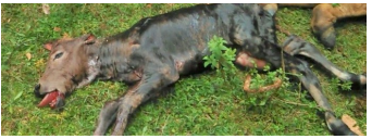
Figure 1B. Simple Interrupted Skin Suture
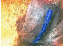
Figure 1C. The Healed Wound on 20th Day
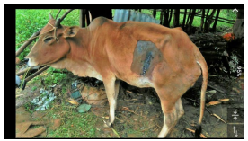
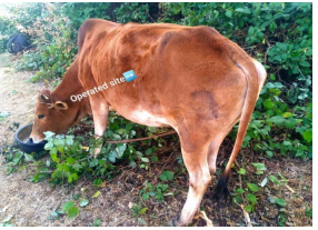
Figure 1D. Completely Healed Heifer (2-Years-Old)
DISCUSSION
Dystocia was caused in this case by a constricted pelvis as a result of the heifer’s immaturity, which may have been avoided with adequate reproductive care. Heifers are more susceptible to dystocia than cows because they are smaller. The heifer was about two-years-old (its pelvic bone had not fully formed) and was housed with the rest of the cattle herd. This resulted in early mating, which made calving difficult. The current case report adhered to the following guidelines11: in that the size of heifers at breeding should attained the average size of mature cow weight, with a minimum of 60%. Otherwise, it is pre-disposed to calving difficulties. This case report was communicated with12 Primiparous cows had a smaller pelvic area, lower live weight, and more difficulties calving, according to the study. Manual delivery was impossible due to the narrowness of the maternal bony pelvis (pelvic cavity). This case had a lot in common with3 who reported that, if the birth canal is too narrow, it is advised to optics for C-Section. Thus, surgical management of the case was the last option. After aseptic preparation of the animal, local analgesic agent is deposited in the form of an inverted L to create a wall of analgesia enclosing the surgical field. This presentation was agreed with Hendrickson13 in the usage of local analgesic. Accordingly, the case was managed surgically while the heifer was on right lateral recumbency. This case study was related with a report of Kolkman14 and Masterson15 who described that recumbent left paralumbar celiotomy approach in a recumbent animal is most suitable in case of cow that is not able to stand for the C-Section throughout the procedure. Following subcutaneous administration of analgesia, surgical intervention was made via left oblique paralumbar fossa. After removal of the fetus, fetal membranes were not loosed, so returned back into the uterus. The uterus was covered with procaine penicillin G and sutured in two layers using absorbable catgut size 2 in an inversion (Lambert) pattern. Abdominal wall was closed using interlock pattern with chromic catgut size (3-0) followed by subcutaneous suturing by chromic catgut (1-0) and Oxytetracycline wound spray smeared on. Finally, the skin was closed routinely. As described by Fesseha et al16 for early recovery and positive outcomes of the surgical procedure, the wound should always be managed and regularly monitored for the fast healing process. Hence, post-operative care was followedup. As result the animal was recovered completely on 20th days of operation. Here, the main goal of C-Section was to save the heifer only because the fetus was already died. In conclusion, mating of immature animals is one of a cause of dystocia. Surgical management (C-Section) is a live saving obstetric method if it is done early. So, it recommended that mating of immature animals should be avoided and every dystocia case should be presented to the veterinarian for early interventions.
CONCLUSION
A 2-year-old heifer with a full-term pregnancy was presented with dystocia in the current case report. The altered vital organs indicators were tachycardia and dyspnea, according to a field examination. However, the temperature of rectum was within the biological range. There was also a ruptured water bag, no straining, vulva edema, and abdominal distension. The case was identified as dystocia due to a constriction of birth canal linked to the dam’s immaturity based on the history and vaginal inspection. After the failure of vaginal birth, C-Section was performed using normal techniques, with aseptic procedures and post-operative care being thoroughly monitored. As a result, the cow was fully recovered. The owner was advised not to mate or inseminate juvenile animals or those who have a pelvis that is either tiny or too narrow. Local attempts should be ignored and it is preferable to consult a veterinarian as soon as possible once it occurs.
ACKNOWLEDGEMENTS
The authors are grateful to Bedeno Woreda Agricultural and Livestock Resource Development Office, Directorate of Animal Health for providing us with computer access and overseeing the work, Bedeno Woreda Veterinary Clinic for providing us with surgical materials and a motorcycle for the success of the work, and our colleague Abdela Abrahim Jibro for arranging surgical materials and capturing all photographs used in this communication belong to the authors while they were performing the surgery, as well as the owner and his entire neighborhood for their help in handling the animal during the treatment.
ETHICAL APPROVAL
The study was carried out under the tight supervision of the Directorate of Animal Health of the Bedeno Agricultural and Livestock Development Office, which concluded that the case report was ethically done and met all animal ethics and welfare criteria. The ethical authorization letter was also delivered to the office for your confirmation.
CONSENT STATEMENT
The authors confirmed that the family member of the animal’s owner was photographed in the case report. The involvement of the owner’s family was limited to handling their animal, and their written and signed agreement was obtained. As a result, we declared that their participation has no bearing on the publication.
CONFLICTS OF INTEREST
It was confirmed that the authors have no potential competing interests in terms of authorship and/or publication of this case report.


