Overview
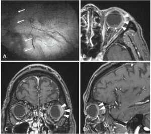
A 74-year-old man visited to our hospital due to severe visual disturbance in his left eye for recent one month. Fundoscopy demonstrated the serous retinal detachment at the nose side of the left eye (Figure 1A, arrows).
Figure 1. Fundoscopy Demonstrated the Serous Retinal Detachment at the Nose Side of the Left Eye (Fig. 1A, Arrows). The MRI on Axial, Coronal, and Sagittal T1 Weighted Images Demonstrated the Enhanced Crescent Shaped Tumor (Fig. 1B,1C,1D, Arrow Heads) with Non-Enhanced Soft Tissue (Fig. 1B, 1D, Arrows), Suggesting Retinal Detachment and Choroidal Metastasis, Respectively.

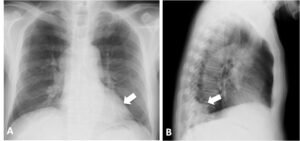
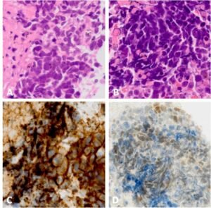
At the same time, chest X-ray demonstrated a mass measuring 4 cm in size at the left lower lung field (Figure 2, arrow) and thoracic computed tomography (CT) depicted the mediastinal lymphadenopathies. On Hematoxylin-Eosin stain, both specimens obtained from right B10 by transbronchial lung biopsy (Figure 3A, 400X) and subcarinal lymphnodes (Figure 3B, 400X) by endobronchial ultrasound-guided transbronchial needle aspiration demonstrated abundant atypical cells with high nuclear cytoplasmic ratio. Those cells were positive for both CD56 (Figure 3C, 400X) and TTF-1 (Figure 3D, 400X), he was thus diagnosed with small cell lung carcinoma (SCLC).
Figure 2. Chest X-Ray Demonstrated the Mass as Large as 4 cm at the Left Lower Lung Field (Fig. 2, Arrow).

Figure 3. On Hematoxylin-Eosin Stain, Biopsied Specimens Obtained from Right B10 (Fig. 3A, 400X) and Subcarinal Lymphnodes (Fig, 3B, 400X) Revealed Abundant Atypical Cells with High Nuclear Cytoplasmic Ratio. Those Cells were Positive for Both CD56 (Fig 3C, 400X) and TTF-1 (Fig. 3D, 400X), Suggesting Small Cell Lung Carcinoma.

For further exploring the reason for loss of visual acuity, contrast enhanced head magnetic resonance imaging (MRI) was performed. The MRI on axial, coronal, and sagittal T1 weighted images demonstrated the enhanced crescent shaped tumor (Figure 1B, 1C, 1D, arrow heads) with non-enhanced soft tissue (Figure 1B, 1D, arrows), which corresponded to retinal detachment and choroidal metastasis, respectively. Previous studies described that the incidence of ocular metastasis in patients with lung carcinoma ranged from 61 to 21%2 in post or antemortem examination. Choroid is the blood-rich tissue where the most preferable area in ocular metastasis. Physicians should be aware of the choroidal metastasis as the initial manifestation of lung carcinoma.3,4


