INTRODUCTION
Duplex collecting system is described as the kidney having two pyelocaliceal systems, superior and inferior. It may have either single, bifid ureter (partial or incomplete duplication) or double ureter draining separately into the bladder (complete duplication).
CLINICAL OBSERVATION
We report the case of a 60-years-old woman, with a history of diabetes mellitus, who presented with a slight lower back pain without other urinary signes. The clinical examination found no abnormalities. The kidney function was normal and the urine culture was negative.
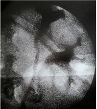
A computed tomography (CT) urogram was performed and it set the diagnosis by showing a bilateral incomplete duplex collecting systems with bifid ureters. It also revealed a modest hydronephrosis in inferiors pyelocaliceal systems without any obstruction (Figure 1). However, we did not find any vesicoureteral reflux at cystography. A cystoscopy was performed revealing two ureteral meatus without bladder abnormalities. Retrograde ureteropyelography confirmed the absence of obstruction on the excretory tracts (Figures 2 and 3). The therapeutic strategy was surgical abstention with a closer clinical follow because of the paucisymptomatic aspect of the clinical picture.
Figure 1. 3D Reconstruction of CT Scan Revealing a Slight Hydronephrosis with Bilateral Duplex Collecting System and Bifidureters
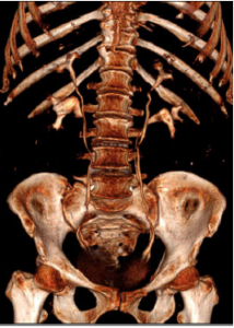
Figure 2. Distal Picture of Retrogradeuretero-Pyelography Showing the Convergence of the Ureters (Y-Junction) at 4 cm from the Ureterovesical Junction
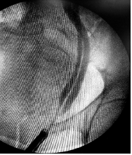
Figure 3. Proximal View of Retrogradeuretero-pyelography Showing Superior and Inferior Pyelocaliceal Systems
DISCUSSION
Renal collecting system duplication is one of the common congenital abnormalities of the urinary tract.1
In the upper part, there are two ureters while lower down the ureters join to form a single ureter. This joining may be intravesical (V-junction) or extravesical (Y-junction).2
Duplication in adults is usually clinically asymptomatic and it is often a chance finding during a radiological examination.3
Ultrasound (US) will reveal evidence of upper tract damage. It may also show the presence of a ureterocoele and an asymmetric dilation of the pyelocalicielles cavities, either of the upper pole (by obstruction) or of the lower pole (most often secondary to the reflux). Visualization of both ureters is difficult at ultrasound in the absence of dilatation. Sectional imaging after injection of contrast makes it possible to visualize them at best. Cystography may demonstrate vesicoureteral reflux, which is common in the lower system with a typical amputation of the upper calyx.4,5
Cystoscopy define the number of ureteral meatus as well as their anatomical situation. It also makes it possible to perform a retrograde ureteropyelographia which will confirm the diagnosis of the double system with bifid ureter and will look for a possible obstacle.
CONCLUSION
The duplication of the upper urinary tract is usually asymptomatic thus requiring no treatment. However, patients should be monitored closely because of the risk of complications such as infection, obstruction or vesicoureteral reflux.
CONSENT
The authors have received written informed consent from the patient.
CONFLICTS OF INTEREST
The authors declare that they have no conflicts of interest.


