Pancreas

Open journal

ISSN 2471-142X

Desmoplastic Reaction in Pancreatic Ductal Adenocarcinoma

Ughur Aghamaliyev*, Yegana Hajiyeva and Felix Rückert

Ughur Aghamaliyev, MD

Department of Surgery Medical Faculty Mannheim University of Heidelberg Theodor-Kutzer-Ufer 1-3 68167 Mannheim, Germany; Tel. +49 1798530153; E-mail: dr.aghamaliyev@gmail.com

INTRODUCTION

Pancreatic Ductal Adenocarcinoma (PDAC) is the fourth leading cause of cancer related death in the USA and Europe. Despite substantial progress of understanding the molecular biology of PDAC, the prognosis remains still poor with a combined overall 5-year-survival rate of less than 7%.1 This high death rate is due to late diagnosis with no effective screening tools for detection of early tumors and the lack of curative treatment methods.2 Accumulating evidence suggests that the aggressive phenotype of PDAC is not only due to epithelial cancer cells, but also to the stromal compartment in which cancer cells are embedded.3,4

The dense desmoplastic reaction is one of the histological hallmarks of PDAC which can often constitute 50-80% of the tumor volume and is comprised of abundant extracellular matrix (ECM), fibroblasts, stellate cells, immune cells, nerve cells, growth factors and cytokines.5 The ECM itself is composed of structural proteins, enzymes and proteins involved in cell communication.5

ACELLULAR COMPONENTS OF THE STROMAL COMPARTMENT

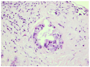
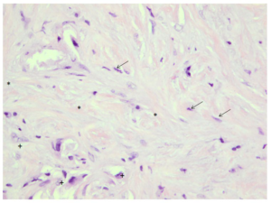
As mentioned above, one of the most important features of PDAC is the development of the desmoplastic reaction around tumor cells which is mainly due to excess ECM production produced by Pancreatic Stellate Cells (PSCs).6 The ECM is composed of variety of fibrous proteins (i.e., collagens), glycoproteins (i.e., fibronectin), proteinases (i.e., matrix metalloproteinase 9) and glycosaminoglycans (i.e., hyaluron).6 Additionally, modulators of the cell matrix interaction such as periostin, tenascin C, SPARC and thrombospondin are found in the PDAC desmoplastic reaction as well.7 (Figures 1A and 1B)

Figure 1: H/E Staining of pancreatic cancer with desmoplastic reaction (1A) and tumor cells (1B) (H&E, 400 ×). Pictures from Prof. Dr. med. Timo Gaiser, Pathology Institute, Medical Faculty Mannheim, University of Heidelberg, Germany.

Figure 1A: Distinct desomplastic reaction in pancreatic cancer (*=Desmoplastic reaction; =Pancreatic stellate cells; +=Single tumor cells) (H&E, 400x).

POJ-1-107Fig1A


Figure 1B
: Picture of pancreatic cancer with stromal reaction (+=Tumor cells) (H&E, 400x).

POJ-1-107Fig1B

One of the predominant proteins within the ECM is type I collagen which has been shown to contribute to the generation of the malignant phenotype.8 Specifically, it was demonstrated in vitro that type I collagen promotes cell adhesion, proliferation and maximal hypokinetic cell migration of cancer cell lines.8 Moreover, 3 dimensional collagen I has been reported to contribute to gemcitabine resistance through MT1-MMP-mediated expression of HMGA2 in pancreatic cancer.9

Increased resistance towards anti-cancer drugs can also be achieved by attachment of PDAC cells to collagen I, IV and fibronectin. This decreases cytotoxicity of anti-cancer drugs such as 5-fluorouracil, cisplatin and doxorubicin. The exact molecular mechanism, however, must still be determined.10

Contact to collagen I was also associated with higher expression of N-Cadherin and activation of c-Jun-NH-2-terminal-kinase-1 in cancer cells, indicating its contribution to metastasis.11 Other studies also showed that collagen I modulates transcription factors of epithelial to mesenchymal transition (EMT), namely Snail and E-Cadherin. This results in increased invasiveness of PDAC cells.12,13

Other ECM proteins believed to participate in tumor cell invasion and metastasis are matrix metalloproteinases (MMPs).14,15 MMPs are a group of zinc-dependent endopeptidases with different substrate specificities.16 Gelatinase subgroups of MMPs that include MMP-2 and MMP-9 are known to degrade type IV collagen. By this, they seem to promote cancer invasion.17 Strong expression of MMP-2 has been observed in the stromal compartment of PDAC.18 A high level of MMP-2 correlated with poor prognosis in pancreatic cancer patients.19 Additionally, suppression of MMP-2 via RNA interference decreased PDAC cell invasiveness and adhesion.20 It is noteworthy that overexpression of MMP-7, another important member of MMP family is involved in subsequent invasion of PDAC cells and PDAC metastasis.21,22 It was shown that MMP-7 can induce cell dissociation and subsequent invasion of cancer cells in pancreatic cancer through activating EGFR mediated MEK-ERK signaling.21

Furthermore Hyaluronic acid (HA), a matrix glycosaminoglycan, is known to be synthesized and secreted by several PDAC cell lines. It is also presented in huge amounts in pancreatic cancer stroma.23 Moreover, enzymatic degradation of HAlet to increased survival in a mouse model of PDAC.24 Therefore HA might also be an interesting therapeutic target in pancreatic cancer.

Recently, periostin, an osteoblast-specific secretory protein has been suggested to promote invasiveness and resistance of PDAC cells.25 This protein is produced by stromal cells rather than PDAC cells.26 Although periostin suppressed the malignant phenotype of tumor cells at low concentrations, it increases migration when expressed at high levels.26

Tenascin-C (TN-C) is an ECM glycoprotein synthesized by PSCs and strongly expressed in pancreatic cancer.27 Although its expression is low in normal adult tissue, the protein level rises dramatically under various physiological and pathological conditions, such as tissue remodeling, neovascularization and inflammation.28 It has also been reported that TNC expression correlates with pancreatic cancer cell differentiation.29 Taking into account that TNC promotes proliferation, migration and adhesion of poorly differentiated pancreatic cancer cell lines, it might play a role in PDAC infiltration and metastasis in vivo.30

Another important ECM glycoprotein, SPARC, is produced by both cancer cells and stromal cells and was shown to increase invasiveness of PDAC cells lines. It also correlates with patient survival. Therefore it seems to have an important pathophysiological role.31,32

One of the most notable extracellular macromolecules involved in PDAC invasion is thrombospondin-1 (TSP-1) which is mainly produced by stromal cells.33,34 PDAC cells might contribute to TSP-1 secretion in relatively low amount. Its expression in cells can be regulated by Transforming growth factorβ1(TGF-β1).34 It was reported that TSP-1 modulates PDAC cell invasiveness by upregulating MMP-1 and TIMP-1.34 Therefore, it would be promising approach to further elucidate contribution of TSP-1 to malignant phenotype of pancreatic cancer.

CELLULAR COMPARTMENTS OF TUMOR STROMA

Fibroblasts

The most prominent cells of the stromal compartment of PDAC are activated fibroblasts, also known as Pancreatic Stellate Cells (PSCs).6 PSCs are also part of the physiological stroma of the pancreas.6 Their homeostatic role is still poorly understood, however they have been shown to contain fat droplets in their cytoplasm; indicating potential role in lipid metabolism.35 In PDAC these cells become activated, obtain myofibroblastic phenotype, express α-smooth muscle actin (SMA) and gain the capacity to produce ECM components collagen I, III and fibronectin in significant amounts.36 These phenotypic changes are induced by several cytokines and growth factors such as TGF-β1, activin A, IL-1, IL-6, platelet-derived growth factor (PDGF) and VEGF.6 In addition, mitogenic factors like PDGF induce cell proliferation of PSCs. On the other hand, fibrogenic mediators like TGF-β1 and connective tissue growth factor (CTGF) induce the synthesis of collagens and fibronectin in activated PSCs.37

It was shown that direct co-culture of PSCs and PDAC cells promote PDAC cell proliferation through activation of Notch signaling pathway.38 Moreover, co-injection of PSCs with PDAC cells has been shown to promote metastasis and angiogenesis in a mouse model of PDAC.39 It was observed that palpable tumors developed earlier in mice injected with PDAC cell lines and PSCs compared to mice injected with cancer cell lines alone. In addition, tumors in mice injected with PDAC cell lines and PSCs together became significantly larger than those in controls.39 But not only cancer growth is affected by PSCs. PSCs produce many factors such as collagen I, laminin, and fibronectin that are thought to promote acquired resistance to anticancer drugs such as 5-fluorouracil, cisplatin and doxorubicin. Therefore PSCs might also take part in chemotherapy resistance.10

Immune cells

It is now widely accepted that PDAC tissue is infiltrated with immune cells, such as T-cells, B-cells, NK cells, neutrophils, and macrophages as well as myeloid-derived suppressor cells (MDSCs).40,41 Higher levels of CD8 positive T-cell infiltration have been shown to correlate with a better survival,40,41  while macrophage and neutrophil infiltration as well as high levels of MDSCs have been reported to be associated with poor survival.40 The infiltration of MDSCs in PDAC tissue leads to the establishment of antigen-specific T-cell tolerance, which might enable cancer cells to escape from immune surveillance.42

Evasion of the immune system is also a well-recognized feature of pancreatic cancer.43 PDAC cells have been shown to evade host immune response by producing granulocte-macrophage colony-stimulating factor to suppress anti-tumor T-cell immunity.43

Recent studies suggest that PSCs may contribute immune evasion as well.44 It has been reported that PSCs negatively modulate immune responses via reducing the migration of CD8 positive T-cells to cancer cells in human PDAC and the KPC mouse model of pancreatic cancer.41 Additionally, PSCs have been shown to activate mast cells in vitro promoting tryptase and IL13 release from the latter. These mast cell-derived factors have been shown to stimulate cancer cell proliferation. Mast cells also induce PSC proliferation which is mediated by IL1.45

Neural cells

In contrast to immune cells, little is known about the neural elements of the desmoplastic reaction. Generally, perineural invasion (PNI) is the process of the cancer cell invasion of nerves and correlates with the extent of desmoplastic reaction in PDAC.46 PNI is a common but not specific feature of PDAC and believed to correlate with poorer prognosis.47 Most probably, it is a result of tumor cells migrating into the adjacent neural tissue where they cannot be removed during routine tumor resection, leading to tumor recurrence.48 Moreover, nerve growth factor has been reported to enhance PDAC cell growth and invasion in vitro and in vivo.49 Although cross-talk between PSCs and neural elements has not yet been widely studied, recently the effects of cancer cells on neurite outgrowth in the presence and absence of PSCs was assessed.50 It was found that nerve invasion index and the Dorsal Root Ganglion (DRG) outgrowth index were significantly increased in the presence of PSCs compared with the absence of PSCs. Additionally, the authors further showed that the interaction between cancer cells and neural cells was likely mediated via a paracrine effect of Sonic Hedgehog ligand binding to the receptor Smo on PSCs with subsequent activation of PSCs. These data indicate that PNI might have a role in pancreatic aggressiveness and metastasis. Thus, further studies on PNI could improve survival prognosis of pancreatic cancer patients and also might lead new therapeutic approaches in PDAC treatment.

Accumulating evidence suggest that several neurotrophins including nerve growth factor (NGF), brain-derived neurotrophic factor (BDNF) and neurotrophin-3 (NT-3) might promote tumor cell invasion and might be key mediators in the PNI pathogenesis.51,52 For instance, NGF may stimulate cancer cell growth and may mediate nerve invasion through its interaction with TrkA, an NGF-specific receptor.53 This may lead to the activation of the p44/42 MAPK signaling pathway and the upregulation of MMP-2, a pro-invasive mediator. It is also important to note that NGF and its receptor TrkA are overexpressed in pancreas cancer cell lines as well as the perineurium of peripheral.53

Members of the glial cell-derived neurotrophic factor (GDNF) family promote pancreatic cancer invasion into peripheral nerves as wel.54 In another study, GDNF up-regulated expression and enzymatic activity of MMP-9, thus promoting pancreatic cancer invasion.55

SIGNALING PATHWAYS IN PANCREATIC DESMOPLASTIC REACTION

Sonic Hedgehog

One of the signaling pathways known to mediate pancreatic desmoplastic reaction is Hedgehog signaling pathway.56 Binding of the Hedgehog ligand (Sonic, Indian, and Desert Hedgehog) to its receptor Patched releases the co-receptor Smoothened from repression and results in translocation of the transcription factor Gli-1 from the cytoplasm to the nucleus where it regulates genes involved in cell differentiation, proliferation, apoptosis, adhesion, and migration.57 In pancreatic cancer Smoothened is highly expressed by PSC, whereas Sonic Hedgehog ligand is expressed only by cancer cells.58 Administration of cyclopamine, a Smoothened antagonist, in genetically engineered pancreatic cancer mouse model resulted in an extension of the overall median survival.59 Moreover, IPI-926, a semisynthetic derivative of cyclopamine, has been shown to improve the delivery of chemotherapeutic agent to the tumours and an extension of the median survival.60 However, the lack of translation of these encouraging preclinical results to the clinical setting arises questions about effectiveness of these models.

TGF-β

Another important signaling pathway in PDAC desmoplastic reaction is TGF-β signaling pathway.61 In short, TGF-β binding to its type II-receptor (TβRII) results in recruitment and phosphorylation of type I receptor (TβRI), followed by activation of SMAD2 and SMAD3. SMAD2 and SMAD3 then bind to SMAD4 and translocate to the nucleus and regulate expression of TGF-β associated genes.62 SMAD4 mutation is seen in an estimated 50% of PDAC tumors63 and its deficiency combined with activated K-ras mutation has been shown to accelerate the activation of PSC and production of ECM.64 In mouse models, overexpression of SMAD7, which negatively regulates TGF-β signaling, has been reported to reduce fibrosis and diminish activation of PSCs.65 Taken together, these finding indicate that TGF-β and SMAD family proteins play an important role in PDAC desmoplastic reaction and might be interesting therapeutic targets.

THERAPEUTIC TARGETING OF DESMOPLASTIC REACTION IN PANCREATIC CANCER

Since accumulating evidence supports the essential role of the desmoplastic reaction in pancreatic cancer, strategies need to be developed to target not only cancer cells, but also the stromal compartment of the tumor.

The Hedgehog signaling pathway is believed to have a crucial role in PSCs activation.56 As mentioned above, inhibition of Smoothened by cyclopamine increased survival rate in mouse models.59 Another Smoothened inhibitor, AZD8542 was reported to reduce tumor volume, metastasis, and Hedgehog downstream signaling activity in an orthotopic model of pancreatic cancer.58 In a recent clinical study, depletion of Sonic Hedgehog has been shown to decrease tumor stroma. On the other hand it increased tumor vascularity, resulting in an overall increase in aggressiveness of the cancer.66

Administration of nab-paclitaxel (combination of nanoparticle albumin bound paclitaxel) alone or in combination with gemcitabine has been reported to deplete the stroma in a patient-tumor-derived subcutaneous xenograft model.67 The exact mechanisms behind these effects are still unknown. However, it is postulated that albumin in nab-paclitaxel is bound by SPARC, leading to accumulation of nab-paclitaxel near tumor cells.68

In tumor bearing KPC mice the enzymatic degradation of HA using PEG-ylated human recombinant PH20 hyaluronidase (PEGPH20) increased gemcitabine delivery to cancer cells and improved median survival.24 Although Phase Ib clinical trial showed only a partial response in patients with advanced pancreatic cancer, a Phase II multi-centre randomized trial has been started to evaluate PEGPH20 as a first line therapy in metastatic pancreatic cancer patients.

Using a subcutaneous mouse model, the effects of olmesartan, an angiotensin II receptor blocker, were analyzed.69 It was shown that olmesartam inhibited the subcutaneous tumor growth in these co-injected mice but not in controls injected only with tumor cells. Moreover, olmesartan decreased expression of α-smooth muscle actin, a marker of activated PSCs, and collagen deposition.69

It is known that activation of CD40 (a member of the TNF-α receptor superfamily) leads to activation of macrophages in tumor stroma. Therefore, the antitumor activity of CP870,893, an agonist CD40 antibody, was investigated in combination with gemcitabinein advanced pancreatic cancer patients. Its combination with gemcitabine was associated with higher anti-tumor activity.70 However, the results were too heterogeneous, particularly in terms of metastatic disease.70 (Table 1)


Table1: Therapeutic targeting of desmoplastic reaction in pancreatic cancer (PEGPH20, PEG-ylated human recombinant PH20 hyaluronidase).

Element

Mechanism Effect Model

References

Cyclopamine

Inhibition of Smoothened Survival rate↑ In mouse models 58
AZD8542 Inhibition of Smoothened Tumor volume↓ metastasis↓ Orthotopic model of pancreatic cancer

58

Nab-paclitaxel

Is bound by SPARC, leading to accumulation of nab-paclitaxel near tumor cells (postulated). Stroma↓ Patient-tumor-derived subcutaneous xenograft mode 67,68
PEGPH20 Enzymatic degradation of HA Gemcitabine delivery↑
Median survival↑
Tumor bearing KPC mice

24

Olmesartan

An angiotensin II Receptor blocker Subcutaneous tumor growth↓α-smooth muscle actin↓ Subcutaneous mouse model 69
CP-870,893 An agonist CD40 antibody Anti-tumor activity↑ Advanced pancreatic cancer patients

70


CONCLUSION

Until recently, targeting cancer cells has been the main approach in pancreatic cancer treatment. However, recent studies suggested that desmoplastic reaction also contributes to the aggressiveness and growth of tumor in pancreatic cancer. Therefore, targeting not only cancer cells, but also desmoplastic reaction components may generate novel therapeutic strategies in pancreatic cancer treatment.

ACKNOWLDEGMENTS

We thank Dr. Yagublu and VAND e.V. (Vereinigung aserbaidschanischer Mediziner in Deutschland) and Vetter-Stiftung for the support of our experimental work. We also want to thank Professor Stefan Post for the support of our work.

CONFLICTS OF INTEREST

We have no conflicts of interest.

FUNDING STATEMENT

This work was supported by the VAND e.V. (Vereinigung aserbaidschanischer Mediziner in Deutschland). The funder had no role in study design, data collection and analysis, decision to publish, or preparation of the manuscript.

1. Siegel RL, Miller KD, Jemal A. Cancer statistics, 2015. CA Cancer J Clin. 2015; 65(1): 5-29. doi: 10.3322/caac.21254

2.Ying H, Dey P,Yao W, et al. Genetics and biology of pancreatic ductal adenocarcinoma. Genes Dev. 2016; 30(4): 355-385. doi: 10.1101/gad.275776.115

3. Seymour AB, Hruban RH, Redston M, et al. Allelotype of pancreatic adenocarcinoma. Cancer Res. 1994; 54(10): 2761- 2764. Website. http://cancerres.aacrjournals.org/content/54/10 /2761.short. Accessed February 29, 2016.

4.Ruckert F, GrutzmannR, PilarskyC. Feedback within the intercellular communication and tumorigenesis in carcinomas. PLoS One. 2012; 7(5): e36719. doi: 10.1371/journal.pone.0036719

5. Chu GC, Kimmelman AC, Hezel AF, DePinho RA. Stromal biology of pancreatic cancer. J Cell Biochem. 2007. 101(4): 887- 907. doi: 10.1002/jcb.21209

6. Apte MV, Park S, Phillips PA, et al. Desmoplastic reaction in pancreatic cancer: role of pancreatic stellate cells. Pancreas. 2004; 29(3): 179-187. Website. http://journals.lww.com/ pancreasjournal/Abstract/2004/10000/Desmoplastic_Reaction_ in_Pancreatic_Cancer_Role.2.aspx. Accessed February 29, 2016.

7. Lunardi S, Muschel RJ, Brunner TB. The stromal compartments in pancreatic cancer: are there any therapeutic targets? Cancer Lett. 2014. 343(2): 147-155. doi: 10.1016/j.canlet.2013.09.039

8. Grzesiak JJ, Bouvet M. The alpha2beta1 integrin mediates the malignant phenotype on type I collagen in pancreatic cancer cell lines. Br J Cancer. 2006; 94(9): 1311-1319. doi: 10.1038/sj.bjc.6603088

9. Dangi-Garimella S, Krantz SB, Barron MR, et al. Three dimensional collagen I promotes gemcitabine resistance in pancreatic cancer through MT1-MMP-mediated expression of HMGA2. Cancer Res. 2011; 71(3): 1019-1028. doi: 10.1158/0008-5472.CAN-10-1855

10. Miyamoto H, Murakami T, Tsuchida K, Sugino H, Miyake H, Tashiro S, et al. Tumor-stroma interaction of human pancreatic cancer: acquired resistance to anticancer drugs and proliferation regulation is dependent on extracellular matrix proteins. Pancreas. 2004; 28(1): 38-44.
Website. http://journals.lww.com/pancreasjournal/Abstract/2004/01000/Tumor_Stroma_ Interaction_of_Human_Pancreatic.6.aspx. Accessed February 29, 2016.

11. Shintani Y, Hollingsworth MA, Wheelock MJ, Johnson KR. Collagen I promotes metastasis in pancreatic cancer by activating c-Jun NH(2)-terminal kinase 1 and up-regulating N-cadherin expression. Cancer Res. 2006; 66(24): 11745-11753. doi: 10.1158/0008-5472.CAN-06-2322

12. Imamichi Y, König A, Gress T, Menke A. Collagen type I-induced Smad-interacting protein 1 expression downregulates E-cadherin in pancreatic cancer. Oncogene. 2007; 26(16): 2381- 2385. doi: 10.1038/sj.onc.1210012

13. Shields MA, Dangi-Garimella S, Krantz SB, Bentrem DJ, Munshi HG. Pancreatic cancer cells respond to type I collagen by inducing snail expression to promote membrane type 1 matrix metalloproteinase-dependent collagen invasion. J Biol Chem. 2011; 286(12): 10495-10504. doi: 10.1074/jbc.M110.195628

14. Yang X, Staren ED, Howard JM, Iwamura T, Bartsch JE, Appert HE. Invasiveness and MMP expression in pancreatic carcinoma. J Surg Res. 2001; 98(1): 33-39. doi: 10.1006/jsre.2001.6150

15. Nagai S, Nakamura M, Yanai K, et al. Gli1 contributes to the invasiveness of pancreatic cancer through matrix metalloproteinase-9 activation. Cancer Sci. 2008. 99(7): 1377- 1384. doi: 10.1111/j.1349-7006.2008.00822.x

16. Murphy G, Docherty AJ. The matrix metalloproteinases and their inhibitors. Am J Respir Cell Mol Biol. 1992; 7(2): 120-125. doi: 10.1165/ajrcmb/7.2.120

17. Liotta LA, Tryggvason K, Garbisa S, Hart I, Foltz CM, Shafie S. Metastatic potential correlates with enzymatic degradation of basement membrane collagen. Nature. 1980; 284(5751): 67-68. doi: 10.1038/284067a0

18. Maatta M, Soini Y, Liakka A, Autio-Harmainen H. Differential expression of matrix metalloproteinase (MMP)- 2, MMP-9, and membrane type 1-MMP in hepatocellular and pancreatic adenocarcinoma: implications for tumor progression and clinical prognosis. Clin Cancer Res. 2000; 6(7): 2726-2734. Website. http://clincancerres.aacrjournals.org/content/6/7/2726. long. Accessed February 29, 2016.

19. Ellenrieder V, Alber B, Lacher U, et al. Role of MTMMPs and MMP-2 in pancreatic cancer progression. Int J Cancer. 2000; 85(1): 14-20. doi: 10.1002/(SICI)1097- 0215(20000101)85:1<_x0031_4:_x003a_AID-IJC3>3.0.CO;2-O

20. Zhi YH, Song MM, Wang PL, Zhang T, Yin ZY. Suppression of matrix metalloproteinase-2 via RNA interference inhibits pancreatic carcinoma cell invasiveness and adhesion. World J Gastroenterol. 2009; 15(9): 1072-1078. doi: 10.3748/wjg.15.1072

21. Tan X, Egami H, Abe M, Nozawa F, Hirota M, Ogawa M. Involvement of MMP-7 in invasion of pancreatic cancer cells through activation of the EGFR mediated MEK-ERK signal transduction pathway. J Clin Pathol. 2005; 58(12): 1242-1248. doi: 10.1136/jcp.2004.025338

22.Li YJ, Wei ZM, Meng YX, Ji XR. Beta-catenin up-regulates the expression of cyclinD1, c-myc and MMP-7 in human pancreatic cancer: relationships with carcinogenesis and metastasis. World J Gastroenterol. 2005; 11(14): 2117-23. doi: 10.3748/wjg.v11.i14.2117

23. Mahlbacher V, Sewing A, Elsässer HP, Kern HF. Hyaluronan is a secretory product of human pancreatic adenocarcinoma cells. Eur J Cell Biol. 1992; 58(1): 28-34.
Website. http://europepmc.org/abstract/med/1644063. Accessed February 29, 2016.

24. Provenzano PP, Cuevas C, Chang AE, Goel VK, Von Hoff DD, Hingorani SR. Enzymatic targeting of the stroma ablates physical barriers to treatment of pancreatic ductal adenocarcinoma. Cancer Cell. 2012; 21(3): 418-429. doi: 10.1016/j.ccr.2012.01.007

25. Baril P, Gangeswaran R, Mahon PC, et al. Periostin promotes invasiveness and resistance of pancreatic cancer cells to hypoxia-induced cell death: role of the beta4 integrin and the PI3k pathway. Oncogene. 2007; 26(14): p. 2082-94. doi: 10.1038/sj.onc.1210009

26. Kanno A, Satoh K, Masamune A, et al. Periostin, secreted from stromal cells, has biphasic effect on cell migration and correlates with the epithelial to mesenchymal transition of human pancreatic cancer cells. Int J Cancer. 2008; 122(12): 2707-2718. doi: 10.1002/ijc.23332

27. Esposito I, Penzel R, Chaib-Harrireche M, et al. Tenascin C and annexin II expression in the process of pancreatic carcinogenesis. J Pathol. 2006; 208(5): 673-85. doi: 10.1002/path.1935

28. Jones FS, Jones PL. The tenascin family of ECM glycoproteins: structure, function, and regulation during embryonic development and tissue remodeling. Dev Dyn. 2000; 218(2): 235-59. doi: 10.1002/(SICI)1097-0177(200006)218:2<_x0032_35:_x003a_AID-DVDY2> 3.0.CO;2-G

29. Juuti A, Nordling S, Louhimo J, Lundin J, Haglund C. Tenascin C expression is upregulated in pancreatic cancer and correlates with differentiation. J Clin Pathol. 2004; 57(11): 1151-1155. doi: 10.1136/jcp.2003.015818

30. Paron I, Berchtold S, Vörös J, et al. Tenascin-C enhances pancreatic cancer cell growth and motility and affects cell adhesion through activation of the integrin pathway. PLoS One. 2011; 6(6): e21684. doi: 10.1371/journal.pone.0021684

31. Infante JR, Matsubayashi H, Sato N, et al. Peritumoral fibroblast SPARCexpression and patient outcome with resectable pancreatic adenocarcinoma. J Clin Oncol. 2007; 25(3): 319-325. doi: 10.1200/JCO.2006.07.8824

32. Mantoni TS, Schendel RR, Rödel F, et al. Stromal SPARCexpression and patient survival after chemoradiation for nonresectable pancreatic adenocarcinoma. Cancer Biol Ther. 2008; 7(11): 1806-1815. doi: 10.4161/cbt.7.11.6846

33.AlboD,BergerDH,VogelJ,TuszynskiGP.Thrombospondin-1 and transforming growth factor beta-1 upregulate plasminogen activator inhibitor type 1 in pancreatic cancer. J Gastrointest Surg. 1999; 3(4): 411-7. doi: 10.1016/S1091-255X(99)80058-4

34. Qian X, Rothman VL, Nicosia RF, Tuszynski GP. Expression of thrombospondin-1 in human pancreatic adenocarcinomas: role in matrix metalloproteinase-9 production. Pathol Oncol Res. 2001; 7(4): p. 251-259. doi: 10.1007/BF03032381

35. Apte MV, Pirola RC, Wilson JS. Pancreatic stellate cells: a starring role in normal and diseased pancreas. Front Physiol. 2012; 3: 344. doi: 10.3389/fphys.2012.00344

36. Bachem MG, Zhou S, Buck K, Schneiderhan W, Siech M. Pancreatic stellate cells–role in pancreas cancer. Langenbecks Arch Surg. 2008; 393(6): p. 891-900. doi: 10.1007/s00423-008- 0279-5

37. Bachem MG, Schünemann M, Ramadani M, et al. Pancreatic carcinoma cells induce fibrosis by stimulating proliferation and matrix synthesis of stellate cells. Gastroenterology. 2005; 128(4): 907-921. doi: 10.1053/j.gastro.2004.12.036

38. Fujita H, Ohuchida K, Mizumoto K, et al. Tumor-stromal interactions with direct cell contacts enhance proliferation of human pancreatic carcinoma cells. Cancer Sci. 2009; 100(12): 2309-2317. doi: 10.1111/j.1349-7006.2009.01317.x

39. Xu Z, Vonlaufen A, Phillips PA, et al. Role of pancreatic stellate cells in pancreatic cancer metastasis. Am J Pathol. 2010; 177(5): 2585-2596. doi: 10.2353/ajpath.2010.090899

40. Ino Y, Yamazaki-Itoh R, Shimada K, et al. Immune cell infiltration as an indicator of the immune microenvironment of pancreatic cancer. Br J Cancer. 2013; 108(4): 914-923. doi: 10.1038/bjc.2013.32

41. Ene-Obong A, Clear AJ, Watt J, et al. Activated pancreatic stellate cells sequester CD8+ T cells to reduce their infiltration of the juxtatumoral compartment of pancreatic ductal adenocarcinoma. Gastroenterology. 2013; 145(5): 1121-1132. doi: 10.1053/j.gastro.2013.07.025

42. Wang RF. Immune suppression by tumor-specific CD4+ regulatory T-cells in cancer. Semin Cancer Biol. 2006; 16(1): 73-79. doi: 10.1016/j.semcancer.2005.07.009

43. Bayne LJ, Beatty GL, Jhala N, et al. Tumor-derived granulocyte-macrophage colony-stimulating factor regulates myeloid inflammation and T cell immunity in pancreatic cancer. Cancer Cell. 2012; 21(6): 822-835. doi: 10.1016/j. ccr.2012.04.025

44. Chen R, Pan S, Ottenhof NA, et al. Stromal galectin-1 expression is associated with long-term survival in resectable pancreatic ductal adenocarcinoma. Cancer Biol Ther. 2012; 13(10): 899-907. doi: 10.4161/cbt.20842

45. Ma Y, Hwang RF, Logsdon CD, Ullrich SE. Dynamic mast cell-stromal cell interactions promote growth of pancreatic cancer. Cancer Res. 2013; 73(13): p. 3927-37. doi: 10.1158/0008- 5472

46. Ceyhan GO, Bergmann F, Kadihasanoglu M, et al. Pancreatic neuropathy and neuropathic pain–a comprehensive pathomorphological study of 546 cases. Gastroenterology. 2009; 136(1): 177-186. doi: 10.1053/j.gastro.2008.09.029

47. Koide N, Yamada T, Shibata R, et al. Establishment of perineural invasion models and analysis of gene expression revealed an invariant chain (CD74) as a possible molecule involved in perineural invasion in pancreatic cancer. Clin Cancer Res. 2006; 12(8): 2419-2426. doi: 10.1158/1078-0432. CCR-05-1852

48. Meduri F, Diana F, Merenda R, et al. Pancreatic cancer and retroperitoneal neural tissue invasion. It simplication for survival following radical surgery. Zentralbl Pathol. 1994; 140(3): 277- 279. Website. http://europepmc.org/abstract/med/7947636. Accessed February 29, 2016.

49. Zhu Z, Kleeff J, Kayed H, et al. Nerve growth factor and enhancement of proliferation, invasion, and tumorigenicity of pancreatic cancer cells. Mol Carcinog. 2002; 35(3): 138-47. doi: 10.1002/mc.10083

50. Li X, Wang Z, Ma Q, et al. Sonic hedgehog paracrine signaling activates stromal cells to promote perineural invasion in pancreatic cancer. Clin Cancer Res. 2014; 20(16): 4326-4338. doi: 10.1158/1078-0432

51. Miknyoczki SJ, Lang D, Huang L, Klein-Szanto AJ, Dionne CA, Ruggeri BA. Neurotrophins and Trk receptors in human pancreaticductal adenocarcinoma: expression patterns and effects on in vitro invasive behavior. Int J Cancer. 1999; 81(3): 417- 427.
doi: 10.1002/(SICI)1097-0215(19990505)81:3<_x0034_17:_x003a_AIDIJC16>3.0.CO;2-6

52. Zhu ZW, Friess H, Wang L, et al. Nerve growth factor exerts differential effects on the growth of human pancreatic cancer cells. Clin Cancer Res. 2001; 7(1): 105-112. Website. http:// clincancerres.aacrjournals.org/content/7/1/105.long. Accessed February 29, 2016.

53. Okada Y, Eibl G, Guha S, Duffy JP, Reber HA, Hines OJ. Nerve growth factor stimulates MMP-2 expression and activity and increases invasion by human pancreatic cancer cells. Clin Exp Metastasis. 2004; 21(4): 285-292. doi: 10.1023/B:CL IN.0000046131.24625.54

54.CeyhanGO,GieseNA,ErkanM, et al.The neurotrophic factor artemin promotes pancreatic cancer invasion. Ann Surg. 2006; 244(2): 274-281. doi: 10.1097/01.sla.0000217642.68697.55

55. Okada Y, Eibl G, Duffy JP, Reber HA, Hines OJ. Glial cellderived neurotrophic factor upregulates the expression and activation of matrix metalloproteinase-9 in human pancreatic cancer. Surgery. 2003; 134(2): 293-299. doi: 10.1067/msy.2003.239

56. Bailey JM, Swanson BJ, Hamada T, et al. Sonic hedgehog promotes desmoplasia in pancreatic cancer. Clin Cancer Res. 2008; 14(19): 5995-6004. doi: 10.1158/1078-0432.CCR-08- 0291

57. Onishi H, Katano M. Hedgehog signaling pathway as a new therapeutic target in pancreatic cancer. World J Gastroenterol. 2014; 20(9): 2335-2342. doi: 10.3748/wjg.v20.i9.2335

58. Hwang RF, Moore TT, Hattersley MM, et al. Inhibition of the hedgehog pathway targets the tumor-associated stroma in pancreatic cancer. Mol Cancer Res. 2012; 10(9): 1147-1157. doi: 10.1158/1541-7786

59. Feldmann G, Habbe N, Dhara S, et al. Hedgehog inhibition prolongs survival in a genetically engineered mouse model of pancreatic cancer. Gut. 2008; 57(10): 1420-1430. doi: 10.1136/gut.2007.148189

60. Olive KP, Jacobetz MA, Davidson CJ, et al. Inhibition of Hedgehog signaling enhances delivery of chemotherapy in a mouse model of pancreatic cancer. Science. 2009; 324(5933): 1457-1461. doi: 10.1126/science.1171362

61. Tahara H, Sato K, Yamazaki Y, et al. Transforming growth factor-alpha activates pancreatic stellate cells and may be involved in matrix metalloproteinase-1 upregulation. Lab Invest. 2013; 93(6): 720-732. doi: 10.1038/labinvest.2013.59

62. Heldin CH, Miyazono K, ten Dijke P. TGF-beta signalling from cell membrane to nucleus through SMAD proteins. Nature. 1997; 390(6659): 465-71. doi: 10.1038/37284

63. Hansel DE, Kern SE, Hruban RH. Molecular pathogenesis of pancreatic cancer. Annu Rev Genomics Hum Genet. 2003; 4: 237-56. doi: 10.1146/annurev.genom.4.070802.110341

64. Bardeesy N, Cheng KH, Berger JH, et al. Smad4 is dispensable for normal pancreas development yet critical in progression and tumor biology of pancreas cancer. Genes Dev. 2006; 20(22): 3130-3146. doi: 10.1101/gad.1478706

65. He J, Sun X, Qian KQ, Liu X, Wang Z, Chen Y. Protection of cerulein-induced pancreatic fibrosis by pancreas-specific expression of Smad7. Biochim Biophys Acta. 2009; 1792(1): 56- 60. doi: 10.1016/j.bbadis.2008.10.010

66. Rhim AD, Oberstein PE, Thomas DH, et al. Stromal elements act to restrain, rather than support, pancreatic ductal adenocarcinoma. Cancer Cell. 2014; 25(6): 735-747. doi: 10.1016/j.ccr.2014.04.021

67. Von Hoff DD, Ramanathan RK, Borad MJ, et al. Gemcitabine plus nab-paclitaxel is an active regimen in patients with advanced pancreatic cancer: a phase I/II trial. J Clin Oncol. 2011; 29(34): 4548-4554. doi: 10.1200/JCO.2011.36.5742

68. Yardley DA. nab-Paclitaxel mechanisms of action and delivery. J Control Release. 2013; 170(3): 365-72. doi: 10.1016/j. jconrel.2013.05.041

69. Masamune A, Hamada S, Kikuta K, et al. The angiotensin II type I receptor blocker olmesartan inhibits the growth of pancreatic cancer by targeting stellate cell activities in mice. Scand J Gastroenterol. 2013; 48(5): 602-609. doi: 10.3109/00365521.2013.777776

70. Beatty GL, Torigian DA, Chiorean EG, et al. A phase I study of an agonist CD40 monoclonal antibody (CP-870,893) in combination with gemcitabine in patients with advanced pancreatic ductal adenocarcinoma. Clin Cancer Res. 2013; 19(22): 6286-6295. doi: 10.1158/1078-0432

LATEST ARTICLES

Unraveling the Mysteries of Type-A Aortic Dissection Using POCUS/Echocardiography

Syeda Rukh*, Sathyanarayana Machani and Milind Awale

doi.

Blood Sample from the Patient

Hypertriglyceridemia-Induced Pancreatitis: A Case Report and Literature Review

Maarten Bulterys, Melvin Willems* and Agnes Meersman

doi.

From Neck Pain to a Life-Threatening Condition: A Case Report

Floris Vandewoude* and Sören Verstraete

doi.

LATEST ARTICLES