Gastro

Open journal

ISSN 2377-8369

Endoscopic Suturing of Esophageal Stent

Dipti Sagar, Subramanyeswara Arekapudi, Sachdev Thomas, Helen Wong and Shreyas Saligram

Shreyas Saligram MD, MRCP

Assistant Professor of Medicine, Department of Gastroenterology, University of California, San Francisco 155 N. Fresno Street, Fresno, CA 93701-2302, USA; VA Central California Health Care systems, Fresno, CA 93703, USA; E-mail: drsaligram@yahoo.com

INTRODUCTION

Migration is the most common complication of placing an esophageal fully covered self-expanding metal stent (FCSEMS). Prior Studies have demonstrated high migration rates of FCSEMS leading to additional invasive procedures.1 We report a case of esophageal FCSEMS placement with endoscopic suturing which prevented migration of the stent and a successful post procedure course.

CASE

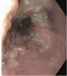
A 68-year-old male presented with complaints of heartburn and progressive dysphagia for solid food. His past medical history was remarkable for coronary artery disease with coronary stent placement in 2008 and 2009 and gastroesophageal reflux disease (GERD). His family history was significant for esophageal cancer in brother diagnosed at the age of 53 years. Physical examination was significant for epigastric tenderness but the rest of the examination was otherwise unremarkable. Labs were significant for iron deficiency anemia: hemoglobulin 10.3 gm/dL, hematocrit 31.1%, MCV 87%, iron 24 mcg/dL, transferrin saturation 7 mcg/dL. A computed tomography (CT) scan of abdomen and pelvis with contrast performed to evaluate iron deficiency anemia showed marked circumferential thickening of the visualized portion of the distal esophagus extending to the gastroesophageal GE junction, which was suspicious for esophageal cancer. There was an enlarged lymph node of the gastro-hepatic ligament suspicious for metastasis. Upper endoscopy was performed which showed a large, fungating and ulcerating mass with no bleeding in the mid and distal esophagus. The mass was partially obstructing and circumferential. It extended from mid esophagus to GE junction (Figure 1). Biopsies were taken with cold forceps for histology, which showed poorly differentiated adenocarcinoma with signet-ring cell type with background of Barrett’s esophagus with high-grade dysplasia and positive Her2/neu overexpression by immunohistochemistry stain.

Figure 1. Esophageal Tumor

Esophageal Tumor

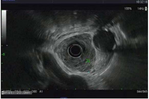
Endoscopic ultrasound showed hypoechoic mass in the middle third of the esophagus and in the lower third of the esophagus. The mass was encountered at 30 cm from the incisors and extended to 40 cm. The lesion was circumferential. The endosonographic borders were poorly defined. There was sonographic evidence suggesting invasion into the adventitia. (Layer 5) Two malignant-appearing lymph nodes were visualized in the lower para-esophageal mediastinum (level 8L). The largest measured was 8 mm by 7 mm, in maximal cross-sectional diameter. The nodes were round, hypoechoic and had well-defined margins. A diagnosis of T3N1MX (stage IIIa) esophageal adenocarcinoma was made (Figure 2).

Figure 2. EUS Examination Showing Tumor Infiltration into Adventitia

EUS Examination Showing Tumor Infiltration into Adventitia

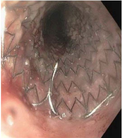
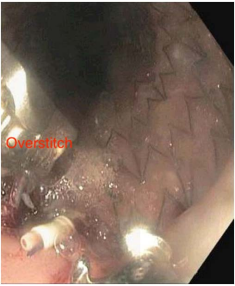
He then underwent upper endoscopy for stent placement for palliation of symptoms of dysphagia from his esophageal adenocarcinoma. The large, fungating and ulcerating esophageal mass was stented with a 23 mm × 150 mm EndoMaxx FCSEMS (Figure 3). The proximal end of the stent was at 28 cm. The stent was then secured with two sutures by an overstitch to prevent migration (Figure 4). There was no immediate complication like post procedure pain, bleeding or perforation from securing the stent with the overstitch.

Figure 3. Esophageal Stent Traversing the Esophageal Tumor

 Esophageal Stent Traversing the Esophageal Tumor

Figure 4. Overstitch used to Secure the Esophageal Stent

Overstitch used to Secure the Esophageal Stent

He was treated with chemotherapy and radiation therapy by oncology for 5 weeks with plans for surgical resection following chemo-radiation. He underwent repeat esophagogastroduodenoscopy (EGD) 8 weeks later when the overstitch was removed by cutting with an endo scissor and the stent was successfully removed.

DISCUSSION

Esophageal stents are frequently placed to palliate symptoms in patients with malignant esophageal strictures.2,3 Esophageal stents can be used to improve symptomatic esophageal lesions from benign esophageal strictures, perforations, fistulae, and leaks.4-7 However, esophageal stents have been associated with various complications including stent migration, which can happen in 40%-60% of patients who undergo this procedure.1 Other complications are severe chest pain, bleeding, fistula formation, ulceration, tissue embedding, and reflux.8,9

Uncovered and partially covered self-expanding metal stents are secure and resist migration due to tissue in-growth. They do not need to be secured with overstitch as they rarely migrate. However, they are permanent stents and will come in the way of future surgery. Also, they are at risk of future stent occlusion due to tumor in-growth and can cause fistula. Therefore, the patients are subjected to additional invasive procedures due to complications from permanent esophageal self-expanding metal stent.

The FCSEMS are temporary stents and rely on radial friction against the esophageal mucosa to hold the stent in place. The nitinol coating is designed to resist tissue ingrowth and this coating allows for the metal stent to be removed after the several weeks of stent placement.3,4 Stent migration represents the most common adverse event of the FCSEMS with migration rates between 30% and 60%.1,9 Techniques to prevent stent migration using endoscopic clips have been described but are ineffective and associated with a high rate of complications.10-13 Esophageal stent fixation by endoscopic suturing with an overstitch is a novel technique that prevents esophageal stent migration in an ex vivo setting.14 Few studies have reported successful use of Overstitch for fixation of FCSEMS to prevent spontaneous migration of esophageal stents. The reported migration rate after securing the stent with overstitch is 8-16%.15-18 The migration despite Overstitch is due to esophageal peristalsis and mostly seen in non-malignant indications for the stent where there is no stricture to keep the stent in place.

OverStitch (Apollo Endosurgery) is the most widely used endoscopic suturing device. The device assembly and use are straightforward and closely resemble surgical suturing. Our patient never experienced peri-procedural complications during the procedure of stent fixation by the overstitch. There was no evidence of procedure-related pain, bleed or perforation. Also, post procedure, there was no evidence of dysphagia or migration during his treatment with chemo-radiation when the risk for migration is high due to tumor shrinkage and stricture resolution. The overstitch is easy to remove along with the stent as was demonstrated in our case. Endoscopic suturing can effectively prevent spontaneous migration of esophageal stents. Although, there is a learning curve for the endoscopists to master the technique of endoscopic suturing with the Overstitch device, this should be the standard of care for placement of esophageal stents.

CONFLICTS OF INTEREST

The authors declare that they have no conflicts of interest.

1. Sharaiha RZ, Kumta NA, Doukides TP, et al. Esophageal stenting with sutures: It’s time to redefine our standards. J Clin Gastroenterol. 2015; 49(6): e57-e60. doi: 10.1097/MCG.0000000000000198

2. Schembre D. Advances in esophageal stenting: The evolution of fully covered stents for malignant and benign disease. Adv Ther. 2010; 27: 413-425. doi: 10.1007/s12325-010-0042-5

3. Langer FB, Schoppmann SF, Prager G, et al. Temporary placement of self-expanding oesophageal stents as bridging for neo-adjuvant therapy. Ann Surg Oncol. 2010; 17: 470-475. doi: 10.1245/s10434-009-0760-6

4. Eloubeidi MA, Lopes TL. Novel removable internally fully covered self-expanding metal esophageal stent: Feasibility, technique of removal, and tissue response in humans. Am J Gastroenterol. 2009; 104: 1374-1381. doi: 10.1038/ajg.2009.133

5. Holm AN, de la Mora Levy JG, Gostout CJ, Topazian MD, Baron TH. Selfexpanding plastic stents in treatment of benign esophageal conditions. Gastrointest Endosc. 2008; 67: 20-25. doi: 10.1016/j.gie.2007.04.031

6. Kim JH, Shin JH, Song HY. Benign strictures of the esophagus and gastric outlet: Interventional management. Korean J Radiol. 2010; 11: 497-506. doi: 10.3348/kjr.2010.11.5.497

7. Swinnen J, Eisendrath P, Rigaux J, et al. Self-expandable metal stents for the treatment of benign upper GI leaks and perforations. Gastrointest Endosc. 2011; 73: 890-899. doi: 10.1016/j.gie.2010.12.019

8. Baron TH. Minimizing endoscopic complications: Endoluminal stents. Gastrointest Endosc Clin N Am. 2007; 17: 83-104. doi: 10.1016/j.giec.2007.01.004

9. Saligram S. Lim D, Pena L, Friedman M, Harris C, Klapman J. Safety and feasibility of esophageal self- expandable metal stent placement without the aid of fluoroscopy. Dis Esophagus. 2017; 30(8): 1-6. doi: 10.1093/dote/dox030

10. Manes G, Corsi F, Pallotta S, Massari A, Foschi D, Trabucchi E. Fixation of a covered self expandable metal stent by means of a polypectomy snare: An easy method to prevent stent migration. Dig Liver Dis. 2008; 40: 791-793. doi: 10.1016/j.dld.2007.10.020

11. Silva RA, Dinis-Ribeiro M, Brandao C, et al. Should we consider endoscopic clipping for prevention of esophageal stent migration? Endoscopy. 2004; 36: 369-370. doi: 10.1055/s-0043-111793

12. Sriram PV, Das G, Rao GV, Reddy DN. Another novel use of endoscopic clipping: To anchor an esophageal endoprosthesis. Endoscopy. 2001; 33: 724-726. doi: 10.1055/s-2001-16207

13. Vanbiervliet G, Filippi J, Karimdjee BS, et al. The role of clips in preventing migration of fully covered metallic esophageal stents: A pilot comparative study. Surg Endosc. 2012; 26: 53-59. doi: 10.1007/s00464-011-1827-6

14. Rieder E, Dunst CM, Martinec DV, et al. Endoscopic suture fixation of gastrointestinal stents: Proof of biomechanical principles and early clinical experience. Endoscopy. 2012; 44: 1121-1126. doi: 10.1055/s-0032-1325730

15. Kantsevoy SV, Bitner M. Esophageal stent fixation with endoscopic suturing device (with video). Gastrointest Endosc. 2012; 76: 1251-1255. doi: 10.1016/j.gie.2012.08.003

16. Sharaiha RZ, Kumta NA, Doukides TP, et al. Esophageal stenting with sutures: Time to redefine our standards? J Clin Gastroenterol.2015; 49(6): e57-e60. doi: 10.1097/MCG.0000000000000198

17. Ngamruengphong S, Sharaiha RZ, Sethi A, et al. Endoscopic suturing for the prevention of stent migration in benign upper gastrointestinal conditions: A comparative multicenter study. Endoscopy. 2016; 48(9): 802-808. doi: 10.1055/s-0042-108567

18. Yang J, Siddiqui AA, Kowalski TE, et al. Esophageal stent fixation with endoscopic suturing device improves clinical outcomes and reduces complications in patients with locally advanced esophageal cancer prior to neoadjuvant therapy: A large multicenter experience. Surg Endosc. 2017; 31(3): 1414-1419. doi: 10.1007/s00464-016-5131-3

LATEST ARTICLES

Unraveling the Mysteries of Type-A Aortic Dissection Using POCUS/Echocardiography

Syeda Rukh*, Sathyanarayana Machani and Milind Awale

doi.

Blood Sample from the Patient

Hypertriglyceridemia-Induced Pancreatitis: A Case Report and Literature Review

Maarten Bulterys, Melvin Willems* and Agnes Meersman

doi.

From Neck Pain to a Life-Threatening Condition: A Case Report

Floris Vandewoude* and Sören Verstraete

doi.

LATEST ARTICLES